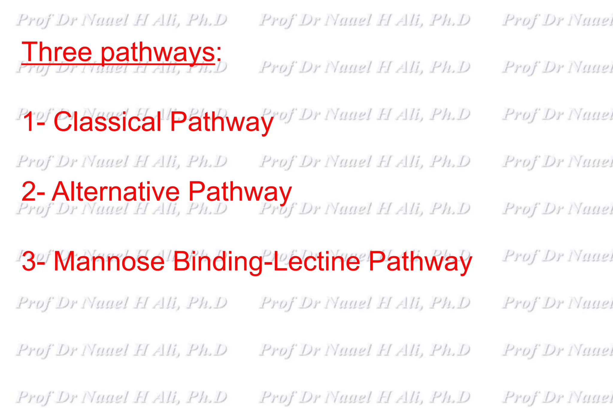
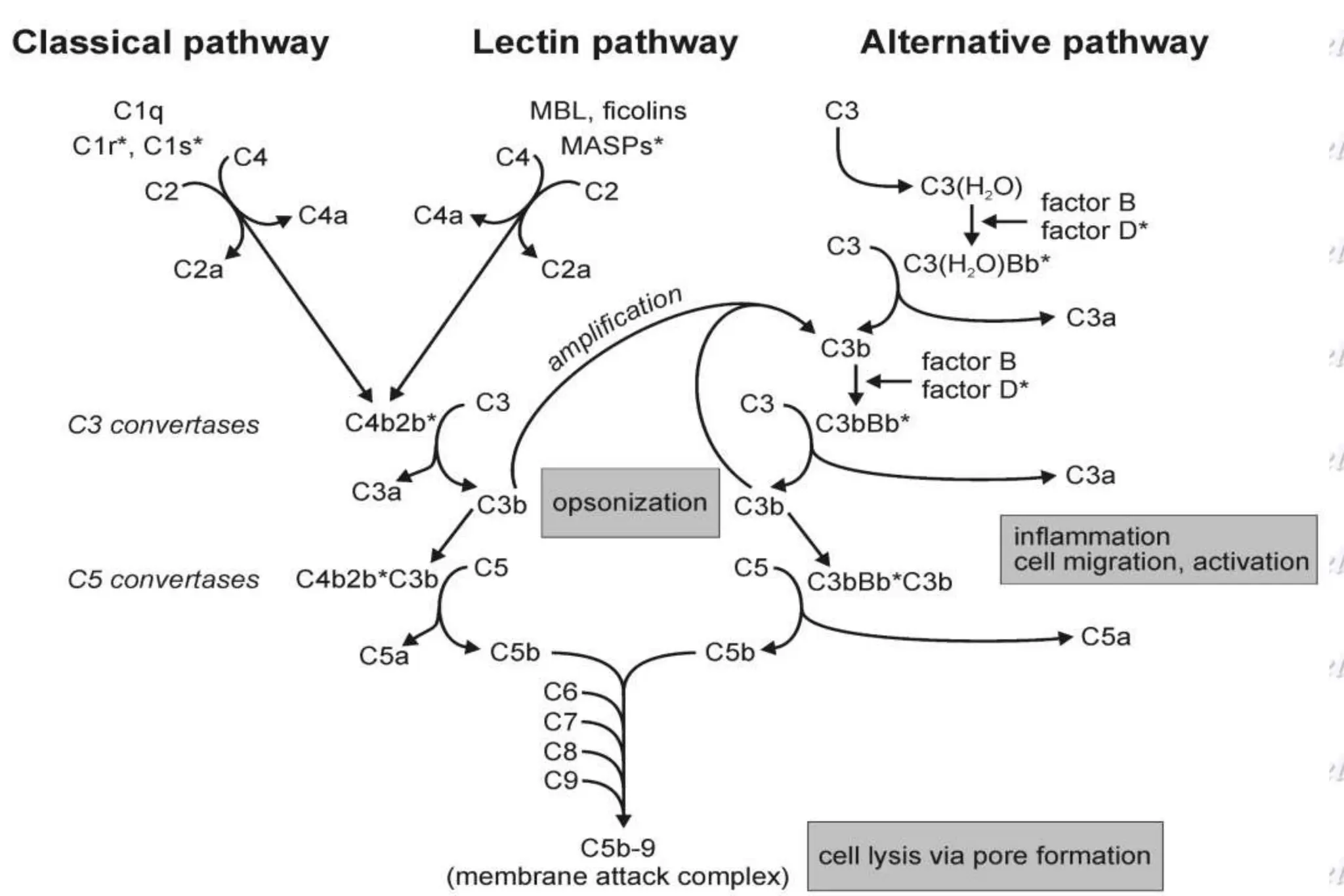
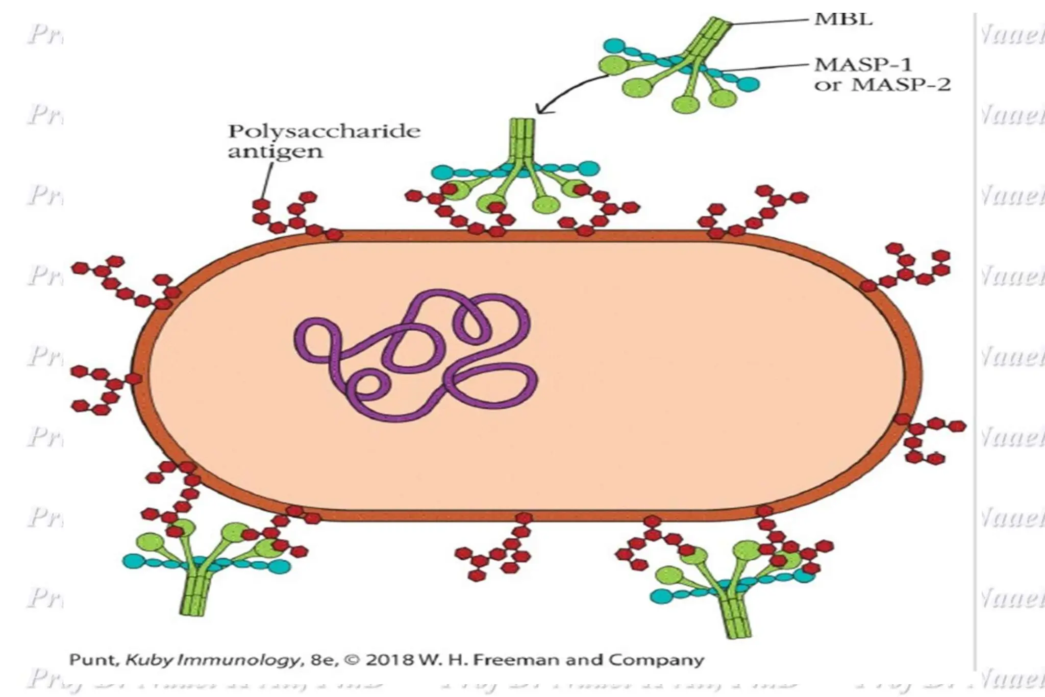
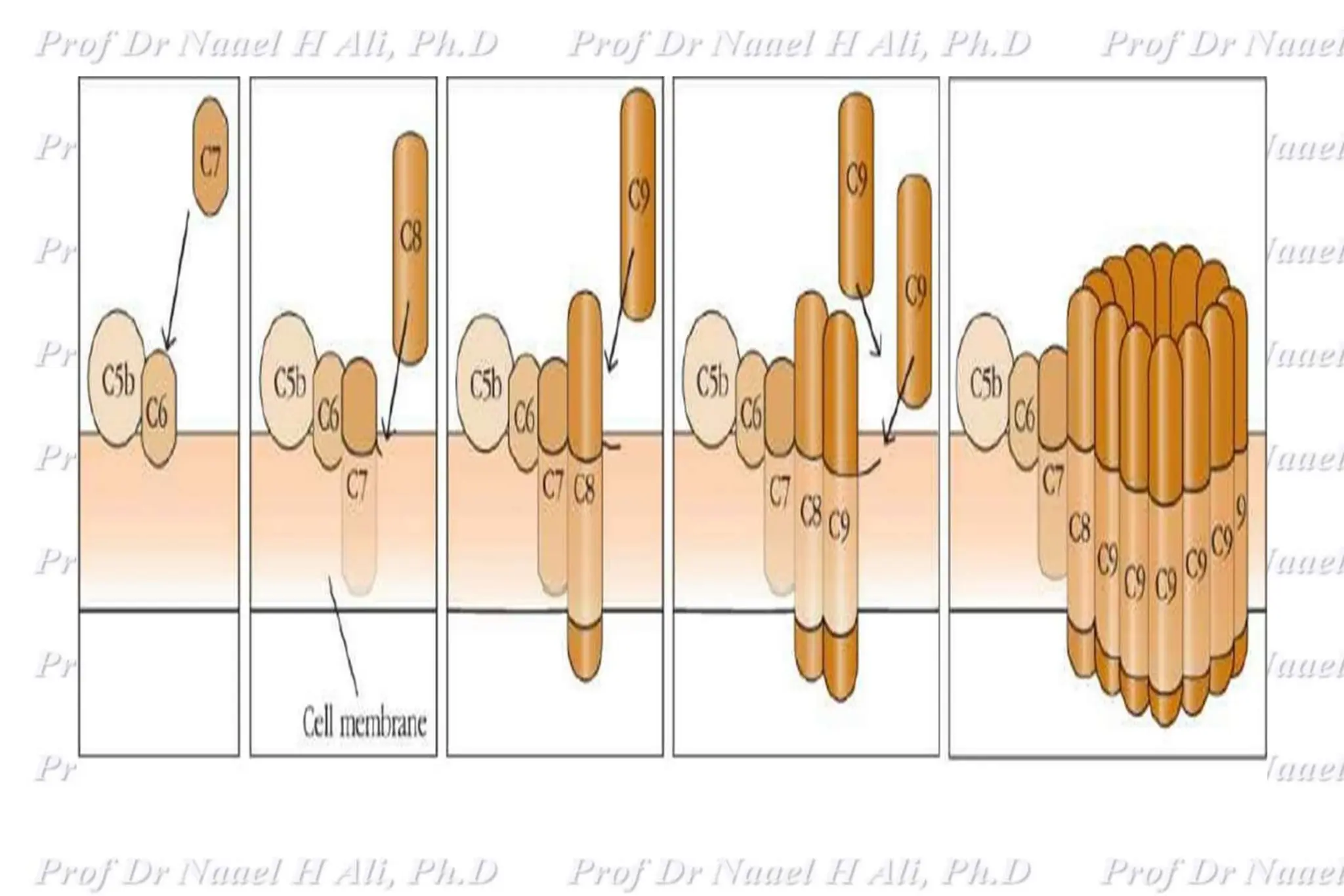
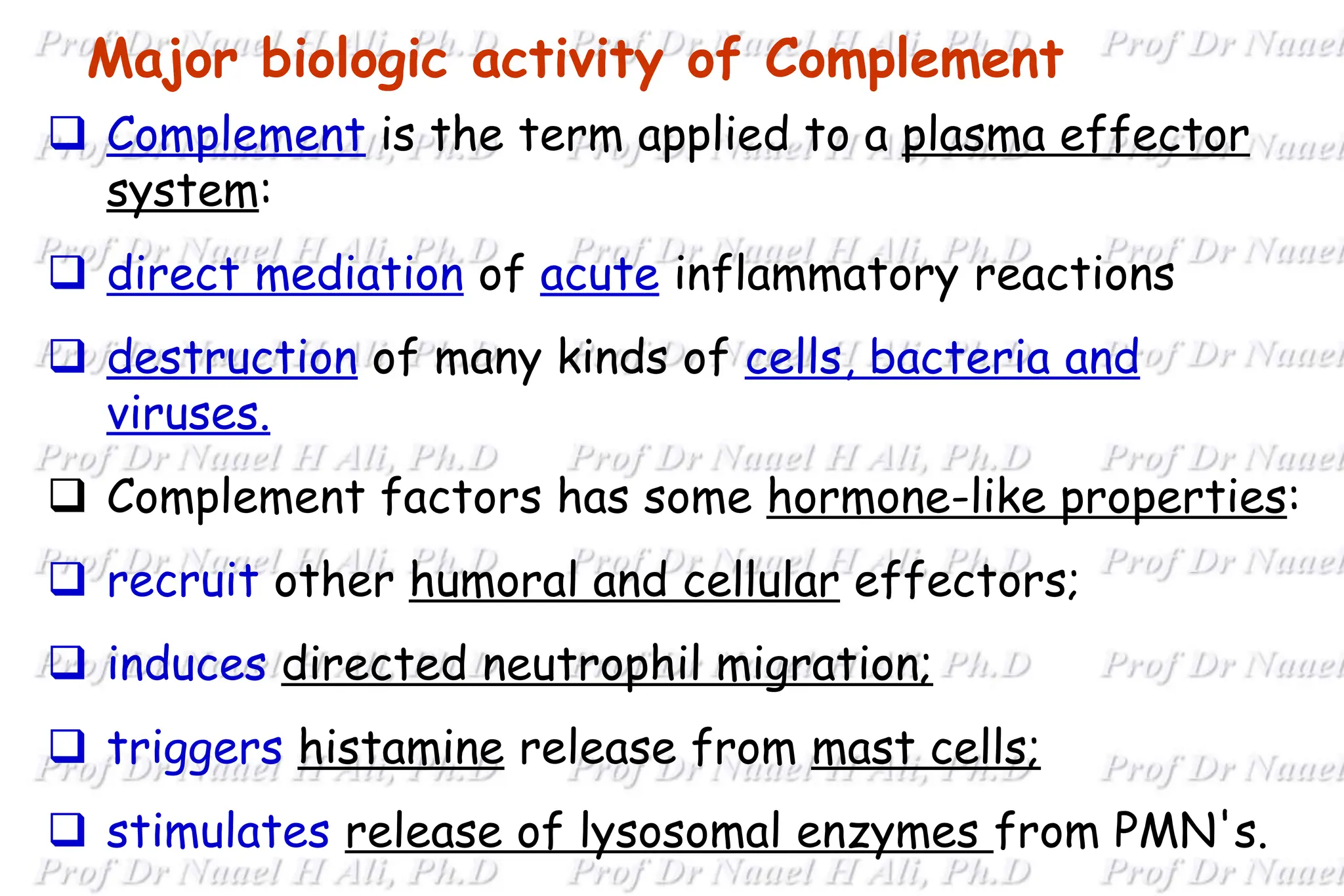
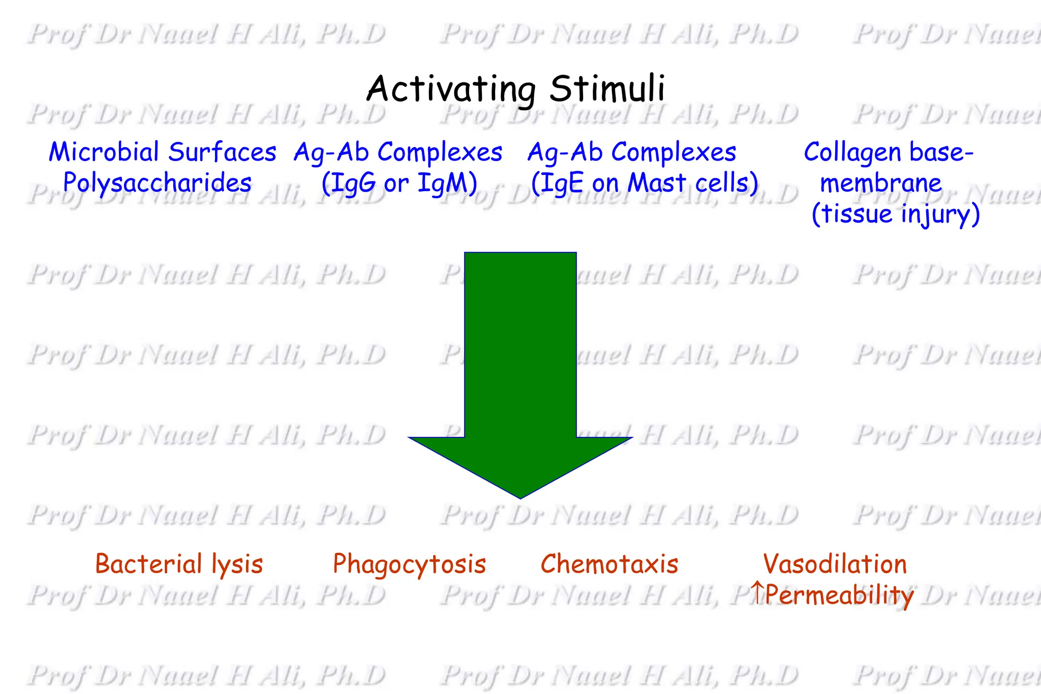
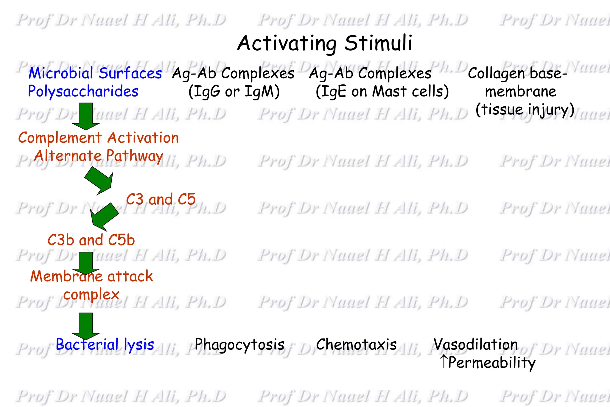
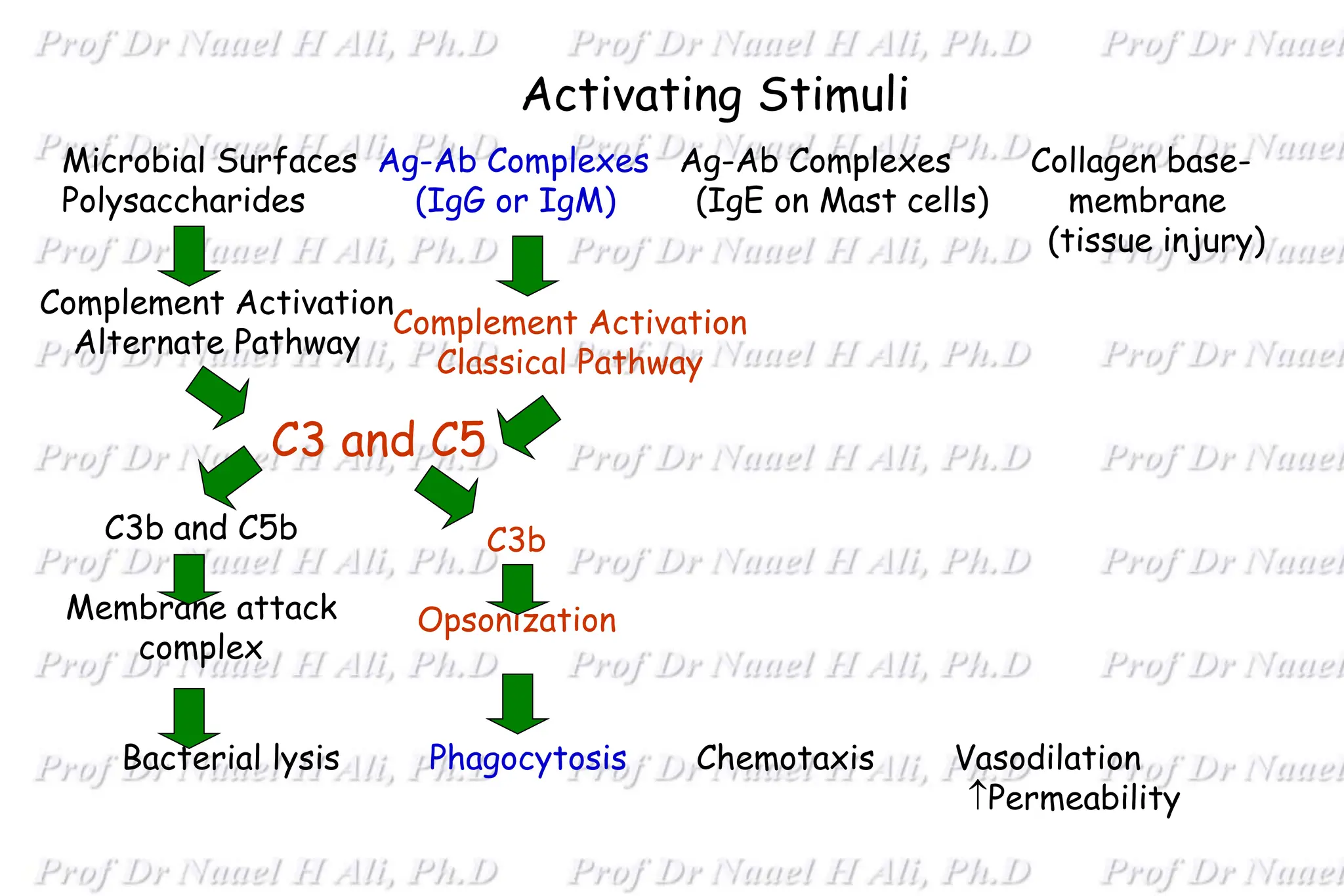
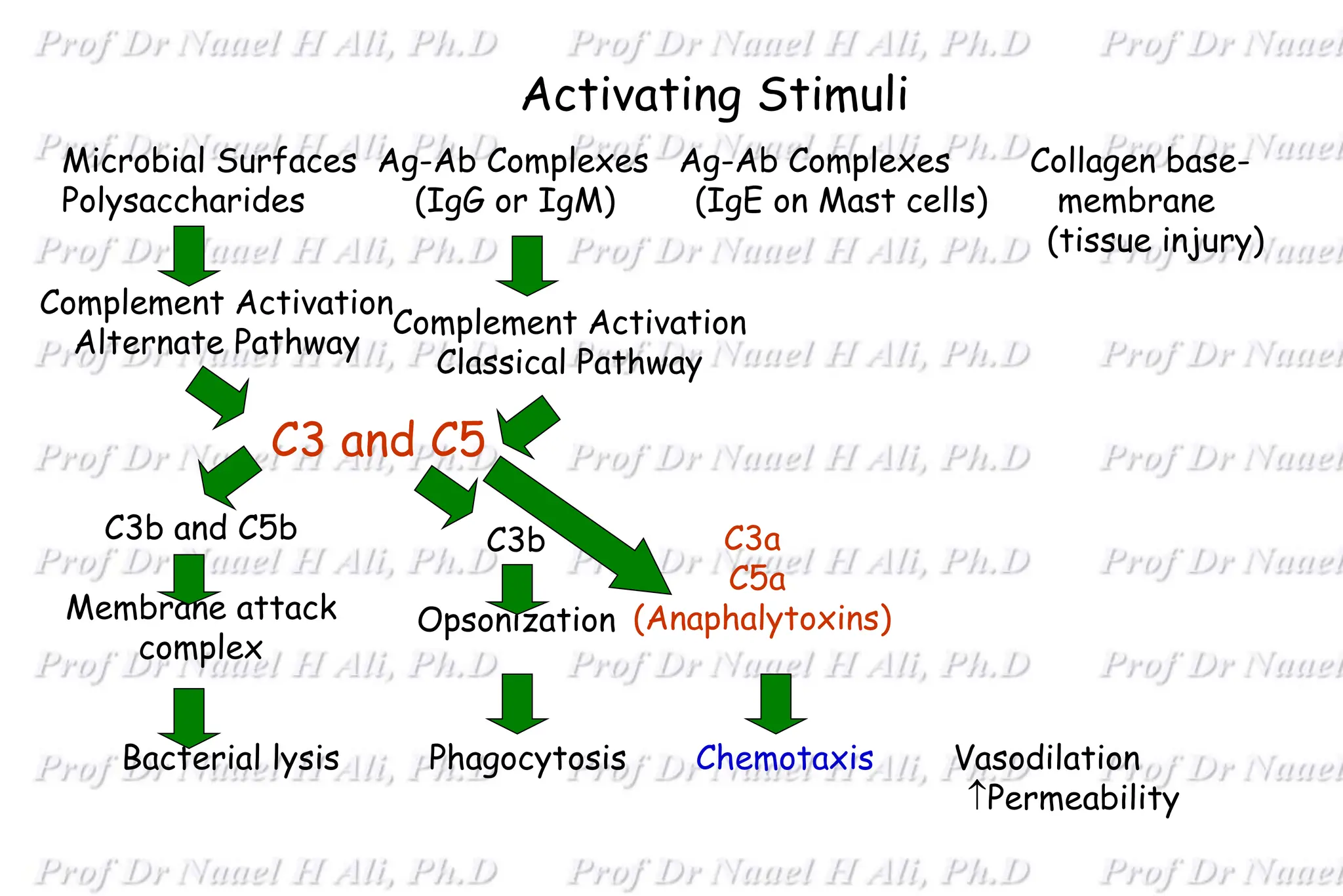
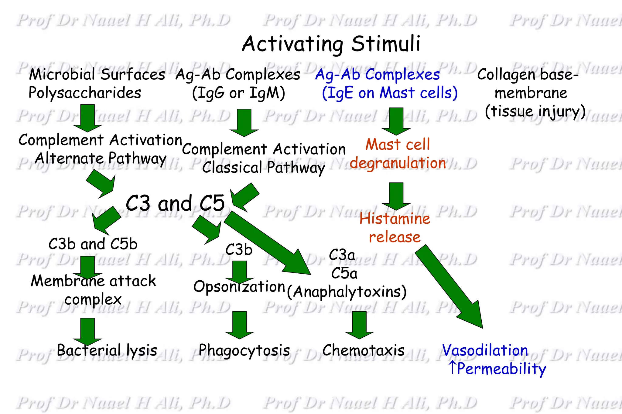
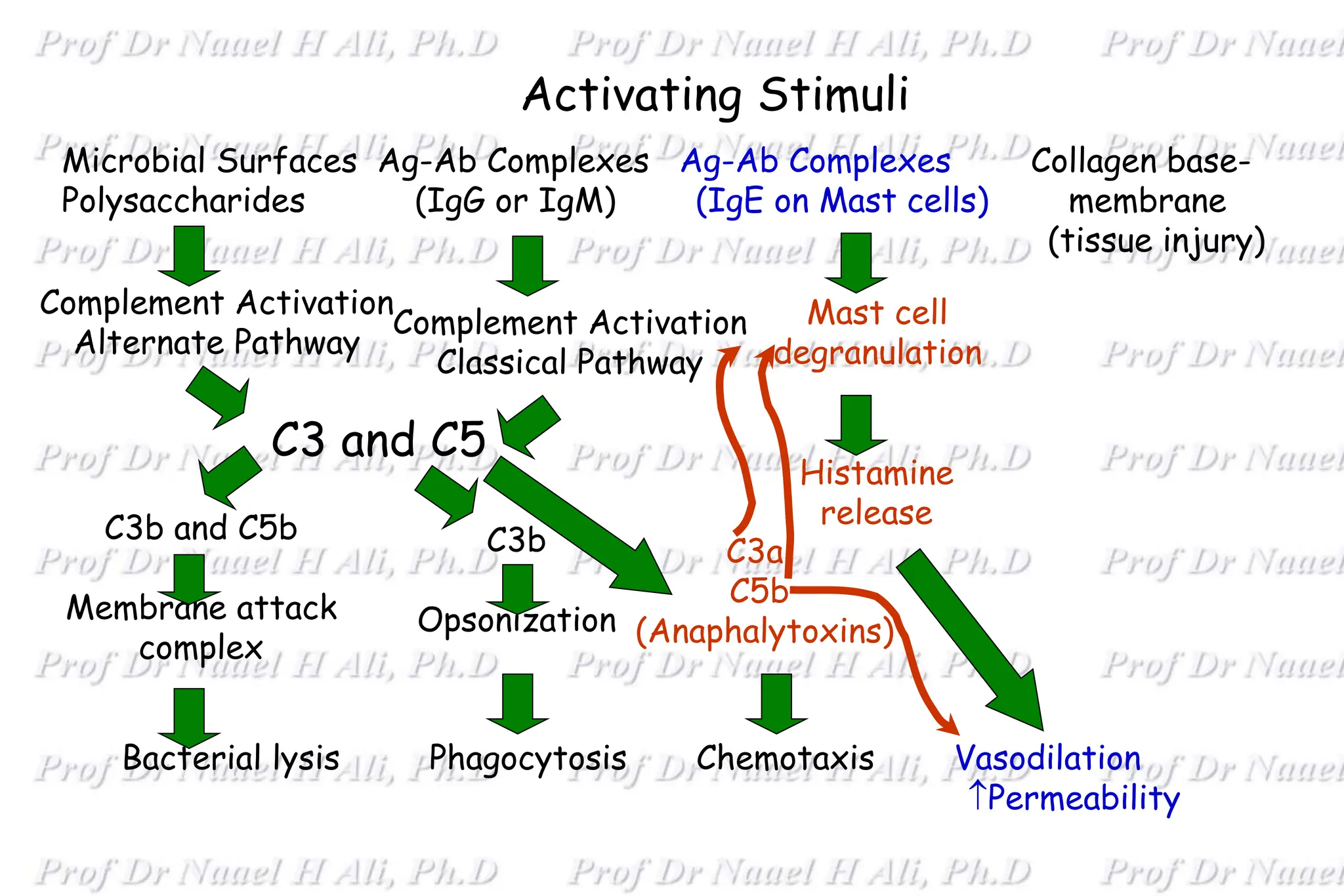
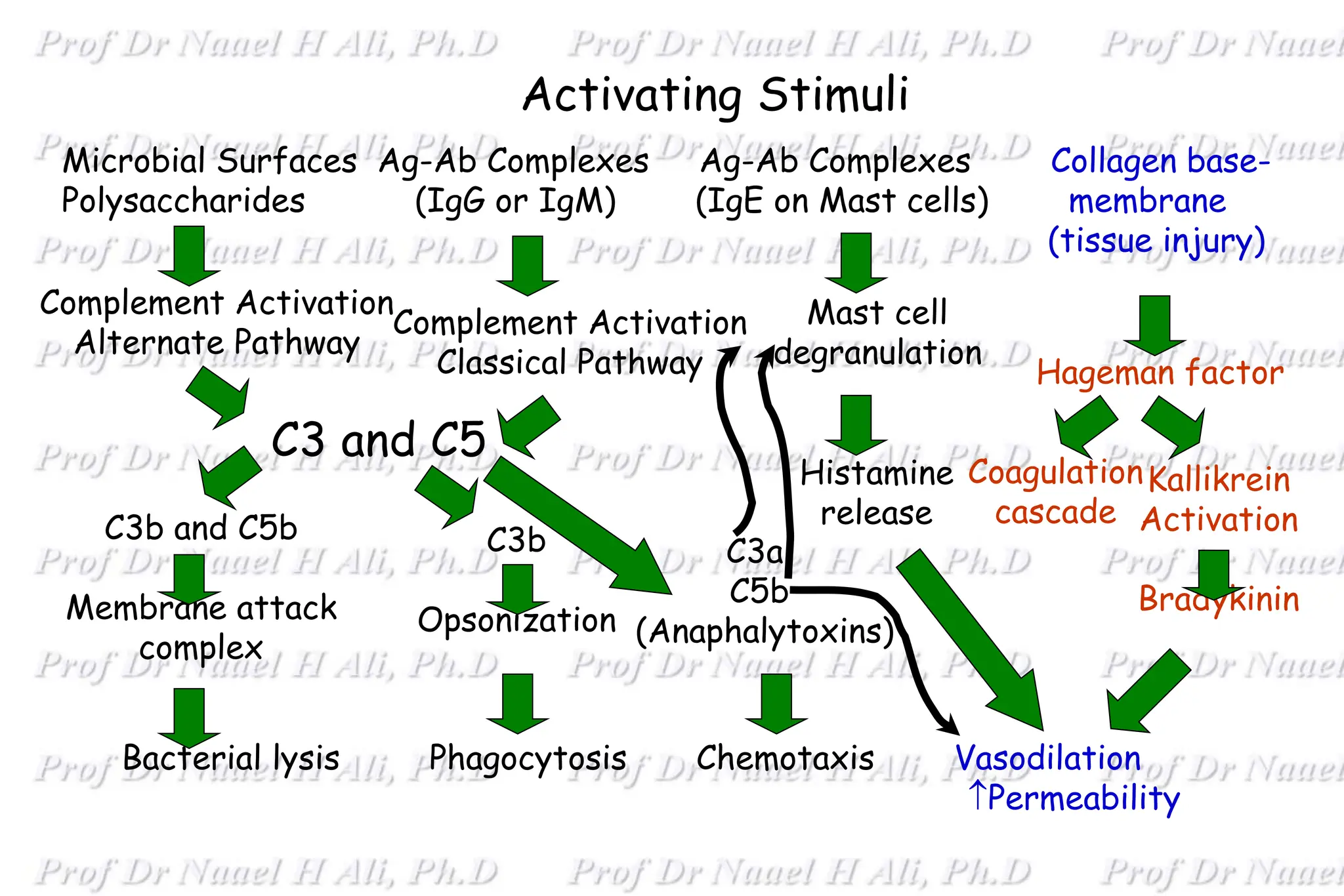
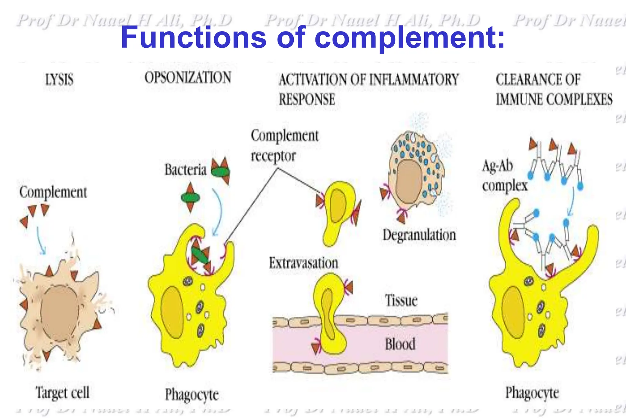
COMPLEMENT SYSTEM
Prof Dr. Naael H Ali, Ph.D
Clinical Immunology
Dep. of Microbiology
College of Medicine
University of Basrah
Introduction
The term complement refers to a set of 50-plus serum proteins
that cooperates with both the innate and the adaptive immune
systems to eliminate pathogens, dying cells, and immune
complexes from the body .
It is named “complement system” because it was first identified
as a heat-labile component of serum that “complemented”
antibodies in the killing of bacteria.
 Normally, these proteins are in an inactive form
(proenzyme), but specific signal can activate the first
protein
Component of Complement system
 Over 50 serum and cell surface proteins:
Complement components
(in serum inactive, activated sequentially as a cascade)
 Complement receptors
(cell surface, recognize activated components)
 Regulatory proteins of complement
(both in serum and cell surface, inhibit activated
components)
Properties of Complements
 Source of complement proteins produced by hepatocytes,
macrophages and intestinal epithelial cells.
 Fibroblasts and intestinal epithelial cells make C1, while the liver
makes C3, C6, and C9.
 Heating at 56 c for 30 minutes can inactivate complements.
 Complete deterioration within a day or two and even in the
refrigerator, considerable loss of activity occurs in 3-4 days.
 Complement may be destroyed by acids and alkalis,
So, all glassware must be chemically clean, although not
necessarily sterile.
 Complements may be preserved by freezing and lyophilization.
 Serum complement levels, especially C3, often drop during
infection (complement is activated faster than it is produced).
Three pathways:
1- Classical Pathway
2- Alternative Pathway
3- Mannose Binding-Lectine Pathway
C1 qrs
Major biologic activity of Complement
 Complement is the term applied to a plasma effector
system:
 direct mediation of acute inflammatory reactions
 destruction of many kinds of cells, bacteria and
viruses.
 Complement factors has some hormone-like properties:
 recruit other humoral and cellular effectors;
 induces directed neutrophil migration;
 triggers histamine release from mast cells;
 stimulates release of lysosomal enzymes from PMN's.
Activating Stimuli
Bacterial lysis Phagocytosis Chemotaxis Vasodilation
Permeability
Microbial Surfaces Ag-Ab Complexes Ag-Ab Complexes Collagen base-
Polysaccharides (IgG or IgM) (IgE on Mast cells) membrane
(tissue injury)
Activating Stimuli
Bacterial lysis Phagocytosis Chemotaxis Vasodilation
Permeability
Microbial Surfaces Ag-Ab Complexes Ag-Ab Complexes Collagen base-
Polysaccharides (IgG or IgM) (IgE on Mast cells) membrane
(tissue injury)
Complement Activation
Alternate Pathway
C3 and C5
C3b and C5b
Membrane attack
complex
Activating Stimuli
Bacterial lysis Phagocytosis Chemotaxis Vasodilation
Permeability
Microbial Surfaces Ag-Ab Complexes Ag-Ab Complexes Collagen base-
Polysaccharides (IgG or IgM) (IgE on Mast cells) membrane
(tissue injury)
Complement Activation
Alternate Pathway
C3b and C5b
Membrane attack
complex
C3 and C5
Complement Activation
Classical Pathway
C3b
Opsonization
C3a
C5a
(Anaphalytoxins)
Mast cell
degranulation
Histamine
release
Activating Stimuli
Bacterial lysis Phagocytosis Chemotaxis Vasodilation
Permeability
Microbial Surfaces Ag-Ab Complexes Ag-Ab Complexes Collagen base-
Polysaccharides (IgG or IgM) (IgE on Mast cells) membrane
(tissue injury)
Complement Activation
Alternate Pathway
C3b and C5b
Membrane attack
complex
C3 and C5
Complement Activation
Classical Pathway
C3b
Opsonization
C3a
C5b
(Anaphalytoxins)
Mast cell
degranulation
Histamine
release
Hageman factor
Coagulation
cascade
Kallikrein
Activation
Bradykinin
Functions of complement:
Diagnostic significant
Increased complement activity
may be seen in:
Cancer
Certain infections
Ulcerative colitis
Decreased complement activity
may be seen in:
Cirrhosis
Glomerulonephritis
Hereditary angioedema
Hepatitis
Kidney transplant rejection
Lupus nephritis
Malnutrition
Systemic lupus erythematosis
Regulation of complement system:
1- CI Inhibitor (C1-INH) Classical pathway .
2- C3a inactivator (C3a-INA; Carboxypeptidase B) It inactivates C3a.
3- Factor H & I: Alternative pathway
4- Decay accelerating factor [DAF] Both.
5- C4 binding protein (C4-BP) and Factor I Alternative .
6- Protein S (vitronectin): MAC pathway.
Complement system proteins and enzymes s

Complement system proteins and enzymes s

  • 1.
    COMPLEMENT SYSTEM Prof Dr.Naael H Ali, Ph.D Clinical Immunology Dep. of Microbiology College of Medicine University of Basrah
  • 2.
    Introduction The term complementrefers to a set of 50-plus serum proteins that cooperates with both the innate and the adaptive immune systems to eliminate pathogens, dying cells, and immune complexes from the body . It is named “complement system” because it was first identified as a heat-labile component of serum that “complemented” antibodies in the killing of bacteria.  Normally, these proteins are in an inactive form (proenzyme), but specific signal can activate the first protein
  • 3.
    Component of Complementsystem  Over 50 serum and cell surface proteins: Complement components (in serum inactive, activated sequentially as a cascade)  Complement receptors (cell surface, recognize activated components)  Regulatory proteins of complement (both in serum and cell surface, inhibit activated components)
  • 4.
    Properties of Complements Source of complement proteins produced by hepatocytes, macrophages and intestinal epithelial cells.  Fibroblasts and intestinal epithelial cells make C1, while the liver makes C3, C6, and C9.  Heating at 56 c for 30 minutes can inactivate complements.  Complete deterioration within a day or two and even in the refrigerator, considerable loss of activity occurs in 3-4 days.  Complement may be destroyed by acids and alkalis, So, all glassware must be chemically clean, although not necessarily sterile.  Complements may be preserved by freezing and lyophilization.  Serum complement levels, especially C3, often drop during infection (complement is activated faster than it is produced).
  • 5.
    Three pathways: 1- ClassicalPathway 2- Alternative Pathway 3- Mannose Binding-Lectine Pathway
  • 7.
  • 10.
    Major biologic activityof Complement  Complement is the term applied to a plasma effector system:  direct mediation of acute inflammatory reactions  destruction of many kinds of cells, bacteria and viruses.  Complement factors has some hormone-like properties:  recruit other humoral and cellular effectors;  induces directed neutrophil migration;  triggers histamine release from mast cells;  stimulates release of lysosomal enzymes from PMN's.
  • 11.
    Activating Stimuli Bacterial lysisPhagocytosis Chemotaxis Vasodilation Permeability Microbial Surfaces Ag-Ab Complexes Ag-Ab Complexes Collagen base- Polysaccharides (IgG or IgM) (IgE on Mast cells) membrane (tissue injury)
  • 12.
    Activating Stimuli Bacterial lysisPhagocytosis Chemotaxis Vasodilation Permeability Microbial Surfaces Ag-Ab Complexes Ag-Ab Complexes Collagen base- Polysaccharides (IgG or IgM) (IgE on Mast cells) membrane (tissue injury) Complement Activation Alternate Pathway C3 and C5 C3b and C5b Membrane attack complex
  • 13.
    Activating Stimuli Bacterial lysisPhagocytosis Chemotaxis Vasodilation Permeability Microbial Surfaces Ag-Ab Complexes Ag-Ab Complexes Collagen base- Polysaccharides (IgG or IgM) (IgE on Mast cells) membrane (tissue injury) Complement Activation Alternate Pathway C3b and C5b Membrane attack complex C3 and C5 Complement Activation Classical Pathway C3b Opsonization
  • 14.
    Activating Stimuli Bacterial lysisPhagocytosis Chemotaxis Vasodilation Permeability Microbial Surfaces Ag-Ab Complexes Ag-Ab Complexes Collagen base- Polysaccharides (IgG or IgM) (IgE on Mast cells) membrane (tissue injury) Complement Activation Alternate Pathway C3b and C5b Membrane attack complex C3 and C5 Complement Activation Classical Pathway C3b Opsonization C3a C5a (Anaphalytoxins)
  • 15.
    Activating Stimuli Bacterial lysisPhagocytosis Chemotaxis Vasodilation Permeability Microbial Surfaces Ag-Ab Complexes Ag-Ab Complexes Collagen base- Polysaccharides (IgG or IgM) (IgE on Mast cells) membrane (tissue injury) Complement Activation Alternate Pathway C3b and C5b Membrane attack complex C3 and C5 Complement Activation Classical Pathway C3b Opsonization C3a C5a (Anaphalytoxins) Mast cell degranulation Histamine release
  • 16.
    Activating Stimuli Bacterial lysisPhagocytosis Chemotaxis Vasodilation Permeability Microbial Surfaces Ag-Ab Complexes Ag-Ab Complexes Collagen base- Polysaccharides (IgG or IgM) (IgE on Mast cells) membrane (tissue injury) Complement Activation Alternate Pathway C3b and C5b Membrane attack complex C3 and C5 Complement Activation Classical Pathway C3b Opsonization C3a C5b (Anaphalytoxins) Mast cell degranulation Histamine release
  • 17.
    Activating Stimuli Bacterial lysisPhagocytosis Chemotaxis Vasodilation Permeability Microbial Surfaces Ag-Ab Complexes Ag-Ab Complexes Collagen base- Polysaccharides (IgG or IgM) (IgE on Mast cells) membrane (tissue injury) Complement Activation Alternate Pathway C3b and C5b Membrane attack complex C3 and C5 Complement Activation Classical Pathway C3b Opsonization C3a C5b (Anaphalytoxins) Mast cell degranulation Histamine release Hageman factor Coagulation cascade Kallikrein Activation Bradykinin
  • 18.
  • 19.
    Diagnostic significant Increased complementactivity may be seen in: Cancer Certain infections Ulcerative colitis Decreased complement activity may be seen in: Cirrhosis Glomerulonephritis Hereditary angioedema Hepatitis Kidney transplant rejection Lupus nephritis Malnutrition Systemic lupus erythematosis
  • 20.
    Regulation of complementsystem: 1- CI Inhibitor (C1-INH) Classical pathway . 2- C3a inactivator (C3a-INA; Carboxypeptidase B) It inactivates C3a. 3- Factor H & I: Alternative pathway 4- Decay accelerating factor [DAF] Both. 5- C4 binding protein (C4-BP) and Factor I Alternative . 6- Protein S (vitronectin): MAC pathway.