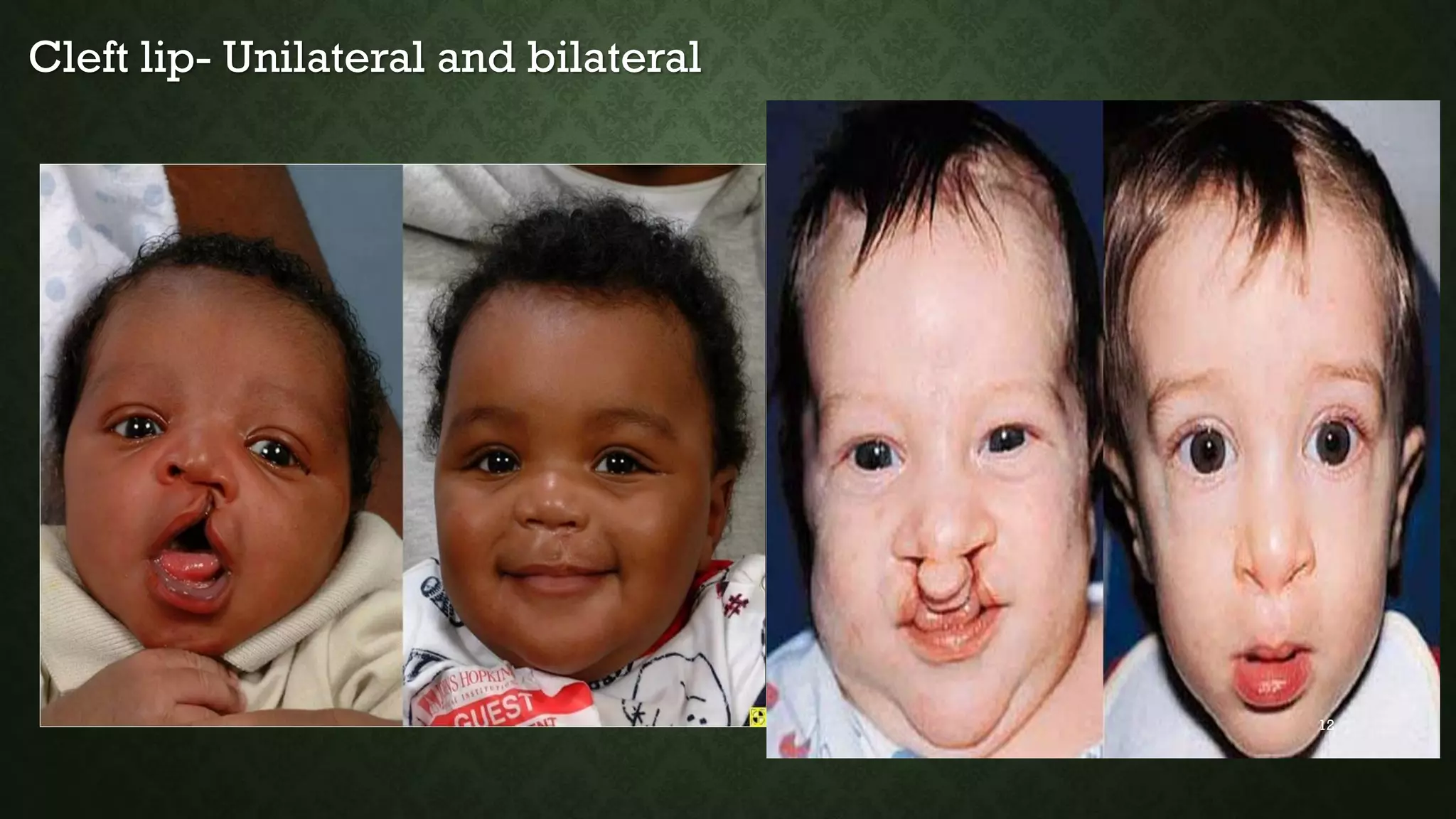
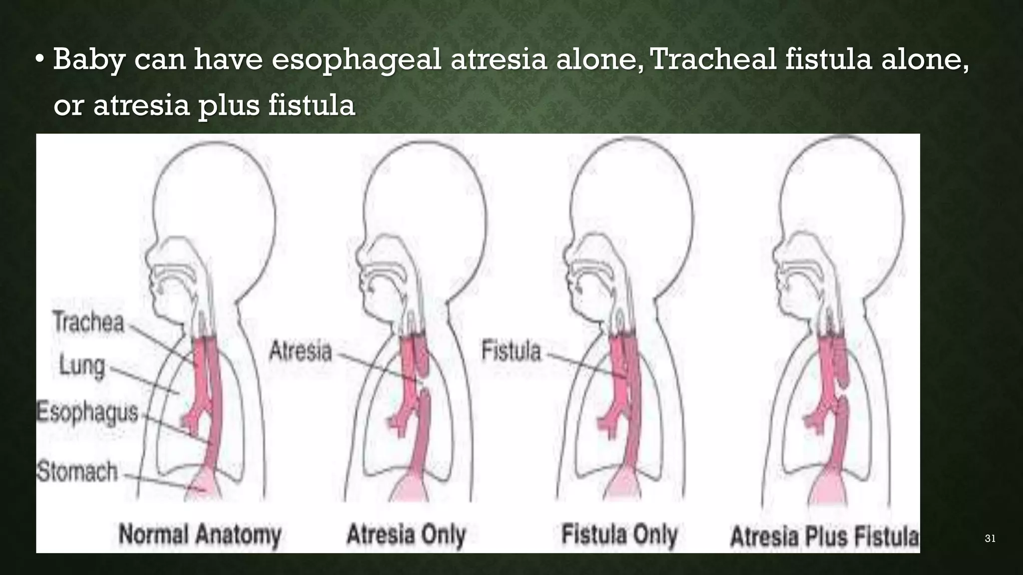
This document discusses common pediatric surgical conditions, including gastrointestinal congenital anomalies present at birth. It identifies anomalies of the mouth (cleft lip and palate), esophagus (esophageal atresia and tracheoesophageal fistula), stomach/duodenum (pyloric stenosis), intestines (imperforate anus, omphalocele, gastroschisis, Hirschsprung's disease, intussusception), and provides descriptions, epidemiology, presentations, diagnoses, and management for each. It also outlines pre and post-operative nursing care for infants with surgical congenital anomalies, focusing on risks like infection, breathing issues, tissue trauma, nutrition and pain management.
This presentation was done by Dr. Julius P. Kessy,MD. An intern Doctor at Dodoma Regional Referral Hospital (DRRH) during pediatrics unit clinical meeting and supervised by Dr. Christina K. Galabawa,MD,Mmed2, Pediatrics and Child Health, University of Dodoma (UDOM) in November, 2017.
Tetanus is explained in very simple wording and style by the help of a scenario. Easy to memorize and present due to related pictures. Helpful for medical students, and knowledge seekers.
cleft lip and palate are the most common type of congenital anomalies. the worldwide prevalence of cleft lip and cleft palate ranges from 0.8 to 2.7 cases per 1000 live births. cleft lip is called cheiloschisis and cleft plate is called palatoschisis. Cleft lip is a gap or indentation in the lip or split continued up to the nostril due to the failure of fusion of the maxillary and medial nasal process.
Cleft palate is the condition in which the two plates of the skull that forms hard palate are not completely joined due to the failure of fusion of the lateral palatine processes, nasal septum and medial palatine process. EMBRYOLOGYPrimary palate forms during the 4-7th week of gestation when two maxillary processes and two medial nasal processes fuse.
Secondary palate forms in 6-9th weeks of gestation when palatal shelves change from vertical to horizontal position and fuse. Tongue migrates Antero-inferiorly.
Cleft lip occurs when an epithelial bridge fails.
Clefts of primary palate occur anterior to incisive foramen and clefts of secondary palate occur posterior to the incisive foramen.
ETIOLOGY Genetic: Non-syndromic inheritance (risk increases with parents or siblings or both affected); chromosome aberrations, associated with other syndromes like Van der Woude syndrome.
Environmental teratogens: Intrauterine exposure to the anticonvulsant phenytoin, alcohol, retinoic acid, maternal smoking, Rubella virus, thalidomide, aminopterin.
Maternal/intrauterine condition: Maternal diabetes mellitus and amniotic band syndrome.
Advanced paternal age
Unknown
CLASSIFICATION Prof. Balakrishnan (1975) classified cleft lip and palate according to the Indian context and divided them into three groups.
Group 1: Only cleft lip, which may be unilateral (right/ left), bilateral, or midline.
Group 2: Only cleft palate, which may be which also can be unilateral (right/left), bilateral, or submucosa.
Group 3: Includes cleft lip, alveolus, and cleft palate, which can be unilateral, bilateral, or midline. LAHSAL system for the classification of cleft lip and/or palate (2005) modified by Royal College of Surgeons Britain: LAHSAL system is a diagrammatic classification of cleft lip and palate. According to this classification, the mouth is divided into six parts. LAHSAL code indicates a complete cleft with a capital letter and an incomplete cleft with a small letter.
CLINICAL FEATURES Cleft lip: Notched vermilion border and may involve alveolar ridge.
Cleft palate: Nasal distortion, exposed nasal cavities.
Misaligned teeth.
Passage of milk through nasal passages during feeding.
Recurrent ear infection.
Speech difficulties.
Poor weight gain and failure to thrive.
DIAGNOSIS Newborn examination at birth
Palpate with a gloved finger or visual examination flashlight
In-utero ultrasonography
PROBLEMS OR COMPLICATIONS OF A CHILD WITH CLEFT LIP AND CLEFT PALATE Immediate Problems:
Feeding difficulty:
Infant with an unrepaired cleft palate will have ...
Management of Orofacial Cleft Dr. Sunil (2).pptx Management of Orofacial Clef...ssuser12303b
Management of Orofacial Cleft Dr. Sunil (2).pptxManagement of Orofacial Cleft Dr. Sunil (2).pptxManagement of Orofacial Cleft Dr. Sunil (2).pptxManagement of Orofacial Cleft Dr. Sunil (2).pptx
The prostate is an exocrine gland of the male mammalian reproductive system
It is a walnut-sized gland that forms part of the male reproductive system and is located in front of the rectum and just below the urinary bladder
Function is to store and secrete a clear, slightly alkaline fluid that constitutes 10-30% of the volume of the seminal fluid that along with the spermatozoa, constitutes semen
A healthy human prostate measures (4cm-vertical, by 3cm-horizontal, 2cm ant-post ).
It surrounds the urethra just below the urinary bladder. It has anterior, median, posterior and two lateral lobes
It’s work is regulated by androgens which are responsible for male sex characteristics
Generalised disease of the prostate due to hormonal derangement which leads to non malignant enlargement of the gland (increase in the number of epithelial cells and stromal tissue)to cause compression of the urethra leading to symptoms (LUTS
Flu Vaccine Alert in Bangalore Karnatakaaddon Scans
As flu season approaches, health officials in Bangalore, Karnataka, are urging residents to get their flu vaccinations. The seasonal flu, while common, can lead to severe health complications, particularly for vulnerable populations such as young children, the elderly, and those with underlying health conditions.
Dr. Vidisha Kumari, a leading epidemiologist in Bangalore, emphasizes the importance of getting vaccinated. "The flu vaccine is our best defense against the influenza virus. It not only protects individuals but also helps prevent the spread of the virus in our communities," he says.
This year, the flu season is expected to coincide with a potential increase in other respiratory illnesses. The Karnataka Health Department has launched an awareness campaign highlighting the significance of flu vaccinations. They have set up multiple vaccination centers across Bangalore, making it convenient for residents to receive their shots.
To encourage widespread vaccination, the government is also collaborating with local schools, workplaces, and community centers to facilitate vaccination drives. Special attention is being given to ensuring that the vaccine is accessible to all, including marginalized communities who may have limited access to healthcare.
Residents are reminded that the flu vaccine is safe and effective. Common side effects are mild and may include soreness at the injection site, mild fever, or muscle aches. These side effects are generally short-lived and far less severe than the flu itself.
Healthcare providers are also stressing the importance of continuing COVID-19 precautions. Wearing masks, practicing good hand hygiene, and maintaining social distancing are still crucial, especially in crowded places.
Protect yourself and your loved ones by getting vaccinated. Together, we can help keep Bangalore healthy and safe this flu season. For more information on vaccination centers and schedules, residents can visit the Karnataka Health Department’s official website or follow their social media pages.
Stay informed, stay safe, and get your flu shot today!
These simplified slides by Dr. Sidra Arshad present an overview of the non-respiratory functions of the respiratory tract.
Learning objectives:
1. Enlist the non-respiratory functions of the respiratory tract
2. Briefly explain how these functions are carried out
3. Discuss the significance of dead space
4. Differentiate between minute ventilation and alveolar ventilation
5. Describe the cough and sneeze reflexes
Study Resources:
1. Chapter 39, Guyton and Hall Textbook of Medical Physiology, 14th edition
2. Chapter 34, Ganong’s Review of Medical Physiology, 26th edition
3. Chapter 17, Human Physiology by Lauralee Sherwood, 9th edition
4. Non-respiratory functions of the lungs https://academic.oup.com/bjaed/article/13/3/98/278874
NVBDCP.pptx Nation vector borne disease control programSapna Thakur
NVBDCP was launched in 2003-2004 . Vector-Borne Disease: Disease that results from an infection transmitted to humans and other animals by blood-feeding arthropods, such as mosquitoes, ticks, and fleas. Examples of vector-borne diseases include Dengue fever, West Nile Virus, Lyme disease, and malaria.
Lung Cancer: Artificial Intelligence, Synergetics, Complex System Analysis, S...Oleg Kshivets
RESULTS: Overall life span (LS) was 2252.1±1742.5 days and cumulative 5-year survival (5YS) reached 73.2%, 10 years – 64.8%, 20 years – 42.5%. 513 LCP lived more than 5 years (LS=3124.6±1525.6 days), 148 LCP – more than 10 years (LS=5054.4±1504.1 days).199 LCP died because of LC (LS=562.7±374.5 days). 5YS of LCP after bi/lobectomies was significantly superior in comparison with LCP after pneumonectomies (78.1% vs.63.7%, P=0.00001 by log-rank test). AT significantly improved 5YS (66.3% vs. 34.8%) (P=0.00000 by log-rank test) only for LCP with N1-2. Cox modeling displayed that 5YS of LCP significantly depended on: phase transition (PT) early-invasive LC in terms of synergetics, PT N0—N12, cell ratio factors (ratio between cancer cells- CC and blood cells subpopulations), G1-3, histology, glucose, AT, blood cell circuit, prothrombin index, heparin tolerance, recalcification time (P=0.000-0.038). Neural networks, genetic algorithm selection and bootstrap simulation revealed relationships between 5YS and PT early-invasive LC (rank=1), PT N0—N12 (rank=2), thrombocytes/CC (3), erythrocytes/CC (4), eosinophils/CC (5), healthy cells/CC (6), lymphocytes/CC (7), segmented neutrophils/CC (8), stick neutrophils/CC (9), monocytes/CC (10); leucocytes/CC (11). Correct prediction of 5YS was 100% by neural networks computing (area under ROC curve=1.0; error=0.0).
CONCLUSIONS: 5YS of LCP after radical procedures significantly depended on: 1) PT early-invasive cancer; 2) PT N0--N12; 3) cell ratio factors; 4) blood cell circuit; 5) biochemical factors; 6) hemostasis system; 7) AT; 8) LC characteristics; 9) LC cell dynamics; 10) surgery type: lobectomy/pneumonectomy; 11) anthropometric data. Optimal diagnosis and treatment strategies for LC are: 1) screening and early detection of LC; 2) availability of experienced thoracic surgeons because of complexity of radical procedures; 3) aggressive en block surgery and adequate lymph node dissection for completeness; 4) precise prediction; 5) adjuvant chemoimmunoradiotherapy for LCP with unfavorable prognosis.
Recomendações da OMS sobre cuidados maternos e neonatais para uma experiência pós-natal positiva.
Em consonância com os ODS – Objetivos do Desenvolvimento Sustentável e a Estratégia Global para a Saúde das Mulheres, Crianças e Adolescentes, e aplicando uma abordagem baseada nos direitos humanos, os esforços de cuidados pós-natais devem expandir-se para além da cobertura e da simples sobrevivência, de modo a incluir cuidados de qualidade.
Estas diretrizes visam melhorar a qualidade dos cuidados pós-natais essenciais e de rotina prestados às mulheres e aos recém-nascidos, com o objetivo final de melhorar a saúde e o bem-estar materno e neonatal.
Uma “experiência pós-natal positiva” é um resultado importante para todas as mulheres que dão à luz e para os seus recém-nascidos, estabelecendo as bases para a melhoria da saúde e do bem-estar a curto e longo prazo. Uma experiência pós-natal positiva é definida como aquela em que as mulheres, pessoas que gestam, os recém-nascidos, os casais, os pais, os cuidadores e as famílias recebem informação consistente, garantia e apoio de profissionais de saúde motivados; e onde um sistema de saúde flexível e com recursos reconheça as necessidades das mulheres e dos bebês e respeite o seu contexto cultural.
Estas diretrizes consolidadas apresentam algumas recomendações novas e já bem fundamentadas sobre cuidados pós-natais de rotina para mulheres e neonatos que recebem cuidados no pós-parto em unidades de saúde ou na comunidade, independentemente dos recursos disponíveis.
É fornecido um conjunto abrangente de recomendações para cuidados durante o período puerperal, com ênfase nos cuidados essenciais que todas as mulheres e recém-nascidos devem receber, e com a devida atenção à qualidade dos cuidados; isto é, a entrega e a experiência do cuidado recebido. Estas diretrizes atualizam e ampliam as recomendações da OMS de 2014 sobre cuidados pós-natais da mãe e do recém-nascido e complementam as atuais diretrizes da OMS sobre a gestão de complicações pós-natais.
O estabelecimento da amamentação e o manejo das principais intercorrências é contemplada.
Recomendamos muito.
Vamos discutir essas recomendações no nosso curso de pós-graduação em Aleitamento no Instituto Ciclos.
Esta publicação só está disponível em inglês até o momento.
Prof. Marcus Renato de Carvalho
www.agostodourado.com
Acute scrotum is a general term referring to an emergency condition affecting the contents or the wall of the scrotum.
There are a number of conditions that present acutely, predominantly with pain and/or swelling
A careful and detailed history and examination, and in some cases, investigations allow differentiation between these diagnoses. A prompt diagnosis is essential as the patient may require urgent surgical intervention
Testicular torsion refers to twisting of the spermatic cord, causing ischaemia of the testicle.
Testicular torsion results from inadequate fixation of the testis to the tunica vaginalis producing ischemia from reduced arterial inflow and venous outflow obstruction.
The prevalence of testicular torsion in adult patients hospitalized with acute scrotal pain is approximately 25 to 50 percent
- Video recording of this lecture in English language: https://youtu.be/lK81BzxMqdo
- Video recording of this lecture in Arabic language: https://youtu.be/Ve4P0COk9OI
- Link to download the book free: https://nephrotube.blogspot.com/p/nephrotube-nephrology-books.html
- Link to NephroTube website: www.NephroTube.com
- Link to NephroTube social media accounts: https://nephrotube.blogspot.com/p/join-nephrotube-on-social-media.html
Ozempic: Preoperative Management of Patients on GLP-1 Receptor Agonists Saeid Safari
Preoperative Management of Patients on GLP-1 Receptor Agonists like Ozempic and Semiglutide
ASA GUIDELINE
NYSORA Guideline
2 Case Reports of Gastric Ultrasound
1. COMMON PEDIATRIC SURGICAL
CONDITIONS
DR EVA VELIKOSHI-INDONGO
(DIP NURSING/MIDWIFERY, RN/RM/RT, BNSc
(HSM), NED (NDP), MNSc, DNSc (UNAM), MBChB
(ZAMBIA)
12/08/2019
1
2. Outline
• To identify different gastrointestinal congenital anomalies that
are present at birth
• Discus some common gastro intestinal congenital anomalies
in the new-born infant.
• Describe management of the GIT congenital anomalies
• Discuss specific pre and post operative nursing care of a child
a surgical congenital anomaly 2
3. 1. Introduction
Congenital anomalies
- structural defects present at birth
- May cause infant mortality
• Terms related to congenital anomalies
• Malformations
- primary error of morphogenesis (abnormal development)
- single or multiple organs may be involved
3
4. • Disruptions
- secondary destruction of normally developed organ
- not heritable (no risk of recurrence in subsequent
pregnancies)
e.g amputation of limbs by amniotic bands
• Deformations
- compression of growing fetus (intra-uterine constraint):
malformed uterus, leiomyoma, multiple fetuses 4
5. • Agenesis
- complete absence of the organ
• Hypoplasia
- underdevelopment of the organ
• Atresia
- absence of opening of the hollow organ (intestine, bile
ducts, esophagus)
5
6. 2. Common GIT anomalies
• Anomalies of the mouth (cleft lip & cleft palate)
• Anomalies of the oesophagus (oesophageal atresia,TEF)
• Anomalies of the stomach and duodenum:
- pyloric stenosis
- duodenal obstruction
- hiatus hernia
- Diaphragmatic hernia
• Anomalies of the intestine:
- Imperforated anus
- Omphalocele
- Gastroschisis
6
7. ….anomalies of intestines cont…..
• Hirschsprung’s disease (congenital aganglionic megacolon)
• Intussusception
NB:Whenever an infant is born with an anomaly, look for other
anomalies as well, from head to toe.
7
8. 2. 1. Anomalies of the mouth
Cleft lip
- A cleft lip is an abnormal slit/opening in the middle of the upper
lip. (Cleft=slit)
- It is the most common birth defect of the head and face.
- It can happen on one side of the lip (unilateral cleft lip) or both
sides of the lip (bilateral cleft lip).
- Can occur alone or with cleft palate
8
9. Cleft palate
-Cleft palate an opening or splits in the roof of the mouth
(palate)
-Can be both palate and upper lip
9
10. Pathophysiology
- The cleft lip and palate defects result from failure of the maxillary
and premaxillary processes to fuse during the 5-8th week of fetal
development.
- Exact cause of failure to fuse is unknown
- Genetics, teratogenic agents (e.g. AED), lack of folic acid, and
environmental factors (alcohol/smoking) may play a role
Epidemiology
-Cleft lip occurs in about 1/1000 live births, common in males.
-Cleft palate occurs in 1/2 500, more often in females.
10
11. Clinical Presentation:
- Cleft lip may be a simple slit in the vermilion line
- may extend up into floor of the nose visible facial disfigurement
- Cleft palate usually has an intact lip no facial disfigurement
- Both cleft lip and cleft palate disfigurement
-Difficulty with feedings chokes on liquids.
-Difficulty swallowing- liquids or coming out though the nose.
-Later in life:
-Nasal speaking voice
11
14. • Diagnosis
-Physical examination and observation
- The examiner inserts a gloved finger into the newborn’s mouth to
feel the palate to determine if it is intact.
14
15. Management
-Surgical repair - Cleft lip/palate/both repair: performed by a
plastic surgeon/pediatric surgeon
-Cleft lip repair is usually done within 3-6 months of age.
-Cleft palate between 6 months to 1 year
may need several repairs later in life
- Multidisciplinary management – speech, hearing (prone to AOM),
dentistry, plastic surgery, orthodontics
15
18. Problems of infant/child with cleft lip/palate
- Difficulty feeding – may be unable to breastfeed or feed from a
normal bottle because they can't seal/grasp with their mouths
effectively
- Dearing problems –more vulnerable to ear infections and a build-
up of fluid in their ears (glue ear)
- Dental problems – maldevelopment of child's teeth, may be at a
higher risk of tooth decay
- Speech problems – if a cleft palate isn't repaired, it can lead to
speech problems such as unclear or nasal-sounding speech when a
child is older
18
19. Nursing care of infant with cleft lip/palate :Pre operative
1. Risk for Aspiration (Breast Milk, Formula, or Mucus) related
to anatomic defect
-Position upright for feedings
-Position on side after feedings.
- Feed slowly and use adaptive equipment as needed (bottle and
nipples).
- Burp frequently
- Keep suction equipment at bedside
- Do not feed the child while resting/sleeping: stimulate child to
wake up.
19
20. 2. Altered Nutrition: Less than body requirements related to
infant’s inability to feed properly.
- If the mother is unable to breastfeed (or prefers not to), initiate
bottle feeding (EBM preferably)
- Hold infant in an upright (or semi-sitting) position for feeding
- Place nipple against the inside cheek toward the back of the
tongue
* May need to use a premature nipple (slightly longer and softer
than regular nipple with a larger opening) or a Brecht feeder (an
oval bottle with a long, soft nipple).
- Feed small amounts slowly
20
21. • Feed in upright position Feeding bottle/nipples (premie
nipples or NUK orthodontic nipples)
21
22. 3. Ineffective Family Coping related to situational crisis of
birth of a child with a defect
- Emotional/psychological support and counselling to help parents
to accept and bond with the infant
- Allow mother/family to hold the infant and facilitate feeding
process
• Encourage parents to participate in caretaking activities (holding,
diapering, feeding).
- Point out positive attributes of infant (hair, eyes, alertness, etc)
- Assess parents and extended family knowledge of the defect,
their degree of anxiety and level of discomfort 22
23. - Provide information about the etiology of cleft lip and palate
defects
- Explain surgical repair procedure and expected outcome
Show pictures of children’s with successful cleft lip repair.
- NB:The role of the nurse here is to be support the family during
to accept and cope, particularly if the child is disfigured!!!!
23
24. Nursing care: Post operative
1. Risk for Infection related to location of surgical procedure
- Assess vital signs every 2 hours
- Assess oral cavity every 2 hours or as needed for tenderness,
reddened areas, lesions, or presence of secretions
- Cleanse suture line with normal saline or sterile water if ordered
- Cleanse the cleft areas by giving 5–15 mL of water after each
feeding
- Apply antibiotic cream to suture line as ordered
- Handwashing and sterile technique when handling with suture line.
24
25. 2. Ineffective Breathing Pattern related to surgical correction of
defect
-Assess respiration status and monitor vital signs at least every 2
hours
- Nurse infant on a cardiorespiratory monitor
- Keep functional suction equipment and bulb syringe at bedside
- Gently suction oropharynx and nasopharynx when needed
- Position change every 2 hours
- Observe, assess the infant and identify problems early
- Report problems. 25
26. 3. Impaired Tissue Integrity and risk for trauma related to
mechanical factors
- Position the infant with cleft lip repair on side or back only
**positioning on abdomen will cause trauma the op. site!!!!**
- Use soft elbow restraints. Remove every 2 hours, but DO NOT
leave the infant unattended.
- Maintain metal bar (Logan bow) or Steri-Strips placed over cleft
lip repair
- Avoid metal utensils or straws after cleft palate repair
- Avoid use of pacifiers disturb suture line. 26
27. • Baby with arms restraints and strip over the suture line
27
28. 4. Pain related to surgical procedures
-Administer analgesics/sedatives as ordered.
-Remove restraints periodically while supervised.
-Provide TLC- cuddling and tactile stimulation.
-Involves parents in infant’s care.
-Apply developmental interventions appropriate for infant’s level
and tolerance.
Toys, mobile phone, music etc. to calm the child
28
29. 5. Altered Nutrition: Less Than Body Requirements related to
inability to feed
- Maintain intravenous infusion as ordered (child is NPO)
- Begin with clear liquids then give half-strength formula or breast
milk as ordered
- Use Asepto syringe or dropper in side of mouth
- Do not allow pacifiers
- Give high-calorie soft diet after cleft palate repair
29
30. • 2.2. ESOPHAGEAL ATRESIA AND TRACHEOESOPHAGEAL
FISTULA (TEF)
• These may occur separately or together.
• Atresia Congenital discontinuity of esophageal lumen
blind pouch in the esophagus.
• TEF- abnormal communication between esophagus and trachea
• Incomplete esophagus in 4-5th wk gestation
• 1:4000 births with 30% premature
30
31. • Baby can have esophageal atresia alone,Tracheal fistula alone,
or atresia plus fistula
31
32. Epidemiology
-1 per 2500 live births.
-50% have 1 or more associated anomalies:
Cardiac, Anorectal , Genitourinary, vertebral/skeletal & others
(VACTREL)
32
33. Pathophysiology
- During development, both the esophagus and trachea originate
from the primitive foregut and this is thought to be a separation
defect.
1) Upper part of esophagus is developed from retropharyngeal
segment and the lower part of the first part of primitive foregut
2) At 4-5 weeks of gestation the laryngeal-tracheal groove is formed.
Two longitudinal furrows develop and separate the respiratory tract
permordium from esophagus
3) Deviation cellular growth of the septum results in formation of
fistula between esophagus and trachea. 33
34. 4) During week 8,the primitive gut failure of recanalization leads to
atresia.
34
35. Types
- Type A: Esophageal atresia without fistula or pure esophageal
atresia (10%)
- Type B: Esophageal atresia with proximal TEF (<1%)
- Type C: Esophageal atresia with distal TEF (85%)
- Type D: Esophageal atresia with proximal & distal TEFs (<1%)
- Type E:TEF without esophageal atresia or H-type fistula (4%)
- Type F : Congenital esophageal stenosis (<1%)
35
37. Clinical manifestations
- Excessive secretion
Excessive drooling (copious, fine, white, frothy, bubbles of
mucus in the mouth and sometimes the nose)
- Intermittent, unexplained cyanosis
- Abdominal distention
- Cough and chokes, fluid/feeds returns through nose and mouth
- Preterm baby (hx of polyhydramnios during pregnancy)
37
38. Diagnosis
- Failure/inability to pass 10F into the stomach through nose or
mouth
- X-ray diagnosis with catheter left in situ
- Esophagogram
- Endoscopy,, bronchoscopy
Others
- Ultrasound scanning
- X-ray of abdomen and chest
- PH of tracheal secretions in acidic
38
40. Managent
- Surgery
1) Establishment of communication between proximal & distal end of
esophagus
- Left cervical esophagostomy and feeding gastrostomy
- Replacement of the gap between proximal and distal esophagus by
isolated vascularized segment of colon or by gastric tube
2) Separation of esophagus from trachea
Thoracotomy & Ligation of tracheoesophageal fistula
* Fistula repair 40
42. 2.3. OMPHALOCELE AND GASTROSCHISIS
- Omphalocele is a protrusion of the abdominal viscera through the
abdominal wall, periumbilical.
- Not an emergency, except if giant omphalocele
- ***have to deal with other congenital anomalies first before
undertaking surgery***
- Gastroschisis is similar, except the protruding viscera is in
another location, usually the right side of the abdomen.
- Emergency surgical repair
42
44. • Epidemiology
Omphalocele Gastroschisis
1:4000
Advanced maternal age
Full-term
Congenital anomalies
~50% VACTREL
Chromosomal abnormalities
e.g. 13, 15, 18, and 21
1:3000
Young mothers (< 20 yrs)
Smokers
Preterm
Low birth weight
GI abnormalities
malrotation
atresia
volvulus
44
45. Preoperative management:
- fluid resuscitation: Gastroschisis > omphalocele Give IV fluids
as prescribed, monitor vital signs, urine output, hematocrit,
electrolytes
- Prevent heat loss: Large surface area exposed
Gastroschisis > omphalocele
• Radiant heater/keep baby warm, cover defect with non-adherent
dressing and warm saline soaked gauze, apply bowel bag.
- Sepsis: Give antibiotics, monitor vital signs
45
46. - preventing trauma to the intra-abdominal organs and exposed
bowel
-Handle baby with care not to twist the bowel at the base
-Insert NGT nasogastric tube to decompress the stomach
Post op
-Nurse in NICU
-Monitor for abdominal compartment syndrome tight
closure/bandage
-Parenteral nutrition if needed
46
47. 2.4. INTUSSUSSEPTION
- Intestine folds (invigilates) into itself like a telescope obstruction
- Usually at ileocecal valve
- Associted with a Meckel diverticulum, polyp, tumor, hematoma
- Can also be spontaneous
Pathology: The intussusceptum (the proximal segment of bowel)
telescopes into the intussuscipiens (a distal segment)
development of venous and lymphatic congestion bowel edema
ischemia, perforation, and peritonitis.
47
49. Epidemiology
- Most common in boys than girls
-Age group: 5 months-3 years 25% of abdominal
emergencies in children up to 6yrs
-associated with cystic fibrosis and celiac disease
49
50. Clinical manifestation
-Presents with intestinal obst. symptoms: -Abdominal distention
- bile-stained or fecal vomiting Acute, colicky, severe abd pain;
- Child screams and cries in pain with knees drawn to chest (Curled
position)
- Bloody, mucoid “currant-jelly” stool
Physical exam:
-Abd: - Sausage-shaped mass, distension
50
52. Nursing care
- IV fluids
- Monitor bowel sounds
-Monitor abdominal distention
- Assess for signs of infection
- Manage pain
- Maintain NGT patency observe draining
52
53. 2.5. IMPERFORATED ANUS (ANAL AGENESIS, ANAL ATRESIA)
-Failure to pass stool within 24 hrs of birth
-Etiology unknown postulations: Cloaca membrane defects during
development
-Incidence: 1 in 5000, more common in males (60%)
-Associated with VACTREL
Clinical manifestation:
-Baby not passing meconium after birth in 24 hours
-Inability to pass thermometer per rectum
- Absence of anal opening
53
54. • Imperforated anus: female and male infants
• Management:
-Surgery- Anoplasty, anorectoplasty
- Colostomy
54
56. Diagnosis
1) Made on appropriate history and Physical examination
-History: Projectile BM/formula , non bilious (without bile) vomitus
Baby feeds well, hungry and wants to be fed again, in spite of
feeding and vomiting. Doesn't appear sick.
-Age: about 2-8 weeks old
Abdominal exam:
-an ‘olive’ mass in the sub-hepatic region of an infant
-Antral peristaltic waves can also be observed 56
58. 2.7. HIRSCHSPRUNGS DISEASE (AGANGLIONIC
MEGACOLON)
- A condition resulting from absent of autonomic parasympathetic
ganglionic cells in part of the large colon.
decreased motility mechanical obstruction
Pathology: inadequate peristalsis proximal dilatation of the
colon proximally to aganglionic site “Megacolon”
partial or complete obstruction
Diagnosis:
- history of bowel patterns: chronic constipation, ribbon like stool
- radiographic contrast studies
- rectal biopsy to check for ganglion cells 58
59. Clinical manifestations
-Newborns: failure to pass meconium, refusal to suck, abdominal
distention and bile stained vomitus
- Older Child: failure to gain weight and stunted growth, abd
distention, constipation , alternating with diarrhea and vomiting
Treatment/Interventions
-Surgical removal of aganglionic bowel with a temporary colostomy
Milder case: dietary modification ( high fibre diet)
stool softeners, isotonic irrigations to prevent impactions (enemas)
59