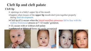
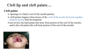
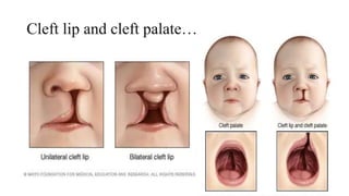
COLLEGE OF HEALTH SCIENCE
DEPARTMENT OF MIDWIFERY
COURSE NEWBORN CARE
FOR POSTBASIC
Cleft lip and cleft palate
1
03-01-2023 BY SHambel N.
Cleft lip and cleft palate
Cleft lip
 openings in a baby's upper lip of the mouth
happen when tissues of the upper lip mouth don't join together properly
during fetal development.
Cleft lip (CL) occurs when the lateral maxillary processes fail to fuse with the
midline frontonasal process at 7–12 weeks’ gestation.
 CL occurs with or without cleft palate
unilateral or bilateral forms are easily recognized
Cleft lip and cleft palate…
Cleft palate
 openings in a baby's roof of the mouth (palate).
 cleft palates happen when tissues of the roof of the mouth don't join together
properly during fetal development.
can involve the hard palate (the bony front portion of the roof of the mouth),
and/or the soft palate (the soft back portion of the roof of the mouth)
Cleft lip and cleft palate…
Risk factors
• Family history. Parents with a family history of cleft lip or cleft palate
face a higher risk of having a baby with a cleft.
• Exposure to certain substances during pregnancy. Cleft lip and
cleft palate may be more likely to occur in pregnant women who
smoke cigarettes, drink alcohol or take certain medications.
• Being obese during pregnancy. There is some evidence that babies
born to obese women may have increased risk of cleft lip and palate.
Causes
The exact cause of cleft lip and cleft palate is not completely
understood
Environmental factors
Medication during pregnancy e.g antiseizure methotrexate, drug used
for treatment of cancer
Folic acid deficiency
Smoking or tobacco using during pregnancy
Signs and symptoms of submucous cleft palate
A split in the lip and roof of the mouth (palate) that affects one or both
sides of the face
A split in the lip that appears as only a small notch in the lip or extends
from the lip through the upper gum and palate into the bottom of the
nose
Difficulty with feedings
Difficulty swallowing, with potential for liquids or foods to come out
the nose
Nasal speaking voice
Chronic ear infections
Diagnosis
• Prenatal ultrasound can sometimes determine if a cleft exists in an
unborn child.
• physical exam of the mouth, nose, and palate confirms the presence of
cleft lip or cleft palate after a child's birth
Management
• Treating cleft lip and cleft palate depends on the extent of the cleft, the
child's age and other special needs or health conditions
• Improve the appearance of the lip and nose
• Close openings between the mouth and nose
• Help to breathe
• Refer for subsequent management (surgery, follow up and speech
rehabilitation) by a team of experts.
Management
• Repair lip within first 3-6 month of age
• Repair palate by the 12 month of age
• Follow up for surgeries between 2-10 years of age
• For children cleft palate , ear tube surgery may placed to reduce the risk of
chronic ear infection
• Surgery to reconstruct appearance
• Feeding strategies e.g bottle feeding -artificial nipples with large openings
and a squeezable bottle
• Speech therapy to correct difficulty with speaking
• Orthodontic adjustment to the teeth
Complications
• Difficulty feeding .
• Ear infections and hearing loss. Babies with cleft palate are
especially at risk of developing middle ear fluid and hearing loss.
• Dental problems. If the cleft extends through the upper gum, tooth
development may be affected.
• Speech difficulties. Because the palate is used in forming sounds, the
development of normal speech can be affected by a cleft palate.
• Challenges of coping with a medical condition--may face social,
emotional and behavioural problems due to differences in appearance
and the stress of intensive medical care.
Management
Gastroschisis
 Is a birth defect where a hole in the abdominal (belly) wall beside the
belly button allows the baby’s intestines to extend outside of the baby’s
body.
The hole can be small or large and sometimes other organs, such as the
stomach and liver, can be found outside of the baby’s body as well.
 Occurs early during pregnancy.
The hole is usually to the right side of the belly button.
 the intestines are not covered in a protective sac and are exposed to the
amniotic fluid, they can become irritated, causing them to shorten, twist,
or swell.
Gastroschisis…
Incidence Rare malformation but incidence remains on the rise.
Latest estimates range between 1.5 and 5 per 10,000 births.
Cause
environmental factors and genital factors
 drugs
nutritional deficiencies
Clinical presentation
 The infant is born with varying amounts of herniated intestine
Prenatal exposure to amniotic fluid results in intestinal edema,
induration, and foreshortened mesentery
Inflammatory peel covers the bowel, leading to intestinal dysmotility
and delayed feeding tolerance.
Up to 10% have an intestinal atresia, and all are malrotated. Other
congenital anomalies are rare
Diagnosis
 Usually identified on prenatal ultrasound and readily apparent at
birth.
P/E
Management
General considerations
Prenatal diagnosis affords prenatal counselling and planned delivery
at a center capable of providing definitive care.
 Safety has been established for both vaginal and cesarean deliveries
Specific measures
Care in the delivery room
The exposed bowel is at risk for injury, hypoperfusion, and fluid and heat loss.
The baby’s abdomen and legs should be placed in a plastic bag.
Alternatively, the viscera may be covered with saline-soaked gauze
Management …
Fluid resuscitation
• Due to exposed viscera, insensible losses begin at birth and remain
significant until abdominal domain is restored.
Thermoregulation
• Hypothermia risk due to evaporative losses occurs rapidly. Immediate
maintain normal temperature.
Prophylactic antibiotics. Broad-spectrum antibiotics are generally
recommended, due to unavoidable seeding of the peritoneum during
delivery and handling of the defect
Management …
Surgical management
• Surgical options include primary closure (when possible) or placement
of silastic silo with staged reduction
• Operative decision is guided by bowel appearance, defect size, and
baby’s general condition.
Nutrition
• Gastroschisis places the newborn in a catabolic state, necessitating
early focus on nutrition.
Gastroschisis with a protective silastic silo in place
Musculoskeletal deformities
1.POLYDACTYL
 This is a name given to a condition where extra digits are on
hand or foot
 Count fingers of hand and foot after birth to confirm refer to
pediatrician for treatment or surgery
 Complex varieties require formal amputation, which is usually
performed at approximately 1 yr of age.
POLYDACTYL…
Symptoms :extra digits
• complete and fully functional
• partially formed, with some bone
• just a small mass of soft tissue (called a nubbin)
Types
• Isolated polydactyly and Syndromic polydactyly
There are three types of isolated polydactyly, classified by the position of
the extra digit:
• Postaxial polydactyly occurs on the outside of the hand or foot, where the
fifth digit is. This is the most common type of polydactyly.
• Preaxial polydactyly occurs on the inside of the hand or foot, where the
thumb or big toe is.
• Central polydactyly occurs in the middle digits of the hand or foot. It’s the
least common type.
Treatment of polydactyly
• Treatment for polydactyly depends on how and where the extra digit is
connected to the hand or foot.
• In most cases, the extra digit is removed in the child’s first two years.
• This gives the child typical use of their hand and allows their feet to
fit into shoes.
2.SYNDACTYLY
Having digits wholly or partly united, either naturally (as in web-
footed animals) or as a malformation.
Syndactyly also occurs in both simple and complex patterns.
Have a tethering effect on the growth of the affected digits.
Surgery for treatment strategies is indicated when the condition is
recognized.
Talipes (club foot)
 Clubfoot is a congenital condition (present at birth) that
causes a baby’s foot to turn inward or downward.
 It can be mild or severe and occur in one or both feet.
 The exact cause is unknown but can be associated with
genetic and environmental factors
Cause
Idiopathic Clubfoot
• E,g talipes equinovarus, idiopathic clubfoot is the most common type of clubfoot and
is present at birth. .
Neurogenic Clubfoot
• underlying neurologic condition. For instance, a child born with spina
bifida , cerebral palsy or a spinal cord compression.
Syndromic Clubfoot
• with the number of other clinical conditions, which relate to an underlying syndrome.
• Examples of syndromes where a clubfoot can occur include arthrogryposis,
constriction band syndrome, tibial hemimelia and diastrophic dwarfism
Symptom
include:
• A foot that turns inward and downward, with toes pointing toward the
opposite foot.
• The clubfoot may be smaller than the other foot (up to ½ inch shorter).
• The heel on the clubfoot may be smaller than normal.
• In severe cases, the clubfoot may be twisted upside down.
• The calf muscle on the leg with the clubfoot will be slightly smaller.
TYPES OF CLUB FOOT
• Talipes equines: The toes are lower than the heel. Common
deformity.
• Talipes Varus :inversion or bending inward
• Talipes valgus :eversion or bending out ward
• Talipes calcanus: The heel is lower than the toes
DIAGNOSIS
Dx –made by examination and x-ray
MANAGEMENT
1. Non surgical correction
-the foot is manipulated into the desired position and plaster cast is
applied (used for 6-8wks) followed by bracing with splints or
corrective shoes for several wks
-casts are changed wkly
2. If the above procedure is failed, surgery will be done
Surgery lengthen the tendons in the foot and may also need to realign
the bones and joints.
Genitourinary system
HYPOSPADIASIS
 is a birth defect in boys in which the opening of the urethra is not located
at the tip of the penis.
 The abnormal opening can form anywhere from just below the end of the
penis to the scrotum.
 There are different degrees of hypospadias; some can be minor and some
more severe
 a urethral opening that is on the ventral surface of the penile shaft
 Affects 1 in 250 male newborn
 There is incomplete development of the prepuce
 Some boys, particularly those with proximal hypospadias, have chordee,
in which there is ventral penile curvature during erection.
C/M – Urethral meatus or opening on the ventral surface of
the penile shaft
DX- Based physical examination and HX
Picture –showing hypospadias
Cause-
 Endocrine disrupting chemicals
 Unknown in some cases
Incidence
 Approximately 60% of cases are distal, 25% are sub coronal or
Midline and 15% are proximal
 Hypospadias is common in boys with multiple congenital
anomalies
 Approximately 10% of boys with hypospadias have an
undescended testes
 Congenital inguinal hernias are also common
Management
 The goal of hypospadias surgery is to correct the functional
and cosmetic deformities .
 Hypospadias repair is recommended for boys with midpenile
and proximal hypospadias
 Bring the urethral opening to the tip of the penis. This allows a
controlled stream of urine while standing.
 Straighten the penis (if chordee is present) to minimize the risk of
painful intercourse later in life.
Complications of untreated hypospadias include:
• Deformity of the urinary stream, either ventral deflection or severe
splaying
• Sexual dysfunction 2o to penile curvature
• Infertility if the urethral meatus is proximal
• Meatal stenois (congenital) → Extremely rare
Postoperative complications of hypospadias repair include:
 Urethrocutaneous fistula
 Hematoma
 Wound infection
 Meatal stenosis
 Urethral diverticulum
 Wound dehiscence
Epispadiasis
 A condition in which the opening (urethral meatus) is on the
dorsal (top) surface of the penis
 It is a congenital malformation, and commonly occurs with
extrophy of the bladder
In boys:
The prepuce is distributed primarily on the ventral aspect of the penile
shaft and the urethral meatus is on the dorsum of the penis
Distal epispadias in boys usually is associated with normal urinary
control and upper urinary tracts
In severe cases of epispadias, both in male and female
The sphincter is incompletely formed
There is total incontinence
DX -Based physical examination and Hx
Mx-surgical repair b/n 6-12 month of age
Nursing care
frequent diaper change and, cleaning the area because of incontinent,
pre-and post-operation care.
IMPERFORATE ANUS
also called an anorectal malformation,
is a rare birth defect that includes the absence of a normal anal opening (anus).
Absence or improper location of the anal opening
Anal canal and sphincter complex are present, but muscles may be attenuated
In absence of normal anal opening, rectum tapers into fistula that may
communicate with genitourinary tract in boys and reproductive tract in girls.
Two types
• high imperforate anus, where rectum ends above the puborectalis sling. There may be
associated recto-urinary fistula in males or rectovaginal fistula in females.
• Low imperforate anus where rectum has traversed the puborectalis sling. This variant may
be associated with or without perineal fistula.
Diagnosis
• Perineal inspection and Absence of meconium passage
• Absence of anal opening
• Failure to pass a rectal thermometer rubber catheter or lubricated little
finger
• Radiology is helpful to detect the extent of atresia.
• The distance between the highest level of the intestinal gas and the
shadow of the coin gives the extent of the atresia.
• Imaging study (X-ray, USG) of the lumbosacral spine and urinary
tract should be done to exclude any other abnormality in this area
Management
• surgery performed to create an opening, or new anus, to allow stool to
pass.
• The surgery may differ, depending on whether the anus ends high or low in
the pelvis.
• If the intestine ends high in the pelvis, treatment usually involves three
procedures:
 colostomy, a stoma — an opening on the abdomen where the intestine is brought
out to the skin — is created. Stool passes out the opening into a special stoma bag
worn on the outside of the body, on the abdomen.
 anoplasty, involves pulling the rectum down to the anus where a new anal opening
is created. If the child has a fistula connecting the intestine to the bladder or vagina,
that connection will be closed.
Several months later after the new anal opening has healed, a third procedure will
be performed to close the stoma.
special stoma bag worn on the outside of the body, on the abdomen.
Management…
• If the intestine ends low in the pelvis, an anal opening often can be
made in a single operation.
• The rectum is pulled down to the anus and a new anal opening is
created, using a minimally invasive technique called laparoscopy. In
these cases, a stoma is not necessary.
• If the anal opening is in the wrong position, it will be closed and
moved to the correct location
• Post operative care

cleft lip.pptx

  • 1.
    COLLEGE OF HEALTHSCIENCE DEPARTMENT OF MIDWIFERY COURSE NEWBORN CARE FOR POSTBASIC Cleft lip and cleft palate 1 03-01-2023 BY SHambel N.
  • 2.
    Cleft lip andcleft palate Cleft lip  openings in a baby's upper lip of the mouth happen when tissues of the upper lip mouth don't join together properly during fetal development. Cleft lip (CL) occurs when the lateral maxillary processes fail to fuse with the midline frontonasal process at 7–12 weeks’ gestation.  CL occurs with or without cleft palate unilateral or bilateral forms are easily recognized
  • 3.
    Cleft lip andcleft palate… Cleft palate  openings in a baby's roof of the mouth (palate).  cleft palates happen when tissues of the roof of the mouth don't join together properly during fetal development. can involve the hard palate (the bony front portion of the roof of the mouth), and/or the soft palate (the soft back portion of the roof of the mouth)
  • 4.
    Cleft lip andcleft palate…
  • 5.
    Risk factors • Familyhistory. Parents with a family history of cleft lip or cleft palate face a higher risk of having a baby with a cleft. • Exposure to certain substances during pregnancy. Cleft lip and cleft palate may be more likely to occur in pregnant women who smoke cigarettes, drink alcohol or take certain medications. • Being obese during pregnancy. There is some evidence that babies born to obese women may have increased risk of cleft lip and palate.
  • 6.
    Causes The exact causeof cleft lip and cleft palate is not completely understood Environmental factors Medication during pregnancy e.g antiseizure methotrexate, drug used for treatment of cancer Folic acid deficiency Smoking or tobacco using during pregnancy
  • 7.
    Signs and symptomsof submucous cleft palate A split in the lip and roof of the mouth (palate) that affects one or both sides of the face A split in the lip that appears as only a small notch in the lip or extends from the lip through the upper gum and palate into the bottom of the nose Difficulty with feedings Difficulty swallowing, with potential for liquids or foods to come out the nose Nasal speaking voice Chronic ear infections
  • 8.
    Diagnosis • Prenatal ultrasoundcan sometimes determine if a cleft exists in an unborn child. • physical exam of the mouth, nose, and palate confirms the presence of cleft lip or cleft palate after a child's birth
  • 9.
    Management • Treating cleftlip and cleft palate depends on the extent of the cleft, the child's age and other special needs or health conditions • Improve the appearance of the lip and nose • Close openings between the mouth and nose • Help to breathe • Refer for subsequent management (surgery, follow up and speech rehabilitation) by a team of experts.
  • 10.
    Management • Repair lipwithin first 3-6 month of age • Repair palate by the 12 month of age • Follow up for surgeries between 2-10 years of age • For children cleft palate , ear tube surgery may placed to reduce the risk of chronic ear infection • Surgery to reconstruct appearance • Feeding strategies e.g bottle feeding -artificial nipples with large openings and a squeezable bottle • Speech therapy to correct difficulty with speaking • Orthodontic adjustment to the teeth
  • 11.
    Complications • Difficulty feeding. • Ear infections and hearing loss. Babies with cleft palate are especially at risk of developing middle ear fluid and hearing loss. • Dental problems. If the cleft extends through the upper gum, tooth development may be affected. • Speech difficulties. Because the palate is used in forming sounds, the development of normal speech can be affected by a cleft palate. • Challenges of coping with a medical condition--may face social, emotional and behavioural problems due to differences in appearance and the stress of intensive medical care.
  • 12.
  • 13.
    Gastroschisis  Is abirth defect where a hole in the abdominal (belly) wall beside the belly button allows the baby’s intestines to extend outside of the baby’s body. The hole can be small or large and sometimes other organs, such as the stomach and liver, can be found outside of the baby’s body as well.  Occurs early during pregnancy. The hole is usually to the right side of the belly button.  the intestines are not covered in a protective sac and are exposed to the amniotic fluid, they can become irritated, causing them to shorten, twist, or swell.
  • 14.
    Gastroschisis… Incidence Rare malformationbut incidence remains on the rise. Latest estimates range between 1.5 and 5 per 10,000 births. Cause environmental factors and genital factors  drugs nutritional deficiencies
  • 15.
    Clinical presentation  Theinfant is born with varying amounts of herniated intestine Prenatal exposure to amniotic fluid results in intestinal edema, induration, and foreshortened mesentery Inflammatory peel covers the bowel, leading to intestinal dysmotility and delayed feeding tolerance. Up to 10% have an intestinal atresia, and all are malrotated. Other congenital anomalies are rare
  • 16.
    Diagnosis  Usually identifiedon prenatal ultrasound and readily apparent at birth. P/E
  • 17.
    Management General considerations Prenatal diagnosisaffords prenatal counselling and planned delivery at a center capable of providing definitive care.  Safety has been established for both vaginal and cesarean deliveries Specific measures Care in the delivery room The exposed bowel is at risk for injury, hypoperfusion, and fluid and heat loss. The baby’s abdomen and legs should be placed in a plastic bag. Alternatively, the viscera may be covered with saline-soaked gauze
  • 18.
    Management … Fluid resuscitation •Due to exposed viscera, insensible losses begin at birth and remain significant until abdominal domain is restored. Thermoregulation • Hypothermia risk due to evaporative losses occurs rapidly. Immediate maintain normal temperature. Prophylactic antibiotics. Broad-spectrum antibiotics are generally recommended, due to unavoidable seeding of the peritoneum during delivery and handling of the defect
  • 19.
    Management … Surgical management •Surgical options include primary closure (when possible) or placement of silastic silo with staged reduction • Operative decision is guided by bowel appearance, defect size, and baby’s general condition. Nutrition • Gastroschisis places the newborn in a catabolic state, necessitating early focus on nutrition.
  • 20.
    Gastroschisis with aprotective silastic silo in place
  • 21.
    Musculoskeletal deformities 1.POLYDACTYL  Thisis a name given to a condition where extra digits are on hand or foot  Count fingers of hand and foot after birth to confirm refer to pediatrician for treatment or surgery  Complex varieties require formal amputation, which is usually performed at approximately 1 yr of age.
  • 22.
    POLYDACTYL… Symptoms :extra digits •complete and fully functional • partially formed, with some bone • just a small mass of soft tissue (called a nubbin) Types • Isolated polydactyly and Syndromic polydactyly There are three types of isolated polydactyly, classified by the position of the extra digit: • Postaxial polydactyly occurs on the outside of the hand or foot, where the fifth digit is. This is the most common type of polydactyly. • Preaxial polydactyly occurs on the inside of the hand or foot, where the thumb or big toe is. • Central polydactyly occurs in the middle digits of the hand or foot. It’s the least common type.
  • 23.
    Treatment of polydactyly •Treatment for polydactyly depends on how and where the extra digit is connected to the hand or foot. • In most cases, the extra digit is removed in the child’s first two years. • This gives the child typical use of their hand and allows their feet to fit into shoes.
  • 24.
    2.SYNDACTYLY Having digits whollyor partly united, either naturally (as in web- footed animals) or as a malformation. Syndactyly also occurs in both simple and complex patterns. Have a tethering effect on the growth of the affected digits. Surgery for treatment strategies is indicated when the condition is recognized.
  • 25.
    Talipes (club foot) Clubfoot is a congenital condition (present at birth) that causes a baby’s foot to turn inward or downward.  It can be mild or severe and occur in one or both feet.  The exact cause is unknown but can be associated with genetic and environmental factors
  • 26.
    Cause Idiopathic Clubfoot • E,gtalipes equinovarus, idiopathic clubfoot is the most common type of clubfoot and is present at birth. . Neurogenic Clubfoot • underlying neurologic condition. For instance, a child born with spina bifida , cerebral palsy or a spinal cord compression. Syndromic Clubfoot • with the number of other clinical conditions, which relate to an underlying syndrome. • Examples of syndromes where a clubfoot can occur include arthrogryposis, constriction band syndrome, tibial hemimelia and diastrophic dwarfism
  • 27.
    Symptom include: • A footthat turns inward and downward, with toes pointing toward the opposite foot. • The clubfoot may be smaller than the other foot (up to ½ inch shorter). • The heel on the clubfoot may be smaller than normal. • In severe cases, the clubfoot may be twisted upside down. • The calf muscle on the leg with the clubfoot will be slightly smaller.
  • 28.
    TYPES OF CLUBFOOT • Talipes equines: The toes are lower than the heel. Common deformity. • Talipes Varus :inversion or bending inward • Talipes valgus :eversion or bending out ward • Talipes calcanus: The heel is lower than the toes
  • 29.
    DIAGNOSIS Dx –made byexamination and x-ray MANAGEMENT 1. Non surgical correction -the foot is manipulated into the desired position and plaster cast is applied (used for 6-8wks) followed by bracing with splints or corrective shoes for several wks -casts are changed wkly 2. If the above procedure is failed, surgery will be done Surgery lengthen the tendons in the foot and may also need to realign the bones and joints.
  • 30.
    Genitourinary system HYPOSPADIASIS  isa birth defect in boys in which the opening of the urethra is not located at the tip of the penis.  The abnormal opening can form anywhere from just below the end of the penis to the scrotum.  There are different degrees of hypospadias; some can be minor and some more severe  a urethral opening that is on the ventral surface of the penile shaft  Affects 1 in 250 male newborn  There is incomplete development of the prepuce  Some boys, particularly those with proximal hypospadias, have chordee, in which there is ventral penile curvature during erection.
  • 31.
    C/M – Urethralmeatus or opening on the ventral surface of the penile shaft DX- Based physical examination and HX Picture –showing hypospadias
  • 32.
    Cause-  Endocrine disruptingchemicals  Unknown in some cases Incidence  Approximately 60% of cases are distal, 25% are sub coronal or Midline and 15% are proximal  Hypospadias is common in boys with multiple congenital anomalies  Approximately 10% of boys with hypospadias have an undescended testes  Congenital inguinal hernias are also common
  • 33.
    Management  The goalof hypospadias surgery is to correct the functional and cosmetic deformities .  Hypospadias repair is recommended for boys with midpenile and proximal hypospadias  Bring the urethral opening to the tip of the penis. This allows a controlled stream of urine while standing.  Straighten the penis (if chordee is present) to minimize the risk of painful intercourse later in life.
  • 34.
    Complications of untreatedhypospadias include: • Deformity of the urinary stream, either ventral deflection or severe splaying • Sexual dysfunction 2o to penile curvature • Infertility if the urethral meatus is proximal • Meatal stenois (congenital) → Extremely rare
  • 35.
    Postoperative complications ofhypospadias repair include:  Urethrocutaneous fistula  Hematoma  Wound infection  Meatal stenosis  Urethral diverticulum  Wound dehiscence
  • 36.
    Epispadiasis  A conditionin which the opening (urethral meatus) is on the dorsal (top) surface of the penis  It is a congenital malformation, and commonly occurs with extrophy of the bladder In boys: The prepuce is distributed primarily on the ventral aspect of the penile shaft and the urethral meatus is on the dorsum of the penis
  • 37.
    Distal epispadias inboys usually is associated with normal urinary control and upper urinary tracts In severe cases of epispadias, both in male and female The sphincter is incompletely formed There is total incontinence DX -Based physical examination and Hx Mx-surgical repair b/n 6-12 month of age Nursing care frequent diaper change and, cleaning the area because of incontinent, pre-and post-operation care.
  • 38.
    IMPERFORATE ANUS also calledan anorectal malformation, is a rare birth defect that includes the absence of a normal anal opening (anus). Absence or improper location of the anal opening Anal canal and sphincter complex are present, but muscles may be attenuated In absence of normal anal opening, rectum tapers into fistula that may communicate with genitourinary tract in boys and reproductive tract in girls. Two types • high imperforate anus, where rectum ends above the puborectalis sling. There may be associated recto-urinary fistula in males or rectovaginal fistula in females. • Low imperforate anus where rectum has traversed the puborectalis sling. This variant may be associated with or without perineal fistula.
  • 39.
    Diagnosis • Perineal inspectionand Absence of meconium passage • Absence of anal opening • Failure to pass a rectal thermometer rubber catheter or lubricated little finger • Radiology is helpful to detect the extent of atresia. • The distance between the highest level of the intestinal gas and the shadow of the coin gives the extent of the atresia. • Imaging study (X-ray, USG) of the lumbosacral spine and urinary tract should be done to exclude any other abnormality in this area
  • 40.
    Management • surgery performedto create an opening, or new anus, to allow stool to pass. • The surgery may differ, depending on whether the anus ends high or low in the pelvis. • If the intestine ends high in the pelvis, treatment usually involves three procedures:  colostomy, a stoma — an opening on the abdomen where the intestine is brought out to the skin — is created. Stool passes out the opening into a special stoma bag worn on the outside of the body, on the abdomen.  anoplasty, involves pulling the rectum down to the anus where a new anal opening is created. If the child has a fistula connecting the intestine to the bladder or vagina, that connection will be closed. Several months later after the new anal opening has healed, a third procedure will be performed to close the stoma.
  • 41.
    special stoma bagworn on the outside of the body, on the abdomen.
  • 42.
    Management… • If theintestine ends low in the pelvis, an anal opening often can be made in a single operation. • The rectum is pulled down to the anus and a new anal opening is created, using a minimally invasive technique called laparoscopy. In these cases, a stoma is not necessary. • If the anal opening is in the wrong position, it will be closed and moved to the correct location • Post operative care

Editor's Notes

  • #25 Tether=restrct
  • #30 brace · n. 1 (braces) Brit. a pair of straps passing over the shoulders and fastening to the top of trousers at the front and back to hold them up. 2 a device supporting a weak or injured part of the body. Ø a wire device fitted in the mouth to straighten the teeth. Ø a strengthening piece of iron or timber in building or carpentry. 3 (also brace and bit) a drilling tool with a crank handle and a socket to hold a bit. 4 a rope attached to the yard of a ship for trimming the sail. 5 (pl. same) a pair of things, especially birds or mammals killed in hunting. 6 either of two connecting marks { and }, used in printing. Ø Music a similar mark connecting staves to be performed at the same time. · v. 1 make stronger or firmer with wood, iron, etc. 2 press (one’s body) firmly against something in order to stay balanced. 3 (brace oneself) prepare for something difficult or unpleasant. – ORIGIN ME: from OFr. bracier ‘embrace’, from brace ‘two arms’, from L. bracchia, pl. of bracchium ‘arm’, from Gk brakhiZn.
  • #33 inguinal hernia/ŋ&wn(ə)l h! niə/ noun a hernia where the intestine bulges through the muscles in the groin
  • #35 Splaying=wider Stream=flow
  • #36 diverticulum /%dVIv@"tIkjUl@m/ · n. (pl. diverticula /-l@/) Anatomy & Zoology 1 a blind tube leading from a cavity or passage. 2 Medicine an abnormal sac or pouch formed at a weak point in the wall of the alimentary tract. – ORIGIN C19: from med. L., var. of L. deverticulum ‘byway’, from devertere ‘turn down or aside’.