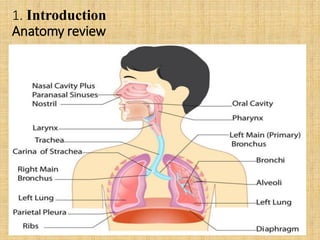
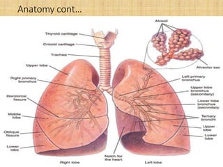
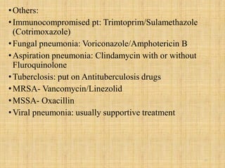
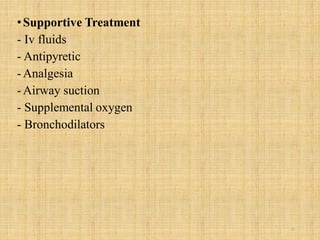
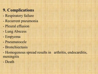
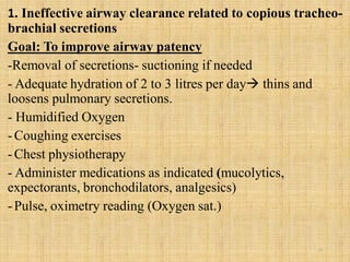
PNEUMONIA
DR EVA VELIKOSHI-INDONGO
•(DIP NURSING/MIDWIFERY, RN/RM/RT, BNSc
(HSM), NED (NDP), MNSc, DNSC (UNAM)
• MBChB (ZAMBIA)
26/02/2020
1
Outline
1. Introduction
2. Epidemiology
3. Classification/types of pneumonia
4. Risk factors
5. Pathophysiology
6. Clinical Manifestation
7. Assessment and diagnosis
8. Management of pneumonia
9. Complications
10. Nursing care plan
2
1. Introduction
Anatomy review
3
Anatomy cont…
4
Pneumonia:
•Definition
-Inflammation of lung parenchyma
-Caused either by bacteria, fungal, viral, mycobacteria
Primary pneumonia is caused by the client’s inhalation or
aspiration of a pathogen
Secondary pneumonia results from lung damage: spread of
an infectious agents from another site in the body, chemical
irritants (including gastric reflux and aspiration, smoke
inhalation)
Pneumonitis general term denoting inflammatory process
in the lung tissue  predispose patient to microbial
invasion. 5
2. Epidemiology
- 40 adults/1000/year
- Up to 10% acute adult admissions
- 8th leading cause of death in USA
- estimated 6 million cases are reported annually;
-hospital discharges attributed to pneumonia in 2009
-HAP accounts for 15% of hospital-acquired infections and
leading cause among those pts with HAP
-Pneumonia and influenza account for nearly 60,000 deaths
annually.
6
3. Classification/Types of Pneumonia
i) 4 major classifications/types
•Community-acquired pneumonia (CAP)
•Hospital acquired Pneumonia (HAP)
•Pneumonia in the immune-compromised host
•Aspiration pneumonia
** Nosocomial pneumonias hospital acquired, ventilator
associated and health care-associated pneumonias (old term)
7
Definition of different types of pneumonias
•Community-acquired pneumonia (CAP): Pneumonia
occurring within 48 hours of hospital admission
 streptococcus pneumoniae, H. influenza, Legionella,
and Pseudomonas aeruginosa.
•Health care–associated pneumonia (HCAP): Pneumonia
occurring in a non-hospitalized patient with extensive health
care contact with one or more of the following:
- Hospitalization for ≥2 days in an acute care facility within 90
days of infection
-Residence in a nursing care institution
-Antibiotic therapy or wound care within 30 days
- Hemodialysis treatment at a hospital or clinic
- Family member with infection due to multidrug-resistant
bacteria 8
•Hospital-acquired pneumonia (HAP): Pneumonia occurring
≥48 hours after hospital admission
Causative agent(s): Enterobacter species, E. coli, influenza,
Klebsiella species, Proteus, S. aureus, S. pneumonie
• Ventilator-associated pneumonia (VAP): A type of HAP that
develops ≥48 hours after endotracheal tube intubation
S. aureus
•Aspiration pneumonia: pulmonary injury resulting from entry
of endogenous or exogenous substances into the lower
airway.
- vomiting and aspiration of gastric or oropharyngeal contents
into the trachea and lungs
 S. pneumonia, H.influenza, and S. aureus. 9
•Pneumonia in the Immunocompromised Host
 includes Pneumocystis pneumonia/ Pneumocystis
jirovecii, fungal pneumonias and Mycobacterium
tuberculosis.
10
•ii) by causative agent
-Viral: Common causative organisms include respiratory
syncytial virus (RSV), influenza, and human parainfluenza
viruses
- Bacteria
Divided into typical and atypical types
Typical bacteria: Gram-positive Streptococcus pneumoniae,
Haemophilus, and Staphylococcus most common bacterial
causes
•Atypical: Mycoplasma pneumonie, Legionella pneumophila,
Chlamydia pneumonie, Mycobacterium tuberculosis,
Coxiella burnetii,
- Fungal: Histoplasma capsulatum, Coccidioides immitis,
Pneumocystis jirovecii (formerly carinii)
11
Causative agents (summary)
•S. pneumoniae 30-75%
•Mycoplasma pneumonia 5-18
•Influenza virus 8
•Other viruses 2-8
•Haemophilus influenza 4-5%
•Legionella spp. 2-5
•Chlamydia spp. 2-3
•S.aureus 1-5
12
•iii) by site/anatomically
-Lobar: involving single lobe
- Broncho pneumonia: smaller lung areas in several lobes
-interstitial: tissues surrounding the alveoli and bronchi
13
4. Risk factors
•Critical illness, ICU, Surgery
•Immunosuppression HIV, Chemotherapy, neutropenia
•Comorbidities e.g. heart or lung disease, DM, renal
diseases
•Malnutrition
•Smoking
•Age <16 or >65 years
•Recent antibiotic therapy
•Endotracheal intubation with mechanical ventilation
•Prolonged immobility
•Hospital procedures e.g. NGT insertion
14
5. Pathophysiology
•Pneumonia arises from normal flora present in patients
whose low/compromised/altered immunity or from
aspiration of flora
•An inflammatory reaction  in the alveoli
exudates that interfere with the diffusion of oxygen and
carbon dioxide
•White blood cells also migrate into the alveoli and fill the
normally air-filled spaces congestion and edema.
•Due to secretions and mucosal edema partial occlusion
of the brochi and alveoli inadequate ventilation
•Hypoventilation may follow causing ventilation-
perfusion mismatch poorly oxygenation of blood
hypoxemia
15
16
6. Clinical manifestations
Mainly related to the causative agent
- Cough dry or with rusty sputum
-High grade fever
-Chills/rigors
- Sputum
- Pleuritic chest pain
- Dyspnoea
- Tachypnoea
- Crackles
- Increased fremitus
- Dullness on percussion
- Increased egophony
17
18
7. Assessment and Diagnosis making
1. Assessment
- History taking and physical examination
Cough: productive/non productive, duration
Chest pain: nature, duration
Sputum: amount, colour
Shortness of breath and its extent
Vital signs: fever, respiratory and pulse rates
Auscultation breathing sounds, percussion abnormalities
2. Diagnosis:
-Sputum studies (Induce sputum if dry cough): MCS, AAFBs
- Chest X-ray (consolidation)
- Pleural fluid aspirate MCS
- FBC WCC/ESR
- Pleural biopsy 19
8. Management of Pneumonia
Antimicrobial Uncomplicated pneumonia
•Amoxycillin 500mg TID po
•Ampicillin 500mg QID po
•Benzyl penicillin 1.2gm QID iv
•Erythromycin 500mg QID po
•Cefuroxime 750mg TID iv
•Cefotaxime 1gm BID
•Immunocompromised pt: Trimtoprim/Sulamethazole
(Cotrimoxazole)
•Fungal pneumonia: Voriconazole/Amphotericin B
•Aspiration pneumonia: Clindamycin with or without
Fluroquinolone
20
•Others:
•Immunocompromised pt: Trimtoprim/Sulamethazole
(Cotrimoxazole)
•Fungal pneumonia: Voriconazole/Amphotericin B
•Aspiration pneumonia: Clindamycin with or without
Fluroquinolone
•Tuberclosis: put on Antituberculosis drugs
•MRSA- Vancomycin/Linezolid
•MSSA- Oxacillin
•Viral pneumonia: usually supportive treatment
21
•Supportive Treatment
- Iv fluids
- Antipyretic
-Analgesia
-Airway suction
- Supplemental oxygen
- Bronchodilators
22
9. Complications
- Respiratory failure
- Recurrent pneumonia
- Pleural effusion
- Lung Abscess
- Empyema
- Pneumatocele
- Bronchiectasis
- Homogenous spread results in arthritis, endocarditis,
meningitis
- Death
23
10. Nursing care plan
24
1. Ineffective airway clearance related to copious tracheo-
brachial secretions
Goal: To improve airway patency
-Removal of secretions- suctioning if needed
- Adequate hydration of 2 to 3 litres per day thins and
loosens pulmonary secretions.
- Humidified Oxygen
-Coughing exercises
-Chest physiotherapy
- Administer medications as indicated (mucolytics,
expectorants, bronchodilators, analgesics)
-Pulse, oximetry reading (Oxygen sat.)
25
2. Fatigue and activity intolerance related to impaired
respiratory function
Goal: To promote rest and conserve energy
•Encourage avoidance of overexertion adequate rest
•Semi-Fowler’s position comfortability, promote rest
and breathing
•Frequent position change enhance secretion clearance,
pulmonary ventilation and perfusion.
26
3. Risk for deficient fluid volume related to fever and a
rapid respiratory rate
Goal: To promote fluid intake
- Increase in fluid intake to at least 2L per day to replace
insensible fluid losses if not contraindicated (CHF)
- Assess vital sign changes: increasing temperature,
prolonged fever, orthostatic hypotension, tachycardia.
- Monitor intake and output
- Provide supplemental IV fluids as necessary.
27
4. Imbalanced nutrition: less than body
requirements
Goal: To maintain nutrition
•Fluids with electrolytes and nutrition-enriched drinks 
provide fluid, calories, and electrolytes.
•Provide small, frequent meals, including dry foods (toast,
crackers) and/or foods that are appealing to patient.
28
5. Deficient knowledge about the treatment regimen and
preventive measures
Goal: To promote patient’s knowledge: Patient and
caregiver will verbalize understanding of condition,
disease process, and prognosis, understanding of the
therapeutic regimen
•Educate patient and family about the cause of pneumonia,
management of symptoms, signs, and symptoms, and the
need for follow-up.
•Educate patient about the factors (risk factors) that may
have contributed to the development of the disease
•Educate patient on the need to complete treatment regimen
29
•Summary
•Introduction to pneumonia: definition, epidemiology
•Classifications of pneumonia: by distribution, etiology,
anatomical, other terms related to pneumonia
classification
•Pathophysiology
•Clinical manifestation: signs and symptoms
•Diagnosis making
•Medical treatment
•Nursing care plans and goals, plus interventions
30
****************************END**********************
31

Pneumonia

  • 1.
    PNEUMONIA DR EVA VELIKOSHI-INDONGO •(DIPNURSING/MIDWIFERY, RN/RM/RT, BNSc (HSM), NED (NDP), MNSc, DNSC (UNAM) • MBChB (ZAMBIA) 26/02/2020 1
  • 2.
    Outline 1. Introduction 2. Epidemiology 3.Classification/types of pneumonia 4. Risk factors 5. Pathophysiology 6. Clinical Manifestation 7. Assessment and diagnosis 8. Management of pneumonia 9. Complications 10. Nursing care plan 2
  • 3.
  • 4.
  • 5.
    Pneumonia: •Definition -Inflammation of lungparenchyma -Caused either by bacteria, fungal, viral, mycobacteria Primary pneumonia is caused by the client’s inhalation or aspiration of a pathogen Secondary pneumonia results from lung damage: spread of an infectious agents from another site in the body, chemical irritants (including gastric reflux and aspiration, smoke inhalation) Pneumonitis general term denoting inflammatory process in the lung tissue  predispose patient to microbial invasion. 5
  • 6.
    2. Epidemiology - 40adults/1000/year - Up to 10% acute adult admissions - 8th leading cause of death in USA - estimated 6 million cases are reported annually; -hospital discharges attributed to pneumonia in 2009 -HAP accounts for 15% of hospital-acquired infections and leading cause among those pts with HAP -Pneumonia and influenza account for nearly 60,000 deaths annually. 6
  • 7.
    3. Classification/Types ofPneumonia i) 4 major classifications/types •Community-acquired pneumonia (CAP) •Hospital acquired Pneumonia (HAP) •Pneumonia in the immune-compromised host •Aspiration pneumonia ** Nosocomial pneumonias hospital acquired, ventilator associated and health care-associated pneumonias (old term) 7
  • 8.
    Definition of differenttypes of pneumonias •Community-acquired pneumonia (CAP): Pneumonia occurring within 48 hours of hospital admission  streptococcus pneumoniae, H. influenza, Legionella, and Pseudomonas aeruginosa. •Health care–associated pneumonia (HCAP): Pneumonia occurring in a non-hospitalized patient with extensive health care contact with one or more of the following: - Hospitalization for ≥2 days in an acute care facility within 90 days of infection -Residence in a nursing care institution -Antibiotic therapy or wound care within 30 days - Hemodialysis treatment at a hospital or clinic - Family member with infection due to multidrug-resistant bacteria 8
  • 9.
    •Hospital-acquired pneumonia (HAP):Pneumonia occurring ≥48 hours after hospital admission Causative agent(s): Enterobacter species, E. coli, influenza, Klebsiella species, Proteus, S. aureus, S. pneumonie • Ventilator-associated pneumonia (VAP): A type of HAP that develops ≥48 hours after endotracheal tube intubation S. aureus •Aspiration pneumonia: pulmonary injury resulting from entry of endogenous or exogenous substances into the lower airway. - vomiting and aspiration of gastric or oropharyngeal contents into the trachea and lungs  S. pneumonia, H.influenza, and S. aureus. 9
  • 10.
    •Pneumonia in theImmunocompromised Host  includes Pneumocystis pneumonia/ Pneumocystis jirovecii, fungal pneumonias and Mycobacterium tuberculosis. 10
  • 11.
    •ii) by causativeagent -Viral: Common causative organisms include respiratory syncytial virus (RSV), influenza, and human parainfluenza viruses - Bacteria Divided into typical and atypical types Typical bacteria: Gram-positive Streptococcus pneumoniae, Haemophilus, and Staphylococcus most common bacterial causes •Atypical: Mycoplasma pneumonie, Legionella pneumophila, Chlamydia pneumonie, Mycobacterium tuberculosis, Coxiella burnetii, - Fungal: Histoplasma capsulatum, Coccidioides immitis, Pneumocystis jirovecii (formerly carinii) 11
  • 12.
    Causative agents (summary) •S.pneumoniae 30-75% •Mycoplasma pneumonia 5-18 •Influenza virus 8 •Other viruses 2-8 •Haemophilus influenza 4-5% •Legionella spp. 2-5 •Chlamydia spp. 2-3 •S.aureus 1-5 12
  • 13.
    •iii) by site/anatomically -Lobar:involving single lobe - Broncho pneumonia: smaller lung areas in several lobes -interstitial: tissues surrounding the alveoli and bronchi 13
  • 14.
    4. Risk factors •Criticalillness, ICU, Surgery •Immunosuppression HIV, Chemotherapy, neutropenia •Comorbidities e.g. heart or lung disease, DM, renal diseases •Malnutrition •Smoking •Age <16 or >65 years •Recent antibiotic therapy •Endotracheal intubation with mechanical ventilation •Prolonged immobility •Hospital procedures e.g. NGT insertion 14
  • 15.
    5. Pathophysiology •Pneumonia arisesfrom normal flora present in patients whose low/compromised/altered immunity or from aspiration of flora •An inflammatory reaction  in the alveoli exudates that interfere with the diffusion of oxygen and carbon dioxide •White blood cells also migrate into the alveoli and fill the normally air-filled spaces congestion and edema. •Due to secretions and mucosal edema partial occlusion of the brochi and alveoli inadequate ventilation •Hypoventilation may follow causing ventilation- perfusion mismatch poorly oxygenation of blood hypoxemia 15
  • 16.
  • 17.
    6. Clinical manifestations Mainlyrelated to the causative agent - Cough dry or with rusty sputum -High grade fever -Chills/rigors - Sputum - Pleuritic chest pain - Dyspnoea - Tachypnoea - Crackles - Increased fremitus - Dullness on percussion - Increased egophony 17
  • 18.
  • 19.
    7. Assessment andDiagnosis making 1. Assessment - History taking and physical examination Cough: productive/non productive, duration Chest pain: nature, duration Sputum: amount, colour Shortness of breath and its extent Vital signs: fever, respiratory and pulse rates Auscultation breathing sounds, percussion abnormalities 2. Diagnosis: -Sputum studies (Induce sputum if dry cough): MCS, AAFBs - Chest X-ray (consolidation) - Pleural fluid aspirate MCS - FBC WCC/ESR - Pleural biopsy 19
  • 20.
    8. Management ofPneumonia Antimicrobial Uncomplicated pneumonia •Amoxycillin 500mg TID po •Ampicillin 500mg QID po •Benzyl penicillin 1.2gm QID iv •Erythromycin 500mg QID po •Cefuroxime 750mg TID iv •Cefotaxime 1gm BID •Immunocompromised pt: Trimtoprim/Sulamethazole (Cotrimoxazole) •Fungal pneumonia: Voriconazole/Amphotericin B •Aspiration pneumonia: Clindamycin with or without Fluroquinolone 20
  • 21.
    •Others: •Immunocompromised pt: Trimtoprim/Sulamethazole (Cotrimoxazole) •Fungalpneumonia: Voriconazole/Amphotericin B •Aspiration pneumonia: Clindamycin with or without Fluroquinolone •Tuberclosis: put on Antituberculosis drugs •MRSA- Vancomycin/Linezolid •MSSA- Oxacillin •Viral pneumonia: usually supportive treatment 21
  • 22.
    •Supportive Treatment - Ivfluids - Antipyretic -Analgesia -Airway suction - Supplemental oxygen - Bronchodilators 22
  • 23.
    9. Complications - Respiratoryfailure - Recurrent pneumonia - Pleural effusion - Lung Abscess - Empyema - Pneumatocele - Bronchiectasis - Homogenous spread results in arthritis, endocarditis, meningitis - Death 23
  • 24.
  • 25.
    1. Ineffective airwayclearance related to copious tracheo- brachial secretions Goal: To improve airway patency -Removal of secretions- suctioning if needed - Adequate hydration of 2 to 3 litres per day thins and loosens pulmonary secretions. - Humidified Oxygen -Coughing exercises -Chest physiotherapy - Administer medications as indicated (mucolytics, expectorants, bronchodilators, analgesics) -Pulse, oximetry reading (Oxygen sat.) 25
  • 26.
    2. Fatigue andactivity intolerance related to impaired respiratory function Goal: To promote rest and conserve energy •Encourage avoidance of overexertion adequate rest •Semi-Fowler’s position comfortability, promote rest and breathing •Frequent position change enhance secretion clearance, pulmonary ventilation and perfusion. 26
  • 27.
    3. Risk fordeficient fluid volume related to fever and a rapid respiratory rate Goal: To promote fluid intake - Increase in fluid intake to at least 2L per day to replace insensible fluid losses if not contraindicated (CHF) - Assess vital sign changes: increasing temperature, prolonged fever, orthostatic hypotension, tachycardia. - Monitor intake and output - Provide supplemental IV fluids as necessary. 27
  • 28.
    4. Imbalanced nutrition:less than body requirements Goal: To maintain nutrition •Fluids with electrolytes and nutrition-enriched drinks  provide fluid, calories, and electrolytes. •Provide small, frequent meals, including dry foods (toast, crackers) and/or foods that are appealing to patient. 28
  • 29.
    5. Deficient knowledgeabout the treatment regimen and preventive measures Goal: To promote patient’s knowledge: Patient and caregiver will verbalize understanding of condition, disease process, and prognosis, understanding of the therapeutic regimen •Educate patient and family about the cause of pneumonia, management of symptoms, signs, and symptoms, and the need for follow-up. •Educate patient about the factors (risk factors) that may have contributed to the development of the disease •Educate patient on the need to complete treatment regimen 29
  • 30.
    •Summary •Introduction to pneumonia:definition, epidemiology •Classifications of pneumonia: by distribution, etiology, anatomical, other terms related to pneumonia classification •Pathophysiology •Clinical manifestation: signs and symptoms •Diagnosis making •Medical treatment •Nursing care plans and goals, plus interventions 30
  • 31.