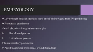
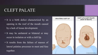
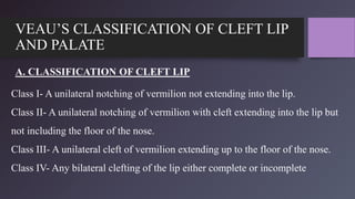
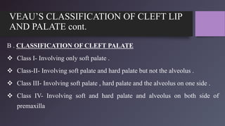
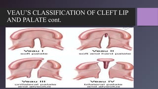
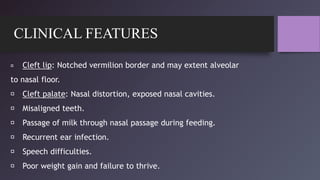
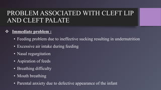
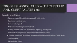
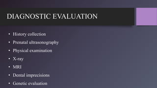
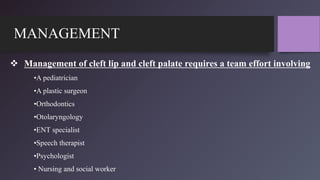
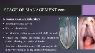
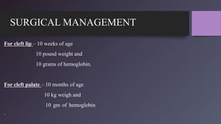
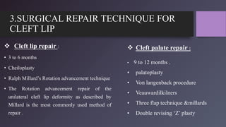
Cleft lip and palate is a birth defect where the tissue of the mouth and palate do not properly fuse together, leaving an opening in the lip and/or roof of the mouth. It affects 1 in 750 births and can cause problems with feeding, speech, dental issues, and facial growth. The document discusses the anatomy, development, causes, classification, and management of cleft lip and palate. Management involves a multi-disciplinary team and stages of surgery to repair the lip, palate, and other issues, as well as other treatments like dental appliances and bone grafts.
Cleft lip and palate abnormalities occur when certain facial structures fail to fuse properly during embryonic development between the fifth and seventh weeks. This can result in openings in the lip and roof of the mouth. Clefts may involve just the lip, just the palate, or both. They can range from mild to severe. Early surgery is aimed at restoring normal anatomy, but long-term management may also include hearing, speech, dental, and orthodontic care.
Cleft lip and palate occur due to the failure of fusion between the medial nasal processes and maxillary processes during embryonic development between the 5th and 7th week of gestation. This results in an opening in the lip and/or roof of the mouth. Cleft lip alone affects 15% of cases, cleft lip and palate affects 45% of cases, and isolated cleft palate affects 40% of cases. The causes are both genetic and environmental, with genetics accounting for 20-30% of non-syndromic clefts. Treatment involves surgical repair of the cleft and management of associated feeding issues, speech/language delays, dental abnormalities, and ear infections.
Cleft palate is an opening in the roof of the mouth caused by a failure of the palatal shelves to fuse during embryonic development. It can occur alone or with cleft lip. Complications include feeding difficulties, ear infections, dental problems, and speech issues. Treatment involves surgery to close the palate, with the goal of minimizing scarring and facial growth impacts. A cleft palate team manages each child's individual care.
This document provides information about cleft lip and cleft palate including causes, risk factors, diagnosis, treatment, and social aspects. It describes how cleft lip occurs when the tissues of the lip do not fully fuse before birth, and cleft palate occurs when the roof of the mouth does not fully close. Treatment often begins in infancy and may include surgery, dental care, speech therapy, and psychological support. The document also discusses cultural beliefs and stigma around cleft conditions as well as organizations providing cleft care in India.
This document provides an overview of cleft lip and palate defects, including their embryology, classification, incidence, and reconstruction. It discusses how clefts occur due to failures in fusion during embryonic development of the lip and palate. Common defects seen in cleft patients are also outlined. The reconstruction of cleft lip and palate is described in stages, including presurgical orthopedics, unilateral and bilateral lip repair techniques, various palatoplasty approaches such as the Bardach and Furlow methods, and bone grafting to reconstruct alveolar defects.
Cleft lip and cleft palate are congenital defects caused by incomplete fusion of facial structures in utero. Cleft lip involves a split in the upper lip, which can be unilateral or bilateral. Cleft palate is a split in the roof of the mouth. Treatment involves surgery to repair the cleft, with lip repair typically in the first year of life and palate repair by 18 months. Nursing management focuses on pre-operative preparation, careful feeding and positioning, and post-operative monitoring of healing and parental education on home care.
The document provides information on cleft lip and cleft palate including definitions, incidence rates, development, classifications, problems associated, and treatment protocols. It defines cleft lip as an opening in the upper lip and cleft palate as an opening in the roof of the mouth. Treatment is a multidisciplinary approach involving surgery to repair the cleft, orthodontics to align teeth and jaws, and speech therapy. Management occurs over many years from infancy through adulthood to address dental, esthetic, speech and other issues.
The Indian Dental Academy is the Leader in continuing dental education , training dentists in all aspects of dentistry and
offering a wide range of dental certified courses in different formats.for more details please visit
www.indiandentalacademy.com
Cleft lip and cleft palate are the most common congenital facial malformations in humans, ranging from slight notches of the lip to complete separation of the lip and palate. They result from failure of fusion of the facial processes during embryonic development. Affected children experience difficulties with feeding, speech, hearing and facial growth. Management involves feeding adaptations, surgical repairs of the lip and palate, dental care and orthodontics from infancy through adolescence to address residual issues and improve function and appearance.
This document discusses cleft lip and cleft palate, which are birth defects where the lip and roof of the mouth do not fully form. Cleft lip involves a separation of the upper lip, while cleft palate involves an opening in the roof of the mouth. The causes may include genetic and environmental factors. Treatment requires a team approach including surgery and other therapies. Dental problems are common in individuals with cleft lip and palate.
Cleft lip and palate is a congenital anomaly caused by failure of fusion during embryonic development. It has a multifactorial etiology involving genetic and environmental factors. It can present as cleft lip alone, cleft palate alone, or a combination of both. Management requires a multidisciplinary approach including surgery to repair the cleft, dental treatment like orthodontics and prosthodontics, and speech therapy. The goal is to close the cleft, improve function, and achieve optimal facial form and appearance.
This document provides information on cleft lip and palate, including definitions, classifications, embryology, problems, and management. It defines cleft lip and palate as an abnormal separation in oral-facial tissue that occurs due to incomplete formation during fetal development. Treatment requires a multidisciplinary approach from prenatal diagnosis through adulthood and aims to address functional, aesthetic, and developmental issues through procedures such as cheilorrhaphy, palatorrhaphy, and alveolar bone grafting. Successful management of cleft lip and palate patients presents ongoing challenges due to the variety of impairments and extended treatment time required.
1. Cleft lip and palate is a common congenital deformity resulting from incomplete fusion of tissues in the face and palate during early embryonic development between 4-8 weeks.
2. Risk factors for cleft lip and palate include family history, certain ethnicities being more susceptible, male sex, environmental exposures like smoking and medications during pregnancy, and advanced parental age.
3. Surgical repair of cleft lip is usually done by 6 months of age, with cleft palate repair by 12 months. Multiple surgeries may be needed. Nursing care focuses on adequate nutrition, positioning, and reducing family anxiety regarding the condition
Surgical management cleft lip and palateNikitha Sree
1) Surgical management of cleft lip and palate is a multidisciplinary approach involving multiple procedures from infancy to adulthood. These include lip repair between 4-6 weeks, palate repair between 12-18 months, alveolar bone grafting at 8-11 years, and orthognathic surgery in late adolescence.
2) Clefts can involve the lip, alveolus, hard and soft palate, and result from failure of fusion between embryonic processes. Classification systems describe the anatomical extent and location of the cleft.
3) Treatment aims to restore form and function, including feeding, speech, dental alignment and facial growth. Key procedures are lip repair to close
This document discusses orofacial clefts, including their formation, classification, incidence, etiology, associated problems, and management through a multidisciplinary team approach. Orofacial clefts result from disturbances in the growth and fusion of facial tissue processes during development. They can involve the lip, palate, or both. Management involves surgery to repair the cleft in stages from infancy through adolescence, along with orthodontic treatment and prosthodontic care to address dental, speech, hearing and psychological issues. The goal of a coordinated team approach is to enable patients with clefts to achieve normal facial and dental development.
The document summarizes the development of the palate and its anomalies. It begins by describing how the face develops from structures around the stomodeum, including the frontonasal process and first pharyngeal arches. It then explains palate development in detail, from the primitive palate formed by the frontonasal process to the definitive palate formed by fusion of the palatine shelves. It classifies palate anomalies and describes cleft lip and cleft palate. It concludes by outlining treatment for cleft palate including surgical management and use of palatal obturators.
Cleft lip and cleft palate are birth defects caused by incomplete fusion of the structures that form the lip and roof of the mouth. They can range from a cleft in only the lip or palate to a complete bilateral cleft. Management involves assessment, feeding assistance, prevention of infection, and surgical repair of the cleft. Nurses play an important role in caring for children with clefts through managing feeding, monitoring airway and nutritional status, providing postoperative care and counseling parents.
Cleft palate is a common congenital craniofacial anomaly that requires a multidisciplinary team for treatment. It can involve the lip, palate, or both and has both genetic and environmental causes. Diagnosis is often made prenatally through ultrasound. Treatment involves a coordinated approach including surgery to repair the cleft, orthodontics, speech therapy, and other interventions throughout development. Successful treatment requires technical skill and knowledge of the abnormal anatomy to achieve functional and aesthetic outcomes.
This document discusses cleft lip and cleft palate, including definitions, causes, types, complications, and management. Cleft lip is a congenital anomaly where the upper lip is split due to failed fusion of the maxillary and medial nasal processes. Cleft palate occurs when the lateral palatine processes fail to fuse, resulting in a split in the roof of the mouth. Causes may include genetic and environmental factors. Complications can include feeding difficulties, infections, speech problems, and impaired development. Surgical repair of cleft lip is usually done between 2-3 months, while cleft palate repair typically occurs between 1-2 years of age.
Cleft lip and cleft palate are birth defects where the tissues of the lip and roof of the mouth do not fully fuse during development in the womb. They occur in approximately 1 in 700 births. Cleft lip involves a split in the upper lip while cleft palate involves an opening in the roof of the mouth. Treatment involves surgery to repair the cleft, with lip surgery typically within the first few months and palate surgery between 6-12 months. Ongoing care from a multidisciplinary team is often required and may include management of feeding issues, speech therapy, dental care and psychological support.
Cleft lip and palate is the second most common birth defect. It results from the failure of facial structures to fuse properly during embryonic development. The document discusses the goals, classification, problems, and multidisciplinary management of cleft lip and palate. Treatment aims to achieve a normalized appearance and function and involves a team of specialists coordinating surgical and orthodontic interventions from infancy through adulthood.
Cleft lip and palate is a birth defect caused by failure of fusion of the facial prominences during embryonic development between 4-12 weeks of gestation. It can involve the lip, alveolus, hard and soft palate. The document discusses the anatomy, embryology, classification, clinical manifestations, diagnostic evaluation, management including surgical techniques, and nursing care of infants with cleft lip and palate. Key aspects of nursing management include addressing feeding difficulties, preparing parents for surgery and their infant's needs, and educating on postoperative care to support healing.
Cleft lip and cleft palate are birth defects where the tissues of the lip and roof of the mouth do not fully fuse during development in the womb. They occur in approximately 1 in 700 births. Cleft lip involves a split in the upper lip while cleft palate involves an opening in the roof of the mouth. Treatment involves surgery to repair the cleft, with cleft lip surgery typically occurring in the first few months of life and cleft palate surgery between 6-12 months. Long term care is multidisciplinary and aims to address issues with feeding, speech, hearing and socialization that can arise.
This document provides an overview of cleft lip and palate disorders including embryology, classification, incidence, clinical presentation, treatment approaches, and secondary management considerations. It discusses the development of the lip and palate, classifications of cleft types, incidence rates, clinical issues such as feeding and speech difficulties, and surgical techniques for cleft lip repair and cleft palate repair in infants and children. Secondary procedures for dental, hearing, nasal, and orthognathic issues are also outlined.
Cleft lip and Cleft palate embryology, features, and management Augustine raj
cleft lip and Cleft palate is one of the most common congenital anomalies encountered in ENT and Pediatrics practice. It is important to be familiar with the clinical features and complications, Surgical procedures, timing of surgery and complications associated with the surgeries. this presentation will give you a simple approach towards the same.
Letter to MREC - application to conduct studyAzreen Aj
Application to conduct study on research title 'Awareness and knowledge of oral cancer and precancer among dental outpatient in Klinik Pergigian Merlimau, Melaka'
The document provides information on cleft lip and cleft palate including definitions, incidence rates, development, classifications, problems associated, and treatment protocols. It defines cleft lip as an opening in the upper lip and cleft palate as an opening in the roof of the mouth. Treatment is a multidisciplinary approach involving surgery to repair the cleft, orthodontics to align teeth and jaws, and speech therapy. Management occurs over many years from infancy through adulthood to address dental, esthetic, speech and other issues.
The Indian Dental Academy is the Leader in continuing dental education , training dentists in all aspects of dentistry and
offering a wide range of dental certified courses in different formats.for more details please visit
www.indiandentalacademy.com
Cleft lip and cleft palate are the most common congenital facial malformations in humans, ranging from slight notches of the lip to complete separation of the lip and palate. They result from failure of fusion of the facial processes during embryonic development. Affected children experience difficulties with feeding, speech, hearing and facial growth. Management involves feeding adaptations, surgical repairs of the lip and palate, dental care and orthodontics from infancy through adolescence to address residual issues and improve function and appearance.
This document discusses cleft lip and cleft palate, which are birth defects where the lip and roof of the mouth do not fully form. Cleft lip involves a separation of the upper lip, while cleft palate involves an opening in the roof of the mouth. The causes may include genetic and environmental factors. Treatment requires a team approach including surgery and other therapies. Dental problems are common in individuals with cleft lip and palate.
Cleft lip and palate is a congenital anomaly caused by failure of fusion during embryonic development. It has a multifactorial etiology involving genetic and environmental factors. It can present as cleft lip alone, cleft palate alone, or a combination of both. Management requires a multidisciplinary approach including surgery to repair the cleft, dental treatment like orthodontics and prosthodontics, and speech therapy. The goal is to close the cleft, improve function, and achieve optimal facial form and appearance.
This document provides information on cleft lip and palate, including definitions, classifications, embryology, problems, and management. It defines cleft lip and palate as an abnormal separation in oral-facial tissue that occurs due to incomplete formation during fetal development. Treatment requires a multidisciplinary approach from prenatal diagnosis through adulthood and aims to address functional, aesthetic, and developmental issues through procedures such as cheilorrhaphy, palatorrhaphy, and alveolar bone grafting. Successful management of cleft lip and palate patients presents ongoing challenges due to the variety of impairments and extended treatment time required.
1. Cleft lip and palate is a common congenital deformity resulting from incomplete fusion of tissues in the face and palate during early embryonic development between 4-8 weeks.
2. Risk factors for cleft lip and palate include family history, certain ethnicities being more susceptible, male sex, environmental exposures like smoking and medications during pregnancy, and advanced parental age.
3. Surgical repair of cleft lip is usually done by 6 months of age, with cleft palate repair by 12 months. Multiple surgeries may be needed. Nursing care focuses on adequate nutrition, positioning, and reducing family anxiety regarding the condition
Surgical management cleft lip and palateNikitha Sree
1) Surgical management of cleft lip and palate is a multidisciplinary approach involving multiple procedures from infancy to adulthood. These include lip repair between 4-6 weeks, palate repair between 12-18 months, alveolar bone grafting at 8-11 years, and orthognathic surgery in late adolescence.
2) Clefts can involve the lip, alveolus, hard and soft palate, and result from failure of fusion between embryonic processes. Classification systems describe the anatomical extent and location of the cleft.
3) Treatment aims to restore form and function, including feeding, speech, dental alignment and facial growth. Key procedures are lip repair to close
This document discusses orofacial clefts, including their formation, classification, incidence, etiology, associated problems, and management through a multidisciplinary team approach. Orofacial clefts result from disturbances in the growth and fusion of facial tissue processes during development. They can involve the lip, palate, or both. Management involves surgery to repair the cleft in stages from infancy through adolescence, along with orthodontic treatment and prosthodontic care to address dental, speech, hearing and psychological issues. The goal of a coordinated team approach is to enable patients with clefts to achieve normal facial and dental development.
The document summarizes the development of the palate and its anomalies. It begins by describing how the face develops from structures around the stomodeum, including the frontonasal process and first pharyngeal arches. It then explains palate development in detail, from the primitive palate formed by the frontonasal process to the definitive palate formed by fusion of the palatine shelves. It classifies palate anomalies and describes cleft lip and cleft palate. It concludes by outlining treatment for cleft palate including surgical management and use of palatal obturators.
Cleft lip and cleft palate are birth defects caused by incomplete fusion of the structures that form the lip and roof of the mouth. They can range from a cleft in only the lip or palate to a complete bilateral cleft. Management involves assessment, feeding assistance, prevention of infection, and surgical repair of the cleft. Nurses play an important role in caring for children with clefts through managing feeding, monitoring airway and nutritional status, providing postoperative care and counseling parents.
Cleft palate is a common congenital craniofacial anomaly that requires a multidisciplinary team for treatment. It can involve the lip, palate, or both and has both genetic and environmental causes. Diagnosis is often made prenatally through ultrasound. Treatment involves a coordinated approach including surgery to repair the cleft, orthodontics, speech therapy, and other interventions throughout development. Successful treatment requires technical skill and knowledge of the abnormal anatomy to achieve functional and aesthetic outcomes.
This document discusses cleft lip and cleft palate, including definitions, causes, types, complications, and management. Cleft lip is a congenital anomaly where the upper lip is split due to failed fusion of the maxillary and medial nasal processes. Cleft palate occurs when the lateral palatine processes fail to fuse, resulting in a split in the roof of the mouth. Causes may include genetic and environmental factors. Complications can include feeding difficulties, infections, speech problems, and impaired development. Surgical repair of cleft lip is usually done between 2-3 months, while cleft palate repair typically occurs between 1-2 years of age.
Cleft lip and cleft palate are birth defects where the tissues of the lip and roof of the mouth do not fully fuse during development in the womb. They occur in approximately 1 in 700 births. Cleft lip involves a split in the upper lip while cleft palate involves an opening in the roof of the mouth. Treatment involves surgery to repair the cleft, with lip surgery typically within the first few months and palate surgery between 6-12 months. Ongoing care from a multidisciplinary team is often required and may include management of feeding issues, speech therapy, dental care and psychological support.
Cleft lip and palate is the second most common birth defect. It results from the failure of facial structures to fuse properly during embryonic development. The document discusses the goals, classification, problems, and multidisciplinary management of cleft lip and palate. Treatment aims to achieve a normalized appearance and function and involves a team of specialists coordinating surgical and orthodontic interventions from infancy through adulthood.
Cleft lip and palate is a birth defect caused by failure of fusion of the facial prominences during embryonic development between 4-12 weeks of gestation. It can involve the lip, alveolus, hard and soft palate. The document discusses the anatomy, embryology, classification, clinical manifestations, diagnostic evaluation, management including surgical techniques, and nursing care of infants with cleft lip and palate. Key aspects of nursing management include addressing feeding difficulties, preparing parents for surgery and their infant's needs, and educating on postoperative care to support healing.
Cleft lip and cleft palate are birth defects where the tissues of the lip and roof of the mouth do not fully fuse during development in the womb. They occur in approximately 1 in 700 births. Cleft lip involves a split in the upper lip while cleft palate involves an opening in the roof of the mouth. Treatment involves surgery to repair the cleft, with cleft lip surgery typically occurring in the first few months of life and cleft palate surgery between 6-12 months. Long term care is multidisciplinary and aims to address issues with feeding, speech, hearing and socialization that can arise.
This document provides an overview of cleft lip and palate disorders including embryology, classification, incidence, clinical presentation, treatment approaches, and secondary management considerations. It discusses the development of the lip and palate, classifications of cleft types, incidence rates, clinical issues such as feeding and speech difficulties, and surgical techniques for cleft lip repair and cleft palate repair in infants and children. Secondary procedures for dental, hearing, nasal, and orthognathic issues are also outlined.
Cleft lip and Cleft palate embryology, features, and management Augustine raj
cleft lip and Cleft palate is one of the most common congenital anomalies encountered in ENT and Pediatrics practice. It is important to be familiar with the clinical features and complications, Surgical procedures, timing of surgery and complications associated with the surgeries. this presentation will give you a simple approach towards the same.
Similar to Presentation on Cleft Lip and Cleft Palate (20)
Letter to MREC - application to conduct studyAzreen Aj
Application to conduct study on research title 'Awareness and knowledge of oral cancer and precancer among dental outpatient in Klinik Pergigian Merlimau, Melaka'
The best massage spa Ajman is Chandrima Spa Ajman, which was founded in 2023 and is exclusively for men 24 hours a day. As of right now, our parent firm has been providing massage services to over 50,000+ clients in Ajman for the past 10 years. It has about 8+ branches. This demonstrates that Chandrima Spa Ajman is among the most reasonably priced spas in Ajman and the ideal place to unwind and rejuvenate. We provide a wide range of Spa massage treatments, including Indian, Pakistani, Kerala, Malayali, and body-to-body massages. Numerous massage techniques are available, including deep tissue, Swedish, Thai, Russian, and hot stone massages. Our massage therapists produce genuinely unique treatments that generate a revitalized sense of inner serenely by fusing modern techniques, the cleanest natural substances, and traditional holistic therapists.
We are one of the top Massage Spa Ajman Our highly skilled, experienced, and certified massage therapists from different corners of the world are committed to serving you with a soothing and relaxing experience. Luxuriate yourself at our spas in Sharjah and Ajman, which are indeed enriched with an ambiance of relaxation and tranquility. We could confidently claim that we are one of the most affordable Spa Ajman and Sharjah as well, where you can book the massage session of your choice for just 99 AED at any time as we are open 24 hours a day, 7 days a week.
Visit : https://massagespaajman.com/
Call : 052 987 1315
Hypertension and it's role of physiotherapy in it.Vishal kr Thakur
This particular slides consist of- what is hypertension,what are it's causes and it's effect on body, risk factors, symptoms,complications, diagnosis and role of physiotherapy in it.
This slide is very helpful for physiotherapy students and also for other medical and healthcare students.
Here is summary of hypertension -
Hypertension, also known as high blood pressure, is a serious medical condition that occurs when blood pressure in the body's arteries is consistently too high. Blood pressure is the force of blood pushing against the walls of blood vessels as the heart pumps it. Hypertension can increase the risk of heart disease, brain disease, kidney disease, and premature death.
Michigan HealthTech Market Map 2024. Includes 7 categories: Policy Makers, Academic Innovation Centers, Digital Health Providers, Healthcare Providers, Payers / Insurance, Device Companies, Life Science Companies, Innovation Accelerators. Developed by the Michigan-Israel Business Accelerator
Healthy Eating Habits:
Understanding Nutrition Labels: Teaches how to read and interpret food labels, focusing on serving sizes, calorie intake, and nutrients to limit or include.
Tips for Healthy Eating: Offers practical advice such as incorporating a variety of foods, practicing moderation, staying hydrated, and eating mindfully.
Benefits of Regular Exercise:
Physical Benefits: Discusses how exercise aids in weight management, muscle and bone health, cardiovascular health, and flexibility.
Mental Benefits: Explains the psychological advantages, including stress reduction, improved mood, and better sleep.
Tips for Staying Active:
Encourages consistency, variety in exercises, setting realistic goals, and finding enjoyable activities to maintain motivation.
Maintaining a Balanced Lifestyle:
Integrating Nutrition and Exercise: Suggests meal planning and incorporating physical activity into daily routines.
Monitoring Progress: Recommends tracking food intake and exercise, regular health check-ups, and provides tips for achieving balance, such as getting sufficient sleep, managing stress, and staying socially active.
R3 Stem Cell Therapy: A New Hope for Women with Ovarian FailureR3 Stem Cell
Discover the groundbreaking advancements in stem cell therapy by R3 Stem Cell, offering new hope for women with ovarian failure. This innovative treatment aims to restore ovarian function, improve fertility, and enhance overall well-being, revolutionizing reproductive health for women worldwide.
TEST BANK For Accounting Information Systems, 3rd Edition by Vernon Richardso...rightmanforbloodline
TEST BANK For Accounting Information Systems, 3rd Edition by Vernon Richardson, Verified Chapters 1 - 18, Complete Newest Version
This particular slides consist of- what is Pneumothorax,what are it's causes and it's effect on body, risk factors, symptoms,complications, diagnosis and role of physiotherapy in it.
This slide is very helpful for physiotherapy students and also for other medical and healthcare students.
Here is a summary of Pneumothorax:
Pneumothorax, also known as a collapsed lung, is a condition that occurs when air leaks into the space between the lung and chest wall. This air buildup puts pressure on the lung, preventing it from expanding fully when you breathe. A pneumothorax can cause a complete or partial collapse of the lung.
LGBTQ+ Adults: Unique Opportunities and Inclusive Approaches to CareVITASAuthor
This webinar helps clinicians understand the unique healthcare needs of the LGBTQ+ community, primarily in relation to end-of-life care. Topics include social and cultural background and challenges, healthcare disparities, advanced care planning, and strategies for reaching the community and improving quality of care.
2. INTRODUCTION
The dictionary meaning of cleft is a crack, fissure, split or gap. Cleft lip and cleft
palate are congenital malformations of the face.
It results from the failure of fusion of the first branchial arch during intrauterine
development.
The cleft can involve the lip, alveolus (gum), hard palate and soft palate and can be
complete or incomplete, unilateral or bilateral.
The complete formation of lip often occurs by 5 to 12 weeks of gestational age.
The formation of palate may occur only by 12 to 14 weeks of gestational age.
3. EMBRYOLOGY
Development of facial structures starts at end of four weeks from five prominence .
Frontonasal prominence
• Nasal placodes – invagination – nasal pits
Medial nasal process
Lateral nasal process
Paired maxillary prominence
Paired mandibular prominence, around stomodeum
5. DEVELOPMENT OF LIPS AND NOSE
2 medial nasal processes fuse in midline – upper lip
Mandibular processes fuse in midline – lower lip
The maxillary and lateral nasal process separated by nasolacrimal groove/ duct
Frontonasal process – bridge of the nose
Medial nasal process – tip pf nose and philtrum of upper lip
Lateral nasal process – ala of the nose.
7. DEVELOPMENT OF LIPS AND NOSE Cont.
Frontonasal Prominence
Nasal Placode
Nasal Pit
Oral Opening
Maxillary
Process
Mandibular
Arch
Nasomedial Process
Nasolateral
Process
Nasolacrimal
Groove
Mandible
Maxillary
Process
Nasolateral
Process
Nasomedial
Processes
Merging
Philtrum
or Lip
Ear Tubercle
Ear
Hyoid bone
Laryngeal
Cartilages
8. DEVELOPMENT OF PALATE
The palate can be divided into the primary and secondary palates.
1. PRIMARY PALATE :
The primary palate consists of the medial portion of the upper lip and the portion of the
alveolar ridge that contains the central and lateral incisors.
Maxillary and medial nasal process merge.
Containing the premaxilla, alveolus and lip is anterior to the incisive foramen
Formation of intermaxillary segment from merge medial nasal prominences.
9. DEVELOPMENT OF PALATE cont.
2. SECONDARY PALATE:
The secondary palate consists of the remaining
portion of the hard palate and all of the soft
palate .
Formed from 2 outgrowth from maxillary
prominences i.e. palatine shelves.
Containing the uvula, palatal musculature,
maxilla, palatine bones and pterygoid plates.
Fuse in midline at 7th week .
11. INCISIVE FORAMEN
Midline landmark between primary
and secondary palate
• Contains the nasopalatine vessels and nerves.
• The maxillary prominences from both sides grow
towards each other and the medial nasal prominence
and fuse together to form the midface.
• The defect in the mesenchymal migration and fusion
of these prominences leads to cleft lip and palate.
12. CLEFT LIP
• A cleft lip occurs when the medial nasal processes fail to fuse with the maxillary process.
• The extent of cleft lip varies from a notch in the vermilion border to a large cleft reaching the
nose floor.
• It is a birth defect which results in a unilateral or bilateral opening in the upper lip between
the mouth and nose.
• It is also known as harelip.
14. CLEFT PALATE
• It is a birth defect characterized by an
opening in the roof of the mouth caused
by a lack of tissue development.
• It may be unilateral or bilateral or may
occur in isolation or with a cleft lip.
• It results from the failure of masses of
lateral palatine processes to meet and fuse
together.
15. INCIDENCE
• It is a birth defect characterized by an opening in the roof of the mouth caused by a lack
of tissue development.
• It may be unilateral or bilateral or may occur in isolation or with a cleft lip.
• It results from the failure of masses of lateral palatine processes to meet and fuse
together.
• Cleft lip births, predominantly seen in males.
• Cleft palate births, mostly seen in females.
• If parents have disorders
16. INCIDENCE Cont.
• Monozygotic twins are more prone to get than the non zygotic twins.
• Cleft lip and cleft palate is higher in Asian and lowest in Africans and Americans.
• Babies with isolated cleft palate have lower birth weight than those with isolated cleft
lip.
• Increasing incidence has been reported with increasing parental age (particularly
mothers).
• Incidence is more in the first born child.
• Malnutrition increases the chance of cleft lip and cleft palate..
17. ETIOLOGY
Genetic factors – it has been estimated that the chances of a child having a cleft lip and cleft palate is 2% when
one of the parent have a cleft lip or cleft palate
Unfavorable maternal factors –
Illness specially viral infection during the fifth and twelfth weeks of gestation, e.g. rubella.
Anemia and Maternal malnutrition and Maternal smoking
Hypoproteinemia
Effect of certain drugs e.g. corticosteroids, aminopterin.
Exposure to radiation during pregnancy.
Physiologic, emotional and traumatic stresses during development.
Defective vascular supply to the affected area.
18. CLASSIFICATION OF CLEFT LIP AND
CLEFT PALATE:
Indian classification : A simple Indian classification is in current use it is called the Nagpur
classification and was first describe by professor C. Balakrishnan in Indian Plastic surgeon.
Group – 1 (Perialveolar) – it includes only cleft lip in right or left side or bilateral and
rarely in midline. It can have subtypes.
Group – 1A – which indicates cleft lip and alveolus.
Group – 2 (postalveolar)– it includes only cleft palate. Submucous cleft may found or as
fistula or bifid uvula.
Group – 3 (combined) – it includes both cleft lip and palate in midline or may be unilateral
or bilateral.
19. VEAU’S CLASSIFICATION OF CLEFT LIP
AND PALATE
A. CLASSIFICATION OF CLEFT LIP
Class I- A unilateral notching of vermilion not extending into the lip.
Class II- A unilateral notching of vermilion with cleft extending into the lip but
not including the floor of the nose.
Class III- A unilateral cleft of vermilion extending up to the floor of the nose.
Class IV- Any bilateral clefting of the lip either complete or incomplete
20. VEAU’S CLASSIFICATION OF CLEFT LIP
AND PALATE cont.
B . CLASSIFICATION OF CLEFT PALATE
Class I- Involving only soft palate .
Class-II- Involving soft palate and hard palate but not the alveolus .
Class III- Involving soft palate , hard palate and the alveolus on one side .
Class IV- Involving soft and hard palate and alveolus on both side of
premaxilla
22. CLINICAL FEATURES
Cleft lip: Notched vermilion border and may extent alveolar
to nasal floor.
Cleft palate: Nasal distortion, exposed nasal cavities.
Misaligned teeth.
Passage of milk through nasal passage during feeding.
Recurrent ear infection.
Speech difficulties.
Poor weight gain and failure to thrive.
23. PROBLEM ASSOCIATED WITH CLEFT LIP
AND CLEFT PALATE
Immediate problem :
• Feeding problem due to ineffective sucking resulting in undernutrition
• Excessive air intake during feeding
• Nasal regurgitation
• Aspiration of feeds
• Breathing difficulty
• Mouth breathing
• Parental anxiety due to defective appearance of the infant
24. PROBLEM ASSOCIATED WITH CLEFT LIP
AND CLEFT PALATE cont.
Long term problem :
• Recurrent ear and throat infection especially otitis media .
•Respiratory tract infection .
•Impairment of speech .
•Malocclusion and misplacement of teeth .
•Hearing problem due to oral malformation especially in cleft palate.
•Impaired body image due to altered shape of face and oral cavity.
•Disturbed parent-child relationship and maladjustment with non acceptance of the infant.
•Nasal distortion
•Exposed nasal cavity
26. MANAGEMENT
Management of cleft lip and cleft palate requires a team effort involving
•A pediatrician
•A plastic surgeon
•Orthodontics
•Otolaryngology
•ENT specialist
•Speech therapist
•Psychologist
• Nursing and social worker
27. TREATMENT PROTOCOL
3-6 weeks : infant orthopedics
10 weeks: primary lip surgery
18 months : secondary palate surgery
9 months – 2 years : under supervision of speech therapist and require orthodontic intervention .
7yrs-8 yrs : correction of incisor malposition.
7 yrs -10 yrs : secondary alveolar bone grafting
8yrs -11 yrs : Maxillary advancement tried with framework appliance
12yrs onward : comprehensive orthodontic treatment with fixed appliance .
18yrs : orthognathic surgery .
28. STAGE OF MANAGEMENT
1. Fabrication of a passive obturator
2. Pre-surgical orthopedics
3. Surgical management of cleft lip
4. Surgical management of cleft palate
29. STAGE OF MANAGEMENT cont.
1. Passive maxillary obturator :
• Intraoral prosthetic device
• Fills the palatal clefts
• Provides false roofing against which child can suck.
• Reduces the feeding difficulties like insufficient
suction , choking , excessive air intake.
• Obturator is fabricated using cold cure acrylic after
selective blocking of all the undesirable undercuts .
30. STAGE OF MANAGEMENT cont.
2. Presurgical orthopedics :
• Nasoalveolar Molding
• Creation of good functioning palate .
• Normalize tongue position
• Help in speech development
• Improve symmetry of nose and cleft of maxilla
• Psychologically boost patient and parents as the
patient get continue supervision .
31. SURGICAL MANAGEMENT
For cleft lip – 10 weeks of age
10 pound weight and
10 grams of hemoglobin.
For cleft palate – 10 months of age
10 kg weigh and
10 gm of hemoglobin
.
32. 3.SURGICAL REPAIR TECHNIQUE FOR
CLEFT LIP
Cleft lip repair :
• 3 to 6 months
• Cheiloplasty
• Ralph Millard’s Rotation advancement technique
• The Rotation advancement repair of the
unilateral cleft lip deformity as described by
Millard is the most commonly used method of
repair .
Cleft palate repair :
• 9 to 12 months .
• palatoplasty
• Von langenback procedure
• Veauwardilkilners
• Three flap technique &millards
• Double revising ‘Z’ plasty
33. NURSING MANAGEMENT OF A CHILD
WITH CLEFT LIP AND CLEFT PALATE
Ineffective airway clearance
related to tracheobronchial
aspiration of feedings as
evidence by abnormal breath
sounds, cyanosis, dyspnea.
• Assess the infant’s respiratory rate, depth, and effort.
• Assess skin color and capillary refill
• Assess for abdominal distention
• Place the infant in an infant seat at a 30° to 45°.
• Position the infant in an upright position during
feeding and elevate the head of the crib 30° after.
• Teach the mother about the feeding technique.
• Provide oral and nasal suctioning as needed.
34. NURSING MANAGEMENT OF A CHILD
WITH CLEFT LIP AND CLEFT PALATE cont.
Imbalanced nutrition, less
than body requirements
related to inability to suck,
repeated infection as
evidence by poor feeding,
underweight, weakness of
the swallowing muscles.
• Assess infant sucking and swallowing ability.
• Monitor daily caloric and fluid intake.
• Record daily weight of the newborn.
• Encourage frequent burping after feeding
• Hold the infant in an upright or a sitting
position while feeding.
• Teach the mother about correct method of
feeding.
• An alternative is for the mother to pump her
breasts and feed the infant with a bottle.
• Instruct mother who bottle feed to use some
cereal to thicken the milk
35. NURSING MANAGEMENT OF A CHILD
WITH CLEFT LIP AND CLEFT PALATE
cont…
Parental anxiety related to
congenital defect of the newborn
as evidence by severe reaction
to the appearance of an infant
with a facial defect, expression
of guilt, blame, and
helplessness, feelings of
inadequacy and uncertainty,
worried and anxious about
impending surgery..
• Assess level of anxiety and need for
information.
• Encourage expression of concerns and
questions about the condition
• Provide an accepting environment and
attitude and handle the infant in a gentle,
caring, way.
• Discuss the surgical procedures for correction
of the defects using pictures and models.
• Allow to view pictures of children with
successful defect repair.
36. NURSING MANAGEMENT OF A CHILD
WITH CLEFT LIP AND CLEFT PALATE cont.
Deficient knowledge related to lack of information about care of newborn,
lack of information on feeding and surgical procedure to correct defect as
evidence by request for information about cause of defects, feeding
techniques, prevention of complications caused by defects.
Risk for aspiration related to anatomical defect.
.