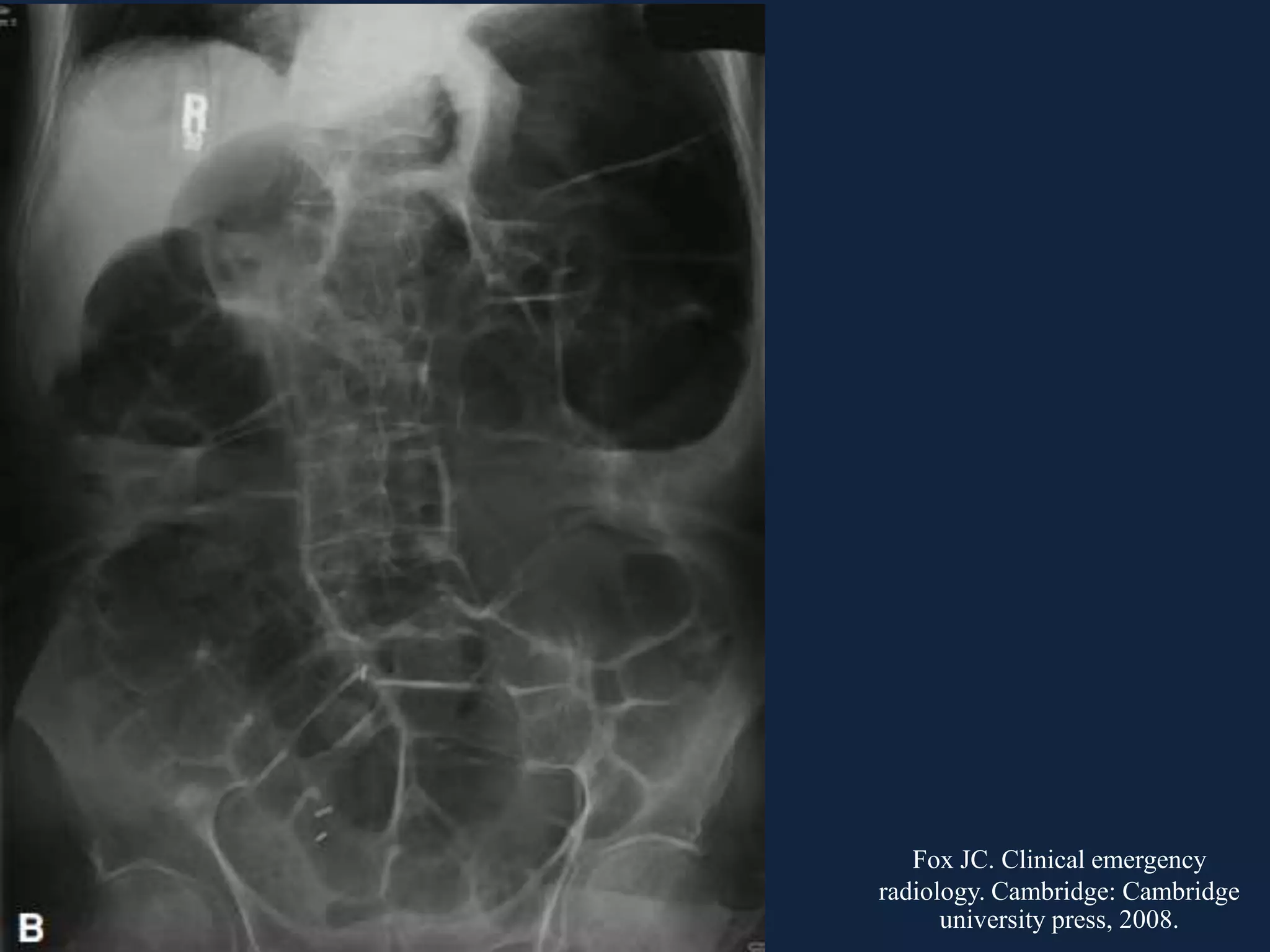
Colonic Obstruction
Facebook: Happy Friday Knight
General Surgical Residency Program
Thailand
Large bowel obstruction = intestinal obstruction
distal to ileocecal valve
Diagnostic Evaluation of
bowel obstruction
• Distinguish mechanical obstruction from ileus
• Determine etiology
• Discriminate partial from complete obstruction
• Discriminate simple from strangulating
obstruction
• Most common mechanical cause = CRC
• Common adynamic cause = acute colonic
pseudoobstruction
Cameron JL, Cameron AM. Current Surgical Therapy. 11th ed. Philadelphia: Elsevier, 2014.
IMAGING
Plain Abdomen
• Bowel obstruction
– Absent bowel gas distal to obstruction
– Different air-fluid level in the same loop
– String of pearl sign: small bubbles between
valvulae conniventes
– Distal large bowel obstruction, especially sigmoid
may be confused with generalized ileus if the
ileocecal valve is incompetent
Plain abdomen
• Ileus
– Diffusely gas-fill bowel including rectum
– Common causes of generalized ileus = abdominal
surgery, severe abdominal illness
– Localized ileus (sentinel loop): local inflammation
Fox JC. Clinical emergency radiology. Cambridge: Cambridge university press, 2008.
CANCER
• Most common symptom of a large bowel
obstruction from malignancy is abdominal
distention
• History: recent weight loss, bowel habit
change, change in stool consistency
• Vomiting is a late sign and can be
stercoraceous
Treatment
• Partial obstruction: NG decompression and
one-stage procedure
• For unresectable disease: stent or colostomy
• For resectable disease:
– Right side: right hemicolectomy
Treatment: Left Side
• Loop colostomy/ileostomy
• Primary resection with end colostomy (Hartmann’s
procedure)
• Primary resection and anastomosis
– Total or subtotal colectomy
– Segmental colectomy
• With intraoperative colonic irrigation
• With manual decompression
• Endoscopic colonic stenting (self-expanding metallic
stent)
– Palliation
– Bridge to surgery
• Colostomy with staged procedure VS
Hartmann’s procedure:
– Colostomy with staged procedure: no more
popular, only decompression, allow surgeon to
evaluate clinical staging, CA rectum
– Hartmann’ s procedure is recommended over the
previous
• Hartmann’s procedure VS primary resection
and anastomosis:
– Primary anastomosis is recommended BUT only in
experienced surgeons and be careful on
anastomotic leakage (2.2 – 12%)
• Total/subtotal colectomy VS segmental
colectomy with intraoperative colonic
irrigation:
– SCOTIA (subtotal colectomy versus on table
irrigation and anastomosis) trial: segmental
colectomy is better
– Intraoperative colonic irrigation: more SSI
– Subtotal colectomy indication:
• Right colon ischemia
• Synchronous proximal malignant
• Intraoperative colonic irrigation VS manual
decompression:
– Both no different morbidity and mortality
– Manual decompression takes less time
• Stent VS colostomy for palliation:
– Stent is recommended due to safe and high success
rate
– Avoid stent in patients with avastin
– Stent: bridge from emergency to elective surgery
• Stent for bridging to surgery VS emergency
surgery:
– Stent is recommended
Self-Expandable Metallic Stent
(SEMS)
• Ideally: 2 cm above and below the stricture
• The waist of the stent should be in the middle
• Fully recoil within 24 – 48 hrs
• Complications:
– Perforation: associated bevacizumab
– Stent migration
– Stent failure
INFLAMMATION
• May be secondary to:
– Acute: diverticulitis, Crohn’s disease, radiation
colitis
– Chronic: from previous diverticulitis, pelvic
radiation, prior bowel anastomosis
Acute Inflammation
• Inflammation of diverticulitis, Crohn’s disease,
radiation colitis
• Can be treated with nonoperative management
if absent of peritonitis: NG, IV resusctitation,
antibiotics
• Surgery: Hartmann’s procedure or loop
colostomy for difficult cases
• Malignancy must always be a concern:
colonoscope
Chronic Stricture
• From stricture, inflammation, and fibrosis
• Previous diverticulitis, pelvic radiation, prior
bowel anastomosis
• Present with chronic constipation and partial
obstructive symptoms
• Should be evaluated for malignancy
• Treatment: elective one-stage resection or stent
COLONIC VOLVULUS
• Volvulus: a torsion on the organ pedicle
• Volvulus of the large bowel results from the
colon’s twisting on its mesentery  producing
symptoms by narrowing of the bowel lumen and
strangulation of the blood vessels
• Third most common
– Sigmoid
– Ileosigmoid
– Cecum
– Transverse colon
– Splenic flexure
Sigmoid Volvulus
• Most common volvulus
• Etiology:
– Sigmoid must be long, floppy, narrow mesenteric
root
– Disproportion between antimesenteric and
mesenteric sites when bowel becomes distended
and elongated
– Antimesenteric border elongates 30% whereas
mesenteric site 10%
Gordon PH, Nivatvongs S. Principles and practice of surgery for the colon, rectum, and anus. 3rd ed. New york:
Informa healthcare USA, 2007.
Pathogenesis
• Torsion clockwise or counterclockwise
• Torsion at least 180 degree (asymtomatic and
physiologic if less than this)
• Closed-loop type of obstruction
Gordon PH, Nivatvongs S. Principles and practice of surgery for the colon, rectum, and anus. 3rd ed. New york:
Informa healthcare USA, 2007.
Clinical Presentation
• Acute fulminating type
– Younger
– Sudden onset and rapid course: early vomiting, diffuse
abdominal pain, signs of gangrene, minimal abdominal
distention
• Subacute progressive type
– More common
– History of chronic constipation
– Abdominal distention is extreme
Diagnosis
• Plain film
• Gastrograffin enema
• CT abdomen: identify strangulation
Coffee bean sign
Fox JC. Clinical emergency
radiology. Cambridge: Cambridge
university press, 2008.
Treatment
• Nonstrangulated
• Strangulated
Strangulated Sigmoid Volvulus
• Sigmoid resection with colostomy and
Hartmann’s procedure is safest
Management of Nonstrangulated
Sigmoid Volvulus
• Decompression
– Rigid sigmoidoscopic
– Colonoscopic and flexible sigmoidoscopic
• Surgical management
– Sigmoidectomy
– Nonresective
• Sigmoid decompression and colopexy
• Mesosigmoidoplasty
• Foley catheter sigmoidostomy
• T-fastener sigmoidopexy
Rigid Sigmoidoscopic Decompression
• Jackknife position
• Lateral decubitus is acceptable
• Carefully insert sigmoidoscope and inspect for
ischemic mucosa or until torsion site is seen
• 40 to 60 cm rectal tube is gently passed and
fixed for 48 hrs
Colonoscopic and Flexible
Sigmoidoscopic Decompression
• Largely replace rigid sigmoidoscope
• Colonoscope itself can pass torsion site
Sigmoidectomy
• Primary anastomosis is safe after successful
decompression
Sigmoid Decompression and Colopexy
• First deflate sigmoid by needle
• Insert rectal tube
• 4 -5 days after: colopexy
– 6 to 8 Strips of Gore-Tex band encircle the bowel
and are sutured to abdominal wall
Gordon PH, Nivatvongs S. Principles and practice of surgery for the colon, rectum, and anus. 3rd ed. New york:
Informa healthcare USA, 2007.
Mesosigmoidoplasty
Gordon PH, Nivatvongs S. Principles and practice of surgery for the colon, rectum, and anus. 3rd ed. New york:
Informa healthcare USA, 2007.
Foley Catheter Sigmoidostomy
• Fix redundant sigmoid to anterior abdominal
wall
• Foley catheter is removed 2 weeks later
T-Fasteners Sigmoidopexy
• Fix sigmoid to anterior abdominal wall with the
aid of colonoscopy
• 3 to 4 T-fastener pulls the sigmoid colon up
against the abdominal wall and is tightened on the
skin over the cotton pledget .These fasteners are
cut at the skin level after 28 days and pass with
stool. By this time, the sigmoid colon should
adhere to the anterior abdominal wall.
• The procedure is performed under mild sedation
Gordon PH, Nivatvongs S. Principles and practice of surgery for the colon, rectum, and anus. 3rd ed. New york:
Informa healthcare USA, 2007.
Ileosigmoid Knotting
• Loop of sigmoid and ileum wrap each other
• Initiated by hyperactive ileum that winds itself
around the pedicle of a passive sigmoid loop
• Majority of patients present with peritonitis
Gordon PH, Nivatvongs S. Principles and practice of surgery for the colon, rectum, and anus. 3rd ed. New york:
Informa healthcare USA, 2007.
Treatment
• Emergency condition
• It cannot reduce via endoscopy
• Viable bowel: untie the knot and resection may
be primary anastomosis
• Gangrenous bowel: En bloc resection with no
anastomosis
Cecal Volvulus
• Younger
• Congenital anatomic variant: incomplete
peritoneal fixation of right colon
• Clinical presentation
– Acute fulminating: acute abdomen
– Acute obstruction: bowel obstruction
– Intermittent or recurrent: subside spontaneously
• Reversed C
• Bird’s beak
• Whirl sign
Fox JC. Clinical emergency
radiology. Cambridge: Cambridge
university press, 2008.
Treatment
• Nonoperative reduction is generally
unsuccessful and hazardous
• Right hemicolectomy with ileostomy for
gangrenous
• Cecostomy with colopexy with peritoneal flap
• Last resort: CT-guided percutaneous
decompression and using SPC set
Gordon PH, Nivatvongs S. Principles and practice of surgery for the colon, rectum, and anus. 3rd ed. New york:
Informa healthcare USA, 2007.
Volvulus of Transverse Colon
Gordon PH, Nivatvongs S. Principles and practice of surgery for the colon, rectum, and anus. 3rd ed. New york:
Informa healthcare USA, 2007.
OGILVIE’S SYNDROME
• Colonic distension in the absence of
mechanical obstruction
• Complication: ischemia and perforation
• Clinical features:
– Abdominal distension
– Abdominal pain 80%
– Nausea and vomiting 60%
– Presence of bowel sound
• ACPO occurs in hospitalized patients with
significant comorbidity and may occurs
postoperative day 4
• Mechanical obstruction should be ruled out by
water-soluble enema
Fox JC. Clinical emergency
radiology. Cambridge: Cambridge
university press, 2008.
Management
• Supportive treatment
• Neostigmine
• Colonoscopy
• Tube cecostomy
• surgery
Supportive Treatment
• NPO
• Correct fluid and electrolyte imbalance: K, Mg,
Ca
• Treat systemic illness
• Stop exacerbating medications: opiates,
anticholinergics, antidiarrheal medication
(loperamide), TCA, antipsychotics, CCB, BB
• NG and rectal tubes
• Ambulation
• Avoid laxatives and enema
Neostigmine
• Acetylcholinesterase inhibitor
• Side effects: abdominal pain, nausea,
excessive salivation, bradyarrhythmia, renal
failure, exacerbate bronchoconstriction
• 2 mg IV with atropine
Colonoscopy
• Should be consider:
– Not improve after 24 hrs
– Mark cecal distention (>12cm) with significant
duration (>6days)
– Cannot tolerate neostigmine
• Colon should NOT be prepped with enema
• After successful colonoscopy, decompression
tube should be placed, flush q4-6hr with
saline, and remove after 72 hrs
Tube Cecostomy
• Can be considered:
– Do not respond to pharmacologic or endoscopic
decompression
– No evidence of ischemia
– Poor surgical candidate
Surgery
• Should only be considered when others
unsuccessful
• Colonic resection
• colostomy
Cameron JL, Cameron AM. Current Surgical Therapy. 11th ed. Philadelphia: Elsevier, 2014.
INTUSSUSCEPTION
• Present with melena or guaiac positive stool
• Less with abdominal pain, nausea, and
vomiting
• Mass may be palpated
• Most useful diagnostic test: CT
– target-shaped area of colon that represents the
intussusceptum in the center surrounded by the
intussuscipiens
Cameron JL, Cameron AM. Current Surgical Therapy. 11th ed. Philadelphia: Elsevier, 2014.
• Unlike children, adult intussusception cause by
malignant focus: 43%
• Treatment: surgical resection along lymphatic
drainage without reduction
– Rt side  Rt hemicolectomy
– Lt side  Hartmann procedure
References
Brunicardi FC et al. Schwartz’s Principles of Surgery. 10th ed. McGraw-Hill
Education, 2015.
Cameron JL, Cameron AM. Current Surgical Therapy. 11th ed. Philadelphia:
Elsevier, 2014.
Fox JC. Clinical emergency radiology. Cambridge: Cambridge university
press, 2008.
Gordon PH, Nivatvongs S. Principles and practice of surgery for the colon,
rectum, and anus. 3rd ed. New york: Informa healthcare USA, 2007.
สุขไชย สาทถาพร และคณะ. ศัลยศาสตร์ทั่วไป เล่ม 21. กรุงเทพ: กรุงเทพเวชสาร, 2558.

Colonic obstruction

  • 1.
    Colonic Obstruction Facebook: HappyFriday Knight General Surgical Residency Program Thailand
  • 2.
    Large bowel obstruction= intestinal obstruction distal to ileocecal valve
  • 3.
    Diagnostic Evaluation of bowelobstruction • Distinguish mechanical obstruction from ileus • Determine etiology • Discriminate partial from complete obstruction • Discriminate simple from strangulating obstruction
  • 4.
    • Most commonmechanical cause = CRC • Common adynamic cause = acute colonic pseudoobstruction
  • 5.
    Cameron JL, CameronAM. Current Surgical Therapy. 11th ed. Philadelphia: Elsevier, 2014.
  • 6.
  • 7.
    Plain Abdomen • Bowelobstruction – Absent bowel gas distal to obstruction – Different air-fluid level in the same loop – String of pearl sign: small bubbles between valvulae conniventes – Distal large bowel obstruction, especially sigmoid may be confused with generalized ileus if the ileocecal valve is incompetent
  • 8.
    Plain abdomen • Ileus –Diffusely gas-fill bowel including rectum – Common causes of generalized ileus = abdominal surgery, severe abdominal illness – Localized ileus (sentinel loop): local inflammation
  • 9.
    Fox JC. Clinicalemergency radiology. Cambridge: Cambridge university press, 2008.
  • 10.
    Fox JC. Clinicalemergency radiology. Cambridge: Cambridge university press, 2008.
  • 11.
  • 12.
    • Most commonsymptom of a large bowel obstruction from malignancy is abdominal distention • History: recent weight loss, bowel habit change, change in stool consistency • Vomiting is a late sign and can be stercoraceous
  • 13.
    Treatment • Partial obstruction:NG decompression and one-stage procedure • For unresectable disease: stent or colostomy • For resectable disease: – Right side: right hemicolectomy
  • 14.
    Treatment: Left Side •Loop colostomy/ileostomy • Primary resection with end colostomy (Hartmann’s procedure) • Primary resection and anastomosis – Total or subtotal colectomy – Segmental colectomy • With intraoperative colonic irrigation • With manual decompression • Endoscopic colonic stenting (self-expanding metallic stent) – Palliation – Bridge to surgery
  • 15.
    • Colostomy withstaged procedure VS Hartmann’s procedure: – Colostomy with staged procedure: no more popular, only decompression, allow surgeon to evaluate clinical staging, CA rectum – Hartmann’ s procedure is recommended over the previous
  • 16.
    • Hartmann’s procedureVS primary resection and anastomosis: – Primary anastomosis is recommended BUT only in experienced surgeons and be careful on anastomotic leakage (2.2 – 12%)
  • 17.
    • Total/subtotal colectomyVS segmental colectomy with intraoperative colonic irrigation: – SCOTIA (subtotal colectomy versus on table irrigation and anastomosis) trial: segmental colectomy is better – Intraoperative colonic irrigation: more SSI – Subtotal colectomy indication: • Right colon ischemia • Synchronous proximal malignant
  • 18.
    • Intraoperative colonicirrigation VS manual decompression: – Both no different morbidity and mortality – Manual decompression takes less time
  • 19.
    • Stent VScolostomy for palliation: – Stent is recommended due to safe and high success rate – Avoid stent in patients with avastin – Stent: bridge from emergency to elective surgery
  • 20.
    • Stent forbridging to surgery VS emergency surgery: – Stent is recommended
  • 21.
    Self-Expandable Metallic Stent (SEMS) •Ideally: 2 cm above and below the stricture • The waist of the stent should be in the middle • Fully recoil within 24 – 48 hrs • Complications: – Perforation: associated bevacizumab – Stent migration – Stent failure
  • 22.
  • 23.
    • May besecondary to: – Acute: diverticulitis, Crohn’s disease, radiation colitis – Chronic: from previous diverticulitis, pelvic radiation, prior bowel anastomosis
  • 24.
    Acute Inflammation • Inflammationof diverticulitis, Crohn’s disease, radiation colitis • Can be treated with nonoperative management if absent of peritonitis: NG, IV resusctitation, antibiotics • Surgery: Hartmann’s procedure or loop colostomy for difficult cases • Malignancy must always be a concern: colonoscope
  • 25.
    Chronic Stricture • Fromstricture, inflammation, and fibrosis • Previous diverticulitis, pelvic radiation, prior bowel anastomosis • Present with chronic constipation and partial obstructive symptoms • Should be evaluated for malignancy • Treatment: elective one-stage resection or stent
  • 26.
  • 27.
    • Volvulus: atorsion on the organ pedicle • Volvulus of the large bowel results from the colon’s twisting on its mesentery  producing symptoms by narrowing of the bowel lumen and strangulation of the blood vessels • Third most common – Sigmoid – Ileosigmoid – Cecum – Transverse colon – Splenic flexure
  • 28.
  • 29.
    • Most commonvolvulus • Etiology: – Sigmoid must be long, floppy, narrow mesenteric root – Disproportion between antimesenteric and mesenteric sites when bowel becomes distended and elongated – Antimesenteric border elongates 30% whereas mesenteric site 10%
  • 30.
    Gordon PH, NivatvongsS. Principles and practice of surgery for the colon, rectum, and anus. 3rd ed. New york: Informa healthcare USA, 2007.
  • 31.
    Pathogenesis • Torsion clockwiseor counterclockwise • Torsion at least 180 degree (asymtomatic and physiologic if less than this) • Closed-loop type of obstruction
  • 32.
    Gordon PH, NivatvongsS. Principles and practice of surgery for the colon, rectum, and anus. 3rd ed. New york: Informa healthcare USA, 2007.
  • 33.
    Clinical Presentation • Acutefulminating type – Younger – Sudden onset and rapid course: early vomiting, diffuse abdominal pain, signs of gangrene, minimal abdominal distention • Subacute progressive type – More common – History of chronic constipation – Abdominal distention is extreme
  • 34.
    Diagnosis • Plain film •Gastrograffin enema • CT abdomen: identify strangulation
  • 35.
    Coffee bean sign FoxJC. Clinical emergency radiology. Cambridge: Cambridge university press, 2008.
  • 37.
  • 38.
    Strangulated Sigmoid Volvulus •Sigmoid resection with colostomy and Hartmann’s procedure is safest
  • 39.
    Management of Nonstrangulated SigmoidVolvulus • Decompression – Rigid sigmoidoscopic – Colonoscopic and flexible sigmoidoscopic • Surgical management – Sigmoidectomy – Nonresective • Sigmoid decompression and colopexy • Mesosigmoidoplasty • Foley catheter sigmoidostomy • T-fastener sigmoidopexy
  • 40.
    Rigid Sigmoidoscopic Decompression •Jackknife position • Lateral decubitus is acceptable • Carefully insert sigmoidoscope and inspect for ischemic mucosa or until torsion site is seen • 40 to 60 cm rectal tube is gently passed and fixed for 48 hrs
  • 41.
    Colonoscopic and Flexible SigmoidoscopicDecompression • Largely replace rigid sigmoidoscope • Colonoscope itself can pass torsion site
  • 42.
    Sigmoidectomy • Primary anastomosisis safe after successful decompression
  • 43.
    Sigmoid Decompression andColopexy • First deflate sigmoid by needle • Insert rectal tube • 4 -5 days after: colopexy – 6 to 8 Strips of Gore-Tex band encircle the bowel and are sutured to abdominal wall
  • 44.
    Gordon PH, NivatvongsS. Principles and practice of surgery for the colon, rectum, and anus. 3rd ed. New york: Informa healthcare USA, 2007.
  • 45.
    Mesosigmoidoplasty Gordon PH, NivatvongsS. Principles and practice of surgery for the colon, rectum, and anus. 3rd ed. New york: Informa healthcare USA, 2007.
  • 46.
    Foley Catheter Sigmoidostomy •Fix redundant sigmoid to anterior abdominal wall • Foley catheter is removed 2 weeks later
  • 47.
    T-Fasteners Sigmoidopexy • Fixsigmoid to anterior abdominal wall with the aid of colonoscopy • 3 to 4 T-fastener pulls the sigmoid colon up against the abdominal wall and is tightened on the skin over the cotton pledget .These fasteners are cut at the skin level after 28 days and pass with stool. By this time, the sigmoid colon should adhere to the anterior abdominal wall. • The procedure is performed under mild sedation
  • 48.
    Gordon PH, NivatvongsS. Principles and practice of surgery for the colon, rectum, and anus. 3rd ed. New york: Informa healthcare USA, 2007.
  • 49.
  • 50.
    • Loop ofsigmoid and ileum wrap each other • Initiated by hyperactive ileum that winds itself around the pedicle of a passive sigmoid loop • Majority of patients present with peritonitis
  • 51.
    Gordon PH, NivatvongsS. Principles and practice of surgery for the colon, rectum, and anus. 3rd ed. New york: Informa healthcare USA, 2007.
  • 52.
    Treatment • Emergency condition •It cannot reduce via endoscopy • Viable bowel: untie the knot and resection may be primary anastomosis • Gangrenous bowel: En bloc resection with no anastomosis
  • 53.
  • 54.
    • Younger • Congenitalanatomic variant: incomplete peritoneal fixation of right colon • Clinical presentation – Acute fulminating: acute abdomen – Acute obstruction: bowel obstruction – Intermittent or recurrent: subside spontaneously
  • 55.
    • Reversed C •Bird’s beak • Whirl sign Fox JC. Clinical emergency radiology. Cambridge: Cambridge university press, 2008.
  • 56.
    Treatment • Nonoperative reductionis generally unsuccessful and hazardous • Right hemicolectomy with ileostomy for gangrenous • Cecostomy with colopexy with peritoneal flap • Last resort: CT-guided percutaneous decompression and using SPC set
  • 57.
    Gordon PH, NivatvongsS. Principles and practice of surgery for the colon, rectum, and anus. 3rd ed. New york: Informa healthcare USA, 2007.
  • 58.
  • 59.
    Gordon PH, NivatvongsS. Principles and practice of surgery for the colon, rectum, and anus. 3rd ed. New york: Informa healthcare USA, 2007.
  • 60.
  • 61.
    • Colonic distensionin the absence of mechanical obstruction • Complication: ischemia and perforation • Clinical features: – Abdominal distension – Abdominal pain 80% – Nausea and vomiting 60% – Presence of bowel sound
  • 62.
    • ACPO occursin hospitalized patients with significant comorbidity and may occurs postoperative day 4 • Mechanical obstruction should be ruled out by water-soluble enema
  • 63.
    Fox JC. Clinicalemergency radiology. Cambridge: Cambridge university press, 2008.
  • 64.
    Management • Supportive treatment •Neostigmine • Colonoscopy • Tube cecostomy • surgery
  • 65.
    Supportive Treatment • NPO •Correct fluid and electrolyte imbalance: K, Mg, Ca • Treat systemic illness • Stop exacerbating medications: opiates, anticholinergics, antidiarrheal medication (loperamide), TCA, antipsychotics, CCB, BB • NG and rectal tubes • Ambulation • Avoid laxatives and enema
  • 66.
    Neostigmine • Acetylcholinesterase inhibitor •Side effects: abdominal pain, nausea, excessive salivation, bradyarrhythmia, renal failure, exacerbate bronchoconstriction • 2 mg IV with atropine
  • 67.
    Colonoscopy • Should beconsider: – Not improve after 24 hrs – Mark cecal distention (>12cm) with significant duration (>6days) – Cannot tolerate neostigmine • Colon should NOT be prepped with enema • After successful colonoscopy, decompression tube should be placed, flush q4-6hr with saline, and remove after 72 hrs
  • 68.
    Tube Cecostomy • Canbe considered: – Do not respond to pharmacologic or endoscopic decompression – No evidence of ischemia – Poor surgical candidate
  • 69.
    Surgery • Should onlybe considered when others unsuccessful • Colonic resection • colostomy
  • 70.
    Cameron JL, CameronAM. Current Surgical Therapy. 11th ed. Philadelphia: Elsevier, 2014.
  • 71.
  • 72.
    • Present withmelena or guaiac positive stool • Less with abdominal pain, nausea, and vomiting • Mass may be palpated • Most useful diagnostic test: CT – target-shaped area of colon that represents the intussusceptum in the center surrounded by the intussuscipiens
  • 73.
    Cameron JL, CameronAM. Current Surgical Therapy. 11th ed. Philadelphia: Elsevier, 2014.
  • 74.
    • Unlike children,adult intussusception cause by malignant focus: 43% • Treatment: surgical resection along lymphatic drainage without reduction – Rt side  Rt hemicolectomy – Lt side  Hartmann procedure
  • 75.
    References Brunicardi FC etal. Schwartz’s Principles of Surgery. 10th ed. McGraw-Hill Education, 2015. Cameron JL, Cameron AM. Current Surgical Therapy. 11th ed. Philadelphia: Elsevier, 2014. Fox JC. Clinical emergency radiology. Cambridge: Cambridge university press, 2008. Gordon PH, Nivatvongs S. Principles and practice of surgery for the colon, rectum, and anus. 3rd ed. New york: Informa healthcare USA, 2007. สุขไชย สาทถาพร และคณะ. ศัลยศาสตร์ทั่วไป เล่ม 21. กรุงเทพ: กรุงเทพเวชสาร, 2558.

Editor's Notes

  • #10 Cecal valvulus = cecum ที่เป็น reverse C
  • #11 Ascending colon dilatation No air in rectum Present of small bowel dilation (IC valve incompetent)
  • #28 Pedicle = stalk
  • #37 ending sharply at the level of the site of torsion (‘‘bird’s beak’’ or ‘‘ace of spades’’ deformity
  • #48 หลักการเดียวกับ PEG
  • #68 หลักการคือ พยายามดูดลมออกและอย่าใส่ลมมาก
  • #75 Treatment: คิดเหมือนเป็น large bowel obs