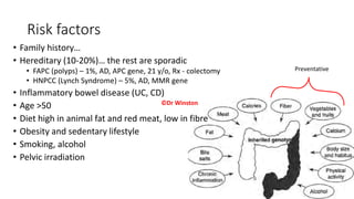
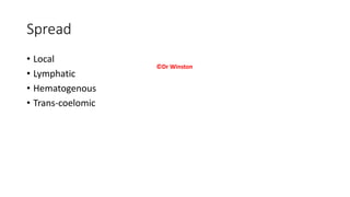
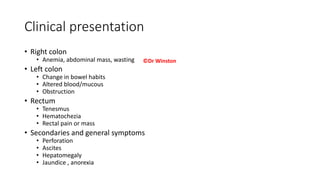
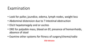
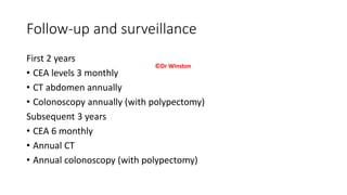
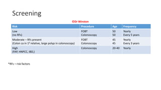
COLORECTAL CANCER
Dr Winston Makanga, M.B.Ch.B., M.Med. (Surg)
Consultant General and Laparoscopic Surgeon
Outline
• Introduction and epidemiology of Colorectal cancer (CRC)
• Surgical anatomy of the colon and rectum
• Classification of colorectal tumors
• Risk factors
• Pathology
• Staging
• Clinical presentation and examination
• Investigation
• Differential diagnosis
• Treatment and follow-up
©Dr Winston
Introduction & epidemiology
• Uncontrolled multiplication of cells in the layers of the colon
• Third commonest cancer worldwide (WHO 2022)
• Third commonest cause of cancer related mortality
• Fourth commonest cancer in Kenya (KEMRI)
• M:F ratio 1:1
©Dr Winston
Surgical Anatomy
©Dr Winston
Surgical anatomy 2 - Mesorectum
©Dr Winston
Classification
• Benign tumors
• Polyps (Inflammatory, PZP, Harmatoma, Adenomatous)
• Lipoma
• Hemangioma
• Papilloma
• Neurofibroma
• Malignant
• Primary
• Carcinoma/epithelial
• Lymphoma
• Carcinoid tumor
• Secondary: Direct invasion from adjacent organs, transcoelomic spread
©Dr Winston
Risk factors
• Family history…
• Hereditary (10-20%)… the rest are sporadic
• FAPC (polyps) – 1%, AD, APC gene, 21 y/o, Rx - colectomy
• HNPCC (Lynch Syndrome) – 5%, AD, MMR gene
• Inflammatory bowel disease (UC, CD)
• Age >50
• Diet high in animal fat and red meat, low in fibre
• Obesity and sedentary lifestyle
• Smoking, alcohol
• Pelvic irradiation
Preventative
©Dr Winston
Pathology
• Most malignancies are epithelial in origin (adenocarcinoma)
• Adenoma to carcinoma sequence ~ 10 years
• Metachronous vs synchronous tumor (5%)
• Macroscopically
• Eccentric mass
• Ulcer
• Annular
• Diffuse infiltrating growth
• Microscopically
• Adenocarcinoma (95%)
• Medullary
• Micropapillary
• Mucinous
• Signet ring
• Others: Lymphoma, GIST, carcinoid
©Dr Winston
Spread
• Local
• Lymphatic
• Hematogenous
• Trans-coelomic
©Dr Winston
Staging
• Tumor
• T1 – Superficial to muscle layer
• T2 – Invading muscle layer
• T3 – Beyond muscle layer to pericolorectal tissue
• T4 – Invading other organs
• Node
• N0 – no nodal metastasis
• N1 – 1-3 nodes
• N2 – 4 or more nodes
• Metastasis
• M0 – no distant metastasis
• M1 – metastasis to distant organ
Stage 1 – T1/2, N0, M0
Stage 2 – T3/4, N0, M0
Stage 3 – Any T, N1/2, M0
Stage 4 – Any T, Any N, M1
©Dr Winston
Clinical presentation
• Right colon
• Anemia, abdominal mass, wasting
• Left colon
• Change in bowel habits
• Altered blood/mucous
• Obstruction
• Rectum
• Tenesmus
• Hematochezia
• Rectal pain or mass
• Secondaries and general symptoms
• Perforation
• Ascites
• Hepatomegaly
• Jaundice , anorexia
©Dr Winston
Examination
• Look for pallor, jaundice, edema, lymph nodes, weight loss
• Abdominal distension due to ? Intestinal obstruction
• Elicit hepatomegaly and or ascites
• DRE for palpable mass, blood on EF, presence of hemorrhoids,
absence of stool
• Examine other systems for fitness of surgery/chemo/radio
©Dr Winston
Investigation
• Blood count
• Fecal occult blood (FOBT, FIT)
• Barium enema – apple core deformity
• Colonoscopy/Sigmoidoscopy
• CT abdomen, CT Colography
• MRI pelvis
©Dr Winston
Differential diagnosis
• Diverticulosis
• Inflamatory bowel disease – UC or CD
• Dysentery
• Ileus/chronic constipation
• Bowel ischemia
• Infectious colitis (amoebic colitis)
©Dr Winston
Treatment
• Mainstay is surgery if tumor is colonic
• Right, extended right colectomy
• Left colectomy
• Large disease in rectum – Neo-adjuvant treatment with chemoradiotherapy
• Surgery for rectal ca
• Local transrectal resections (TEM, TAMIS)
• Anterior or low anterior resection
• Abdomino-perineal resection (APR)
• Adjuvant chemotherapy
• Oxaliplatin, 5 Fluorouracil, Leukovorin, capecitabine
• Second-line chemo – irinotecan, taxanes
• Targeted therapy: bevacizumab
• Palliation
• Local resection, bypass, colostomy
• Irradiation and ? Palliative chemotherapy
• Ascitic drainage, analgesics
©Dr Winston
Anatomical basis of
types of colectomy
©Dr Winston
Local surgery for small rectal cancer
©Dr Winston
Follow-up and surveillance
First 2 years
• CEA levels 3 monthly
• CT abdomen annually
• Colonoscopy annually (with polypectomy)
Subsequent 3 years
• CEA 6 monthly
• Annual CT
• Annual colonoscopy (with polypectomy)
©Dr Winston
Screening
Risk Procedure Age Frequency
Low
(no RFs)
FOBT
Colonoscopy
50
50
Yearly
Every 5 years
Moderate – RFs present
(Colon ca in 10 relative, large polyp in colonoscopy)
FOBT
Colonoscopy
45
45
Yearly
Every 3 years
High
(FAP, HNPCC, IBD,)
Colonoscopy 20-40 Yearly
*RFs – risk factors
©Dr Winston
Review
• Epidemiology of CRC
• Surgical anatomy of the colon and rectum
• Classification of colorectal tumors
• Risk factors
• Pathology
• Staging
• Clinical presentation and examination
• Investigation
• Differential diagnosis
• Treatment and follow-up
©Dr Winston
Thank you

Colon Cancer.pptx

  • 1.
    COLORECTAL CANCER Dr WinstonMakanga, M.B.Ch.B., M.Med. (Surg) Consultant General and Laparoscopic Surgeon
  • 2.
    Outline • Introduction andepidemiology of Colorectal cancer (CRC) • Surgical anatomy of the colon and rectum • Classification of colorectal tumors • Risk factors • Pathology • Staging • Clinical presentation and examination • Investigation • Differential diagnosis • Treatment and follow-up ©Dr Winston
  • 3.
    Introduction & epidemiology •Uncontrolled multiplication of cells in the layers of the colon • Third commonest cancer worldwide (WHO 2022) • Third commonest cause of cancer related mortality • Fourth commonest cancer in Kenya (KEMRI) • M:F ratio 1:1 ©Dr Winston
  • 4.
  • 5.
    Surgical anatomy 2- Mesorectum ©Dr Winston
  • 6.
    Classification • Benign tumors •Polyps (Inflammatory, PZP, Harmatoma, Adenomatous) • Lipoma • Hemangioma • Papilloma • Neurofibroma • Malignant • Primary • Carcinoma/epithelial • Lymphoma • Carcinoid tumor • Secondary: Direct invasion from adjacent organs, transcoelomic spread ©Dr Winston
  • 7.
    Risk factors • Familyhistory… • Hereditary (10-20%)… the rest are sporadic • FAPC (polyps) – 1%, AD, APC gene, 21 y/o, Rx - colectomy • HNPCC (Lynch Syndrome) – 5%, AD, MMR gene • Inflammatory bowel disease (UC, CD) • Age >50 • Diet high in animal fat and red meat, low in fibre • Obesity and sedentary lifestyle • Smoking, alcohol • Pelvic irradiation Preventative ©Dr Winston
  • 8.
    Pathology • Most malignanciesare epithelial in origin (adenocarcinoma) • Adenoma to carcinoma sequence ~ 10 years • Metachronous vs synchronous tumor (5%) • Macroscopically • Eccentric mass • Ulcer • Annular • Diffuse infiltrating growth • Microscopically • Adenocarcinoma (95%) • Medullary • Micropapillary • Mucinous • Signet ring • Others: Lymphoma, GIST, carcinoid ©Dr Winston
  • 9.
    Spread • Local • Lymphatic •Hematogenous • Trans-coelomic ©Dr Winston
  • 10.
    Staging • Tumor • T1– Superficial to muscle layer • T2 – Invading muscle layer • T3 – Beyond muscle layer to pericolorectal tissue • T4 – Invading other organs • Node • N0 – no nodal metastasis • N1 – 1-3 nodes • N2 – 4 or more nodes • Metastasis • M0 – no distant metastasis • M1 – metastasis to distant organ Stage 1 – T1/2, N0, M0 Stage 2 – T3/4, N0, M0 Stage 3 – Any T, N1/2, M0 Stage 4 – Any T, Any N, M1 ©Dr Winston
  • 11.
    Clinical presentation • Rightcolon • Anemia, abdominal mass, wasting • Left colon • Change in bowel habits • Altered blood/mucous • Obstruction • Rectum • Tenesmus • Hematochezia • Rectal pain or mass • Secondaries and general symptoms • Perforation • Ascites • Hepatomegaly • Jaundice , anorexia ©Dr Winston
  • 12.
    Examination • Look forpallor, jaundice, edema, lymph nodes, weight loss • Abdominal distension due to ? Intestinal obstruction • Elicit hepatomegaly and or ascites • DRE for palpable mass, blood on EF, presence of hemorrhoids, absence of stool • Examine other systems for fitness of surgery/chemo/radio ©Dr Winston
  • 13.
    Investigation • Blood count •Fecal occult blood (FOBT, FIT) • Barium enema – apple core deformity • Colonoscopy/Sigmoidoscopy • CT abdomen, CT Colography • MRI pelvis ©Dr Winston
  • 14.
    Differential diagnosis • Diverticulosis •Inflamatory bowel disease – UC or CD • Dysentery • Ileus/chronic constipation • Bowel ischemia • Infectious colitis (amoebic colitis) ©Dr Winston
  • 15.
    Treatment • Mainstay issurgery if tumor is colonic • Right, extended right colectomy • Left colectomy • Large disease in rectum – Neo-adjuvant treatment with chemoradiotherapy • Surgery for rectal ca • Local transrectal resections (TEM, TAMIS) • Anterior or low anterior resection • Abdomino-perineal resection (APR) • Adjuvant chemotherapy • Oxaliplatin, 5 Fluorouracil, Leukovorin, capecitabine • Second-line chemo – irinotecan, taxanes • Targeted therapy: bevacizumab • Palliation • Local resection, bypass, colostomy • Irradiation and ? Palliative chemotherapy • Ascitic drainage, analgesics ©Dr Winston
  • 16.
    Anatomical basis of typesof colectomy ©Dr Winston
  • 17.
    Local surgery forsmall rectal cancer ©Dr Winston
  • 18.
    Follow-up and surveillance First2 years • CEA levels 3 monthly • CT abdomen annually • Colonoscopy annually (with polypectomy) Subsequent 3 years • CEA 6 monthly • Annual CT • Annual colonoscopy (with polypectomy) ©Dr Winston
  • 19.
    Screening Risk Procedure AgeFrequency Low (no RFs) FOBT Colonoscopy 50 50 Yearly Every 5 years Moderate – RFs present (Colon ca in 10 relative, large polyp in colonoscopy) FOBT Colonoscopy 45 45 Yearly Every 3 years High (FAP, HNPCC, IBD,) Colonoscopy 20-40 Yearly *RFs – risk factors ©Dr Winston
  • 20.
    Review • Epidemiology ofCRC • Surgical anatomy of the colon and rectum • Classification of colorectal tumors • Risk factors • Pathology • Staging • Clinical presentation and examination • Investigation • Differential diagnosis • Treatment and follow-up ©Dr Winston
  • 21.