Downloaded 106 times


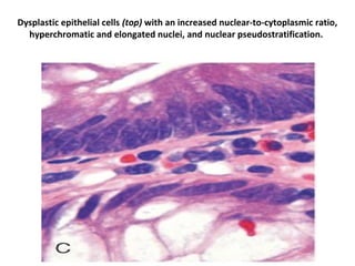


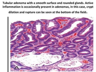


Neoplastic polyps can be benign or malignant. Adenomas are benign epithelial tumors that have the potential to become cancerous over time. There are several types of adenomas classified by their histological features, including tubular, villous, and tubulovillous. Large or villous adenomas have a higher risk of already containing cancer. Removal of adenomas is important as nearly all colon cancers develop from these polyps. Risk factors for the adenoma containing high-grade dysplasia or cancer include large size over 1 cm, villous histology, presence of high-grade dysplasia, and having multiple polyps.


