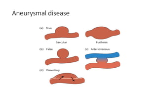
ARTERIAL VASO-OCCLUSIVE
DISEASE
Dr Winston Makanga, M.B.Ch.B, M.Med (Surg)
Consultant General and Laparoscopic Surgeon
MBChB 6 Thursday, November 17, 2022
Objectives of the lecture
• Surgical anatomy
• Physiology and functions
• Epidemiology
• Etiology
• Work-up (screening, diagnosis, staging)
• Treatment
Scope of diseases
• Trauma and its sequelae (blunt vs trauma)
• Pseudoaneurysm
• Intimal flap
• Laceration (partial vs complete)
• Aneurysmal disease
• Aorta
• Popliteal
• Occlusive disease (coronary, mesenteric, peripheral, cerebral)
• Atherosclerosis
• Raynaud’s disease
• Buergers disease
• HIV vasculopathy
• Embolism
Arterial trauma
• Laceration
• Transection
• Contusion
• Pseudoaneurysm
• Extrinsic compression
• AV fistula
Aneurysmal disease
Pathology of vaso-occlusive disease
• Acute ischemia: thrombotic or embolic
• Sudden formation of hemostatic plug over a pre-existing ruptured atheroma
• Chronic ischemia: atherosclerosis
• Progressive deposition of lipids in the intima with reactive inflammation
• Macrovascular disease: affects large named vessels
• Microvascular disease: affects small <0.3 mm vessels
Etiology
• Smoking
• Diabetes mellitus
• Hypertension
• HIV
• Connective tissue disease
• Age
• Obesity and sedentary lifestyle
• Dyslipidemia
Acute Presentation
• Pain
• Parasthesia
• Pulslessness
• Paralysis
• Coldness
Fontaine Classification of chronic disease
I. Assymptomatic
II. Intermittent claudication
III. Rest pain
IV. Tissue damage (ulcer/gangrene)
Classification by length involved
• Class A <3cm
• Class B 3 – 15 cm
• Class C > 15 cm
• Class D Complete occlusion
Clinical evaluation
• History
• Duration of pain/claudication distance
• Prior Rx for cardiac(angina)/aortic, cerebrovascular dx (TIA, CVA), amputation
• Coexistance of connective tissue disease – SLE
• Risk factor assessment: smoking, DM, HIV, dyslipidemia, ergot poisoning
• Examination
• CVS: pulse rate, rhythm, murmurs, abdominal pulsations and bruits
• Limb: skin, temperature, ABPI, pulses, cap refill, venous guttering, bruits
• Buergers test, exercise test
Investigation
• General blood tests
• RBS, HbA1c, urine sugar, UEC, Lipid levels, HIV test, CRP, ASA, Rh
• Cardiac assessment
• CXR, ECG, echo
• Specific limb/vascular exam
• Doppler (with quantitative ABPI)
• Duplex scan
• Angiography (conventional, DSA, CT or MR Angiography)
Differential diagnosis
• Venous claudication
• Neurogenic claudication
• Chronic compartment syndrome
• Bakers cyst
• Cord compression
• Arthritis
Treatment options
• General reduction of cardiovascular risk:
• Cessation of smoking, lipid lowering, control DM & HTN, antiplatelet meds
• Angioplasty
• Balloon dilatation
• Endovascular stenting
• Endaterectomy
• Grafting
• Acute obstruction
• Thrombolysis
• Thrombectomy
• Embolectomy
• Class 4: Amputations, debridement
Questions, comments

Arterial dse MB6.pptx

  • 1.
    ARTERIAL VASO-OCCLUSIVE DISEASE Dr WinstonMakanga, M.B.Ch.B, M.Med (Surg) Consultant General and Laparoscopic Surgeon MBChB 6 Thursday, November 17, 2022
  • 2.
    Objectives of thelecture • Surgical anatomy • Physiology and functions • Epidemiology • Etiology • Work-up (screening, diagnosis, staging) • Treatment
  • 3.
    Scope of diseases •Trauma and its sequelae (blunt vs trauma) • Pseudoaneurysm • Intimal flap • Laceration (partial vs complete) • Aneurysmal disease • Aorta • Popliteal • Occlusive disease (coronary, mesenteric, peripheral, cerebral) • Atherosclerosis • Raynaud’s disease • Buergers disease • HIV vasculopathy • Embolism
  • 4.
    Arterial trauma • Laceration •Transection • Contusion • Pseudoaneurysm • Extrinsic compression • AV fistula
  • 5.
  • 6.
    Pathology of vaso-occlusivedisease • Acute ischemia: thrombotic or embolic • Sudden formation of hemostatic plug over a pre-existing ruptured atheroma • Chronic ischemia: atherosclerosis • Progressive deposition of lipids in the intima with reactive inflammation • Macrovascular disease: affects large named vessels • Microvascular disease: affects small <0.3 mm vessels
  • 7.
    Etiology • Smoking • Diabetesmellitus • Hypertension • HIV • Connective tissue disease • Age • Obesity and sedentary lifestyle • Dyslipidemia
  • 8.
    Acute Presentation • Pain •Parasthesia • Pulslessness • Paralysis • Coldness
  • 9.
    Fontaine Classification ofchronic disease I. Assymptomatic II. Intermittent claudication III. Rest pain IV. Tissue damage (ulcer/gangrene)
  • 10.
    Classification by lengthinvolved • Class A <3cm • Class B 3 – 15 cm • Class C > 15 cm • Class D Complete occlusion
  • 11.
    Clinical evaluation • History •Duration of pain/claudication distance • Prior Rx for cardiac(angina)/aortic, cerebrovascular dx (TIA, CVA), amputation • Coexistance of connective tissue disease – SLE • Risk factor assessment: smoking, DM, HIV, dyslipidemia, ergot poisoning • Examination • CVS: pulse rate, rhythm, murmurs, abdominal pulsations and bruits • Limb: skin, temperature, ABPI, pulses, cap refill, venous guttering, bruits • Buergers test, exercise test
  • 12.
    Investigation • General bloodtests • RBS, HbA1c, urine sugar, UEC, Lipid levels, HIV test, CRP, ASA, Rh • Cardiac assessment • CXR, ECG, echo • Specific limb/vascular exam • Doppler (with quantitative ABPI) • Duplex scan • Angiography (conventional, DSA, CT or MR Angiography)
  • 14.
    Differential diagnosis • Venousclaudication • Neurogenic claudication • Chronic compartment syndrome • Bakers cyst • Cord compression • Arthritis
  • 15.
    Treatment options • Generalreduction of cardiovascular risk: • Cessation of smoking, lipid lowering, control DM & HTN, antiplatelet meds • Angioplasty • Balloon dilatation • Endovascular stenting • Endaterectomy • Grafting • Acute obstruction • Thrombolysis • Thrombectomy • Embolectomy • Class 4: Amputations, debridement
  • 17.