Clostridium Difficile
Infection(CDI)
OBJECTIVES:
• Introduction.
• Microbiology and Pathophysiology.
• Transmission.
• Risk factor.
• Clinical manifestation.
• Diagnosis.
• Treatment.
INTRODUCTION
CDI:
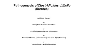
• Clostridioides difficile infection (CDI) is one of the most common
hospital-acquired infections and is an increasingly frequent
cause of morbidity and mortality among older adult hospitalized
patients.
• CDI is also increasingly diagnosed in younger patients and in
the community.
• C. difficile colonizes the human intestinal tract after the normal
gut flora has been disrupted (frequently in association with
antibiotic therapy) and it is the causative organism of antibiotic-
associated colitis including pseudomembranous colitis.
MICROBIOLOGY AND PATHOPHYSIOLOGY
• C. difficile is an anaerobic gram-positive, spore-forming, toxin-
producing bacillus first described in 1935.
• It was named "difficult clostridium" because of difficulty related
to its isolation and growth on conventional media.
• C. difficile can exist in spore and vegetative forms. Outside the
colon, it survives in spore form; spores are resistant to heat,
acid, and antibiotics.
• Once spores are in the intestine, they convert to their fully
functional vegetative, toxin-producing forms and become
susceptible to killing by antimicrobial agents.
• Toxins:
• C. difficile releases two potent exotoxins that mediate colitis
and diarrhea: toxin A and toxin B.
• The organism is rarely invasive, and nontoxigenic strains do not
cause CDI.
• C. difficile toxins contain a series of contiguous repeated units
at the carboxyl terminus. For toxin A, this region is important for
toxin A carbohydrate receptor binding to facilitate intracellular
transport as well as for antibody binding in enzyme-linked
immunosorbent assays.
• In vivo, stool toxin levels correlate with disease severity
• Toxin A (enterotoxin) causes inflammation leading to intestinal
fluid secretion and mucosal injury
• Toxin B (cytotoxin) is a major factor for the virulence of C.
difficile and its 10 times more potent than toxin A on a molar
basis for mediating colonic mucosal damage.
Pathogenesis ofClostridioides difficile
diarrhea:
TRANSMISSION
• Patients with C. difficile carriage are a reservoir for
environmental contamination in the presence or absence of
clinical infection.
• C difficile is highly transmissible via the fecal-oral route by
ingestion of spores.
• The organism can be cultured readily from the hospital
environment, including items and inert surfaces in patient
rooms as well as the hands, clothing, and stethoscopes of health
care workers.
RISK FACTORS
Antibiotic use is the most widely recognized and modifiable risk
factor for CDI.
Other established risk factors include:
• advanced age and severe comorbid illness.
• Hospitalization and gastric acid suppression.
• enteral feeding and gastrointestinal surgery.
• Obesity or Cirrhosis.
• inflammatory bowel disease.
• cancer chemotherapy (may be related to the antimicrobial effect of
chemotherapeutic agents and/or their immunosuppressive effects).
Risk factors for recurrent CDI include:
• age >65 years, severe underlying medical
disorders, need for ongoing therapy with
concomitant antibiotics during treatment for CDI.
• serum creatinine ≥1.2 mg/dL, and lack of an
antibody-mediated immune response to C.
difficile toxins especially toxin B.
Risk factors for complications associated with CDI:
• abnormal blood tests:
(white blood cell count <4000 cells/microL or ≥20,000 cells/microL,
albumin <25 g/L, blood urea nitrogen >7 mmol/L, and C-reactive
protein ≥150 mg/L), and abnormal vital signs (heart rate >90/minute,
respiratory rate >20/minute)
• perforation, toxic megacolon, colectomy, admission to intensive care
unit, death, include older age
• CDI can also occur in the absence of any risk factors.
CLINCAL MANIFISTATION
• CDI can cause a spectrum of manifestations ranging from an
asymptomatic carriage to fulminant disease with toxic
megacolon
• 1-Asymptomatic carriage:
• Asymptomatic individuals do not warrant screening for C.
difficile carriage, and individuals with asymptomatic carriage
of C. difficile do not warrant treatment or contact precautions.
• Its occurs in up to 20 percent of hospitalized adults; these
patients shed C. difficile in stool but do not have diarrhea or
other clinical symptoms.
2-Nonsevere disease:
• Watery diarrhea (≥3 loose stools in 24 hours) is the cardinal
symptom of CDI. Other manifestations include lower abdominal
pain and cramping, low-grade fever, nausea, and anorexia
• Diarrhea may be associated with mucus or occult blood, but
melena or hematochezia are rare.
3-Severe disease:
• Clinical manifestations of severe colitis include diarrhea, lower
quadrant or diffuse abdominal pain, abdominal distention, fever,
hypovolemia, lactic acidosis, hypoalbuminemia, elevated creatinine
concentration, and marked leukocytosis.
• Criteria proposed for severe CDI (based on expert opinion) include
white blood cell count >15,000 cells/microL or serum creatinine ≥1.5
mg/dL; prospectively validated severity scores for CDI are needed.
Peripheral eosinopenia has also been proposed as an indicator of CDI
severity .
4-Fulminant colitis:
• previously referred to as severe, complicated CDI, may be
characterized by hypotension or shock, ileus, or megacolon.
• Severe hypotension progressing to multisystem organ failure may
occur in the setting of fulminant CDI and/or in the setting of bowel
perforation with peritonitis.
• Megacolon should be suspected in patients with severe systemic
toxicity together with radiographic evidence of large bowel dilatation
(>7 cm diameter in the colon and/or >12 cm diameter in the cecum).
• Megacolon may be complicated by bowel perforation; manifestations
include abdominal rigidity, involuntary guarding, diminished bowel
sounds, rebound tenderness, and severe localized tenderness in the
left or right lower quadrants; abdominal radiographs may
demonstrate free abdominal air
5-Unusual presentations:
• Unusual manifestations of C. difficile infection include protein-
losing enteropathy and extracolonic involvement.
• A-Protein-losing enteropathy:
• Inflammation of the bowel wall allows leakage of albumin into
the lumen, causing colonic loss of albumin with inadequate
compensatory hepatic synthesis. As a result, serum albumin
levels may drop below 2.0 g/dL (20 g/L). Ascites and peripheral
edema may be observed.
• The protein losing enteropathy responds to appropriate
medical therapy of the infection.
• B-Extracolonic involvement:
• Rare cases of C. difficile appendicitis, small bowel enteritis, and
extraintestinal involvement have been described.
• Appendicitis due to CDI has been described in a few case
reports.
• Small bowel involvement with C. difficile enteritis is rare but may
may occur in older adults and/or patients with multiple
comorbidities.
• In some cases, patients have had prior colectomy with ileostomy;
manifestations may include increased ileostomy output, and it may
be possible to visualize pseudomembranous (raised white or tan
plaques attached to epithelium) on the exposed ileal mucosa at the
stoma.
• Such patients may be at increased risk for fulminant disease with a
high mortality rate.
DIAGNOSIS
TREATMENT
THANK YOU

Clostridium Difficile Infection(CDI).pptx

  • 1.
  • 2.
    OBJECTIVES: • Introduction. • Microbiologyand Pathophysiology. • Transmission. • Risk factor. • Clinical manifestation. • Diagnosis. • Treatment.
  • 3.
  • 4.
    CDI: • Clostridioides difficileinfection (CDI) is one of the most common hospital-acquired infections and is an increasingly frequent cause of morbidity and mortality among older adult hospitalized patients. • CDI is also increasingly diagnosed in younger patients and in the community.
  • 5.
    • C. difficilecolonizes the human intestinal tract after the normal gut flora has been disrupted (frequently in association with antibiotic therapy) and it is the causative organism of antibiotic- associated colitis including pseudomembranous colitis.
  • 6.
  • 7.
    • C. difficileis an anaerobic gram-positive, spore-forming, toxin- producing bacillus first described in 1935. • It was named "difficult clostridium" because of difficulty related to its isolation and growth on conventional media.
  • 8.
    • C. difficilecan exist in spore and vegetative forms. Outside the colon, it survives in spore form; spores are resistant to heat, acid, and antibiotics. • Once spores are in the intestine, they convert to their fully functional vegetative, toxin-producing forms and become susceptible to killing by antimicrobial agents.
  • 9.
    • Toxins: • C.difficile releases two potent exotoxins that mediate colitis and diarrhea: toxin A and toxin B. • The organism is rarely invasive, and nontoxigenic strains do not cause CDI.
  • 10.
    • C. difficiletoxins contain a series of contiguous repeated units at the carboxyl terminus. For toxin A, this region is important for toxin A carbohydrate receptor binding to facilitate intracellular transport as well as for antibody binding in enzyme-linked immunosorbent assays.
  • 11.
    • In vivo,stool toxin levels correlate with disease severity • Toxin A (enterotoxin) causes inflammation leading to intestinal fluid secretion and mucosal injury • Toxin B (cytotoxin) is a major factor for the virulence of C. difficile and its 10 times more potent than toxin A on a molar basis for mediating colonic mucosal damage.
  • 12.
  • 13.
  • 14.
    • Patients withC. difficile carriage are a reservoir for environmental contamination in the presence or absence of clinical infection. • C difficile is highly transmissible via the fecal-oral route by ingestion of spores. • The organism can be cultured readily from the hospital environment, including items and inert surfaces in patient rooms as well as the hands, clothing, and stethoscopes of health care workers.
  • 15.
  • 16.
    Antibiotic use isthe most widely recognized and modifiable risk factor for CDI. Other established risk factors include: • advanced age and severe comorbid illness. • Hospitalization and gastric acid suppression. • enteral feeding and gastrointestinal surgery. • Obesity or Cirrhosis. • inflammatory bowel disease. • cancer chemotherapy (may be related to the antimicrobial effect of chemotherapeutic agents and/or their immunosuppressive effects).
  • 17.
    Risk factors forrecurrent CDI include: • age >65 years, severe underlying medical disorders, need for ongoing therapy with concomitant antibiotics during treatment for CDI. • serum creatinine ≥1.2 mg/dL, and lack of an antibody-mediated immune response to C. difficile toxins especially toxin B.
  • 18.
    Risk factors forcomplications associated with CDI: • abnormal blood tests: (white blood cell count <4000 cells/microL or ≥20,000 cells/microL, albumin <25 g/L, blood urea nitrogen >7 mmol/L, and C-reactive protein ≥150 mg/L), and abnormal vital signs (heart rate >90/minute, respiratory rate >20/minute) • perforation, toxic megacolon, colectomy, admission to intensive care unit, death, include older age • CDI can also occur in the absence of any risk factors.
  • 20.
  • 21.
    • CDI cancause a spectrum of manifestations ranging from an asymptomatic carriage to fulminant disease with toxic megacolon • 1-Asymptomatic carriage: • Asymptomatic individuals do not warrant screening for C. difficile carriage, and individuals with asymptomatic carriage of C. difficile do not warrant treatment or contact precautions. • Its occurs in up to 20 percent of hospitalized adults; these patients shed C. difficile in stool but do not have diarrhea or other clinical symptoms.
  • 22.
    2-Nonsevere disease: • Waterydiarrhea (≥3 loose stools in 24 hours) is the cardinal symptom of CDI. Other manifestations include lower abdominal pain and cramping, low-grade fever, nausea, and anorexia • Diarrhea may be associated with mucus or occult blood, but melena or hematochezia are rare.
  • 23.
    3-Severe disease: • Clinicalmanifestations of severe colitis include diarrhea, lower quadrant or diffuse abdominal pain, abdominal distention, fever, hypovolemia, lactic acidosis, hypoalbuminemia, elevated creatinine concentration, and marked leukocytosis. • Criteria proposed for severe CDI (based on expert opinion) include white blood cell count >15,000 cells/microL or serum creatinine ≥1.5 mg/dL; prospectively validated severity scores for CDI are needed. Peripheral eosinopenia has also been proposed as an indicator of CDI severity .
  • 24.
    4-Fulminant colitis: • previouslyreferred to as severe, complicated CDI, may be characterized by hypotension or shock, ileus, or megacolon. • Severe hypotension progressing to multisystem organ failure may occur in the setting of fulminant CDI and/or in the setting of bowel perforation with peritonitis.
  • 25.
    • Megacolon shouldbe suspected in patients with severe systemic toxicity together with radiographic evidence of large bowel dilatation (>7 cm diameter in the colon and/or >12 cm diameter in the cecum). • Megacolon may be complicated by bowel perforation; manifestations include abdominal rigidity, involuntary guarding, diminished bowel sounds, rebound tenderness, and severe localized tenderness in the left or right lower quadrants; abdominal radiographs may demonstrate free abdominal air
  • 26.
    5-Unusual presentations: • Unusualmanifestations of C. difficile infection include protein- losing enteropathy and extracolonic involvement.
  • 27.
    • A-Protein-losing enteropathy: •Inflammation of the bowel wall allows leakage of albumin into the lumen, causing colonic loss of albumin with inadequate compensatory hepatic synthesis. As a result, serum albumin levels may drop below 2.0 g/dL (20 g/L). Ascites and peripheral edema may be observed. • The protein losing enteropathy responds to appropriate medical therapy of the infection.
  • 28.
    • B-Extracolonic involvement: •Rare cases of C. difficile appendicitis, small bowel enteritis, and extraintestinal involvement have been described. • Appendicitis due to CDI has been described in a few case reports. • Small bowel involvement with C. difficile enteritis is rare but may may occur in older adults and/or patients with multiple comorbidities.
  • 29.
    • In somecases, patients have had prior colectomy with ileostomy; manifestations may include increased ileostomy output, and it may be possible to visualize pseudomembranous (raised white or tan plaques attached to epithelium) on the exposed ileal mucosa at the stoma. • Such patients may be at increased risk for fulminant disease with a high mortality rate.
  • 30.
  • 33.
  • 39.