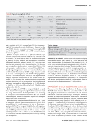
This document provides guidelines for the diagnosis, treatment and prevention of Clostridium difficile infections (CDI). It summarizes key recommendations with evidence grading. For diagnosis, it recommends nucleic acid amplification tests over toxin enzyme immunoassays, and only testing diarrheal stool samples. It stratifies treatment based on disease severity into mild-moderate (treat with metronidazole), severe (vancomycin with/without metronidazole), and complicated (vancomycin orally and rectally with intravenous metronidazole). It also covers recurrent CDI treatment, managing CDI in patients with comorbidities, and infection control practices like contact precautions and environmental disinfection. The guidelines
![The American Journal of GASTROENTEROLOGY VOLUME 108 | APRIL 2013 www.amjgastro.com
484 Surawicz et al.
pouch, or colostomy. Metronidazole may also fail to treat a diverted
segment of downstream colon because metronidazole is rapidly ab-
sorbedbythesmallintestinewithonly6–15%ofdrugexcretedinthe
stool. Moreover, there are data to suggest that IV metronidazole will
also enter the colon lumen following secretion across the inflamed
colonic mucosa, and CDI patients who respond to treatment have a
dramatic fall in the fecal concentrations of the antibiotic following
initiation of therapy. When CDI is documented in an excluded seg-
ment of diverted colon, administration of vancomycin by enema is
recommended to guarantee that treatment will reach the affected
area, using vancomycin enemas of 500mg in 100–500ml of normal
saline every 6h for CDI (58), volume depending on length of seg-
ment to be treated. The duration of enema therapy should continue
until the patient has significant improvement.
Recommendation
13. The use of anti-peristaltic agents to control diarrhea from con-
firmed or suspected CDI should be limited or avoided, as they
may obscure symptoms and precipitate complicated disease.
Use of anti-peristaltic agents in the setting of CDI must always
be accompanied by medical therapy for CDI. (Strong recom-
mendation, low-quality evidence)
Summary of the evidence. The IDSA/SHEA guidelines included a
C-III recommendation to “avoid [the] use of antiperistaltic agents,
as they may obscure symptoms and precipitate toxic megacolon”
(3). A literature review of 55 patients with CDI who were exposed
to such agents found that 17 patients developed colonic dilatation
and 5 died (59). All of these adverse outcomes, however, occurred
in patients with CDI who initially received treatment with anti-
peristaltic agents alone. All 23 patients in this review who received
antiperistaltic agents only in combination with CDI antimicrobial
therapy survived. For patients with mild-to-moderate CDI whose
antimicrobial treatment is well underway, the use of these drugs
to control the most debilitating symptom of CDI should be
further studied in prospective trials.
in this study, it is reasonable to persist with metronidazole mono-
therapy for patients with mild-to-moderate CDI for at least 7 days
unless signs or symptoms consistent with severe CDI or metro-
nidazole intolerance develop at any point during therapy and
escalating to vancomycin at standard dosing for patients who
do not respond in 5–7 days or who develop signs or symptoms
of severe CDI. We recommend discontinuing metronidazole
because the side effects (nausea, vomiting, and taste disturbances)
may be mistaken for patients with signs of ileus due to worsen-
ing CDI, and because there is insufficient evidence to support
the practice of continuing metronidazole for mild-to-moderate
CDI when a decision to escalate therapy to vancomycin has
been made.
The use of very high doses of vancomycin (500mg orally four
times daily) was included in the IDSA/SHEA treatment guide-
lines for management of severe complicated CDI as defined by the
treating physician (3). As a result, it has become common practice
to use higher doses of vancomycin if patients are failing to respond
to the standard recommended dose of 125mg four times daily. A
trial of 46 patients randomized to 500 or 125mg of vancomycin
four times daily for the initial treatment of CDI showed no differ-
ence in duration of diarrhea, relapse rate, or microbiological cure
(carriage of C. difficile at the end of therapy) (56). Moreover, fecal
levels of vancomycin in patients with CDI with this dose achieve
levels that are a minimum of 10 times the minimal inhibitory con-
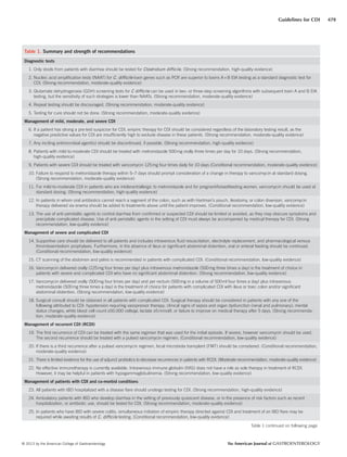
centration reported for C. difficile strains (57). Given the high cost
of vancomycin therapy, there is insufficient evidence to support
the use of doses >125mg four times daily for patients with mild-
to-moderate CDI, particularly for outpatients. Drug costs are in
Table 4.
Recommendation
11. For mild-to-moderate CDI in patients who are intolerant/
allergic to metronidazole and for pregnant/breastfeeding
women, vancomycin should be used at standard dosing.
(Strong recommendation, high-quality evidence)
Summary of the evidence. Metronidazole treatment should be
avoided in pregnancy and breast feeding. First trimester exposure
to metronidazole is not recommended in FDA guidelines because
of concern regarding ready placental transmission and case re-
ports describing facial anomalies following exposure. Metronida-
zole and its active metabolites are readily detected in breast milk
and in the plasma of infants.
Recommendation
12. In patients in whom oral antibiotics cannot reach a segment of
the colon, such as with Hartman’s pouch, ileostomy, or colon
diversion, vancomycin therapy delivered via enema should be
added to treatments above until the patient improves. (Condi-
tional recommendation, low-quality evidence)
Summary of the evidence. Oral vancomycin cannot reach seg-
ments of colon that are not in continuity with the gastrointestinal
tract, such as the patient with an upstream ileostomy, Hartman’s
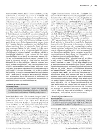
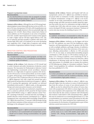
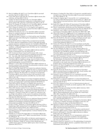
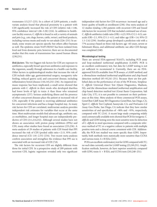
Table 4. Cost of antibiotic therapy for C. difficile infection
Cost per dose Regimen
Cost per 10-day
regimen
Metronidazole
500mg
$0.73 500mg three
times a day
$22.00
Vancomycin
125mg pills
$17.00 125mg four
times a day
$680.00
Vancomycin
125mg
IV compounded
for oral
$2.50–
$10.00
125mg four
times a day
$100.00–$400.00
Fidaxomicin
200mg
$140.00 200mg twice
a day
$2,800.00
IV, intravenous.
Vancomycin IV form can be compounded for oral use as well as used for
enema therapy.](https://image.slidesharecdn.com/acgguidelinecdifficileapril2013-170131235414/85/Acg-guideline-cdifficile_april_2013-7-320.jpg)


![CASE_PRESENTATION_ON_subdural_hematoma(SDH)[1 FINAL PPT]-1.pptx](https://cdn.slidesharecdn.com/ss_thumbnails/casepresentationonsubduralhematomasdh1finalppt-1-260129172522-d405d375-thumbnail.jpg?width=640&height=640&fit=bounds)


