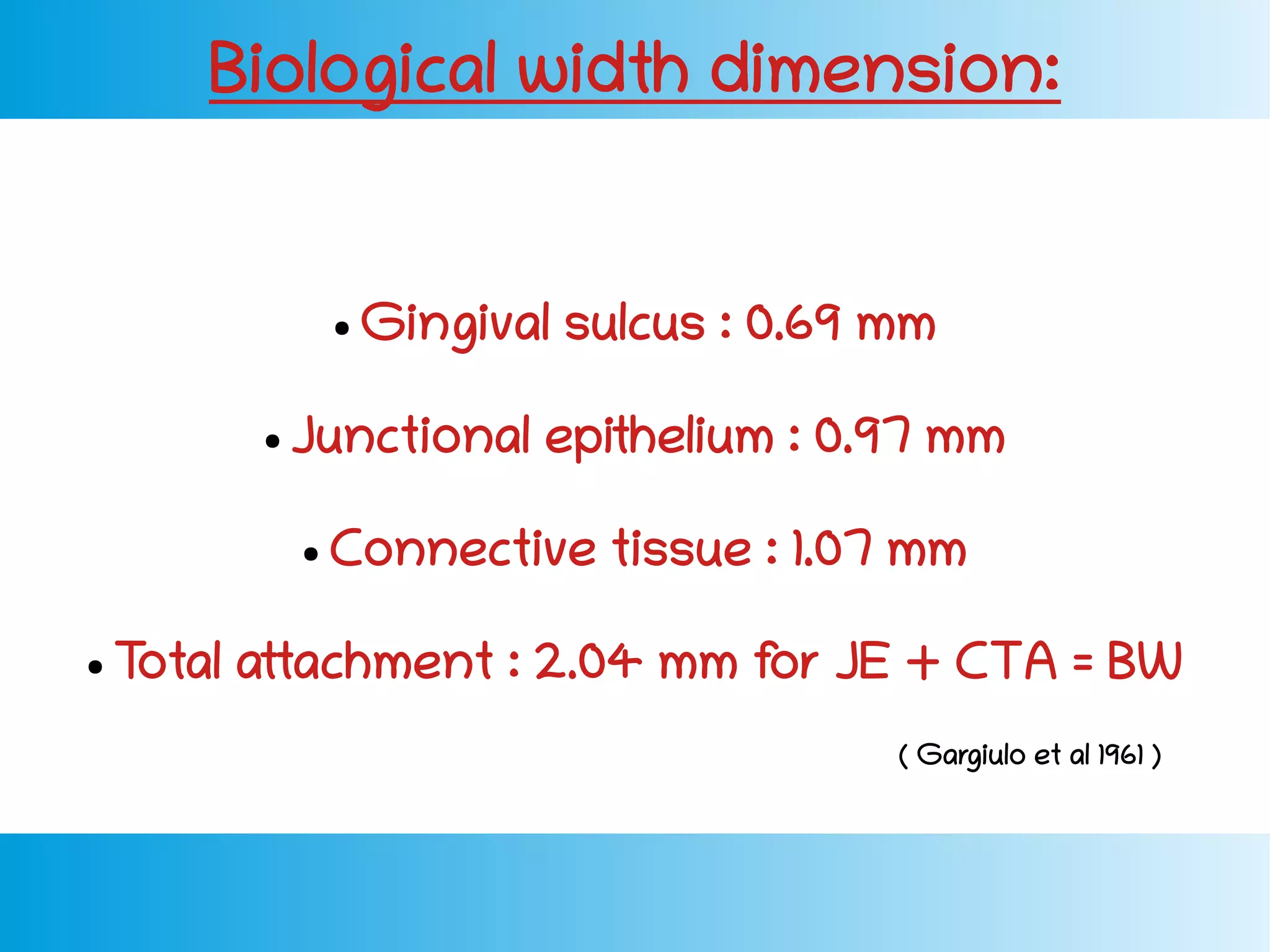
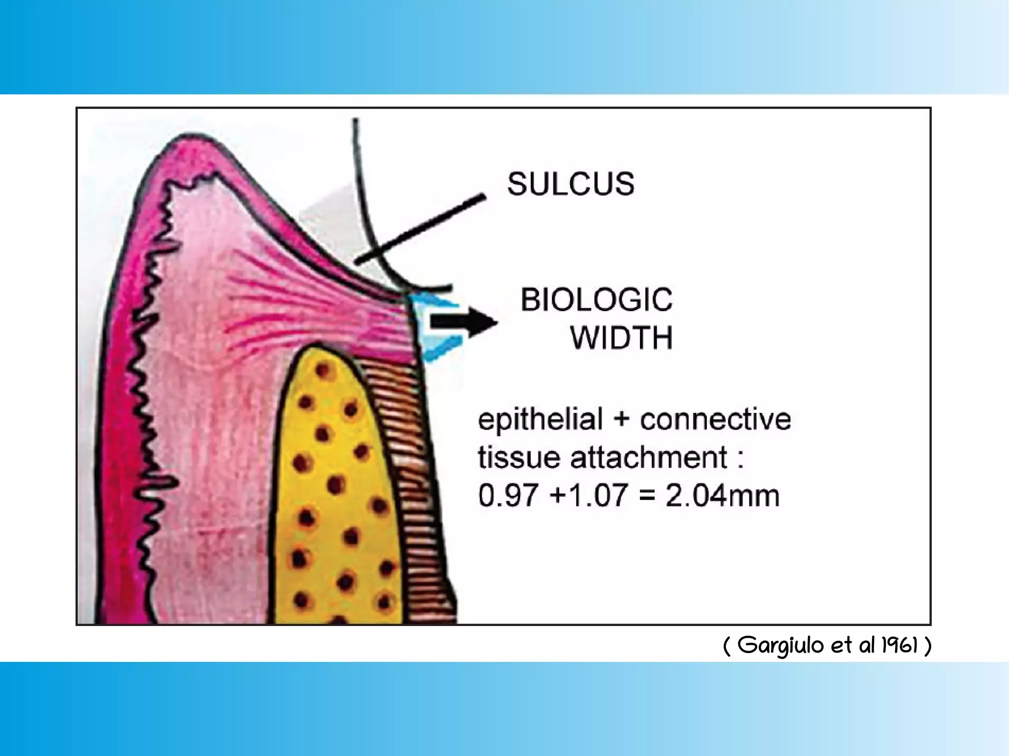
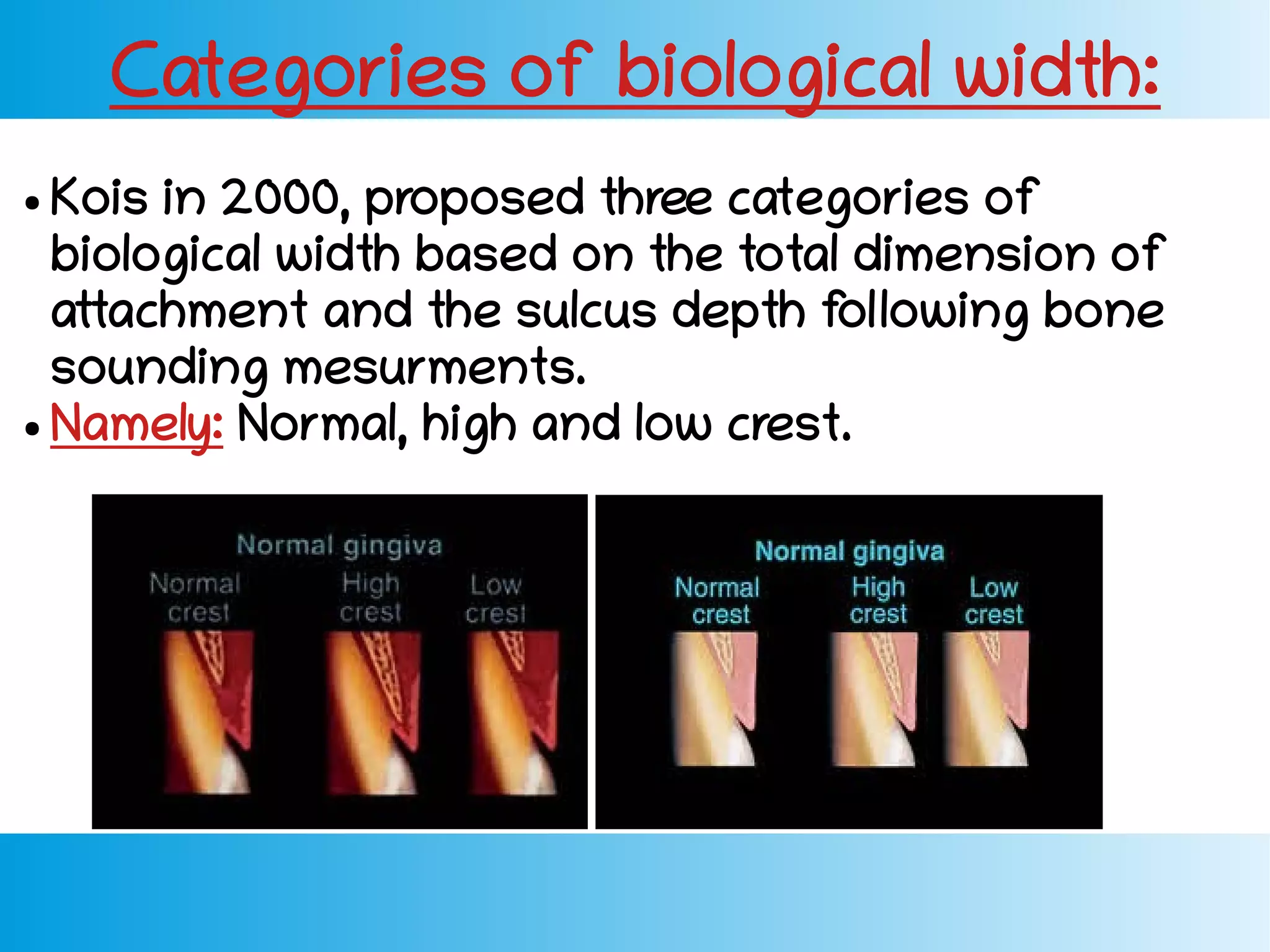
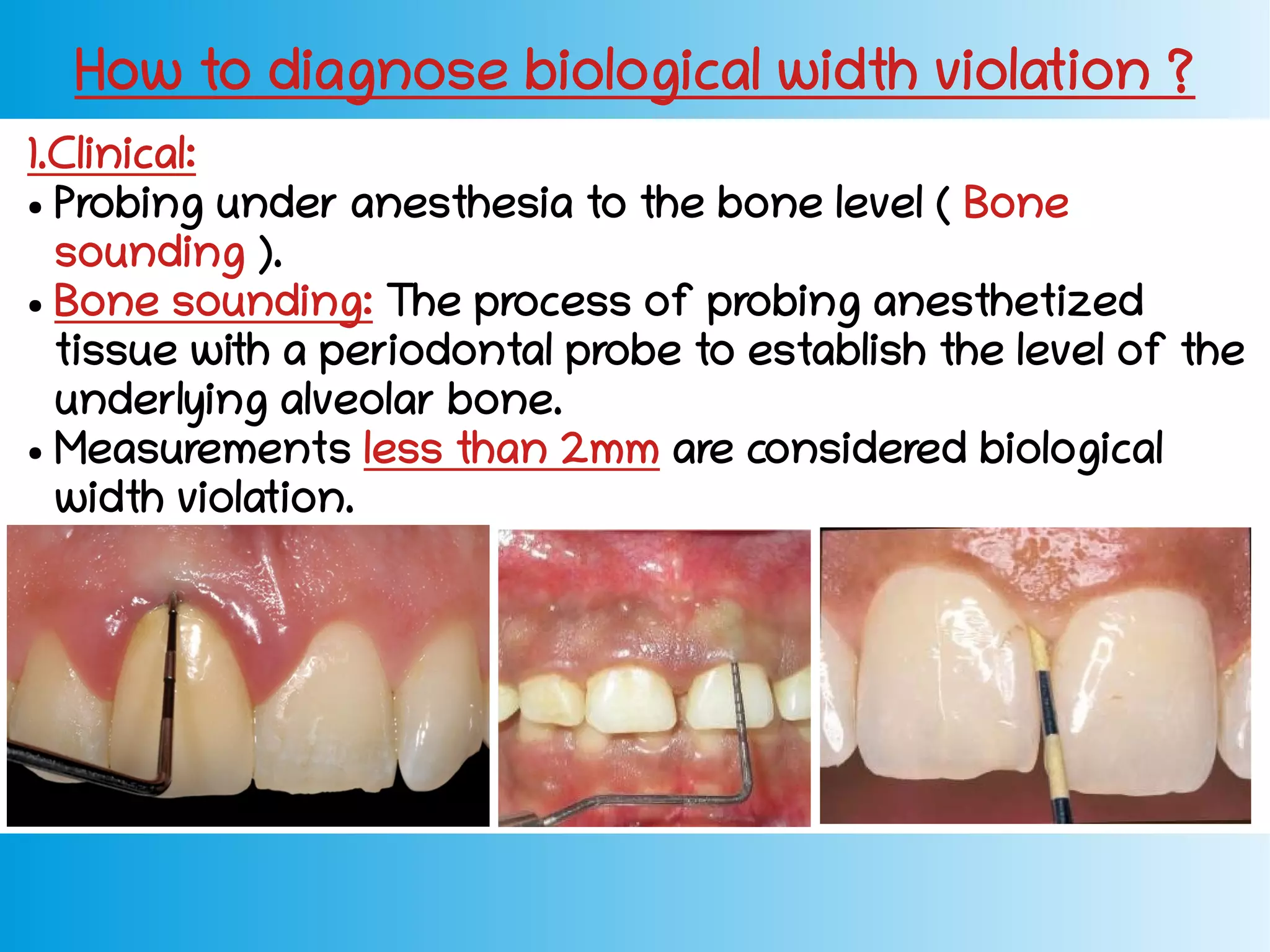
BIOLOGICAL WIDTH
Dr. Dina M. Fahim
Definition:
● The physiologic dimension of the junctional
epithelium and connective tissue attachment
above the crest of alveolar bone.
● The dimension of soft tissue which is attached
to the portion of the tooth coronal to the crest
of the alveolar bone.
( Gargiulo et al 1961 )
The junctional epithelium + The connective tissue
attachment = Biological width
( Gargiulo et al 1961 )
Biological width dimension:
● Gingival sulcus : 0.69 mm
● Junctional epithelium : 0.97 mm
● Connective tissue : 1.07 mm
● Total attachment : 2.04 mm for JE + CTA = BW
( Gargiulo et al 1961 )
Features Tooth Implant
Probing depth ● 2-3 mm ● 3-5 mm
Connective
tissue
● Low collagen.
● High fibroblast.
● High collagen.
● Low fibroblast.
Vascular
supply
● More. ● Less.
Periodontal
Ligament
● Present. ● Absence.
CT fibers ● Perpendicular to
tooth.
● Parallel to implant.
Junctional
Epithelium
● Origin:Reduced
enamel epithelium.
● High regenerative
capability.
● Origin: Adjacent
oral epithelium.
● Low regenerative
capability.
Tooth VS Implant biological width:
Functional role of biological width:
● Preserve healthy gingival tissues.
● Removal of irritation that might damage
the periodontium.
● Controlling the gingival form around
restorations.
● Kois in 2000, proposed three categories of
biological width based on the total dimension of
attachment and the sulcus depth following bone
sounding mesurments.
● Namely: Normal, high and low crest.
Categories of biological width:
Normal
crest
High crest Low
crest
Mid facial 3mm Less than 3mm More than 3mm
Proximal Range from
3mm to 4.5mm
Less than 3mm More than
4.5mm
Occurrence 85% of time. 2% of time. 13% of time.
( Kois 2000 )
Crest positions:
( Normal Crest ) ( High Crest )
( Low Crest )
Biological width violation:
When the biological width is encroached with
caries or restoration, the periodontium reacts by
recreating room between the alveolar bone and
the restorative margin to allow space for tissue
reattachment.
Possible causes of biological width violation:
● Poor oral hygiene ( plaque and calculus ).
● Defective restorative margins.
● Over contoured crowns.
Biological width violation is seen clinically as:
● Gingival inflammation.
● Bleeding on probing.
● Increased probing depth.
● Bone loss.
● Gingival recession.
● Gingival hyperplasia (subgingivally placed
restorations).
How to diagnose biological width violation ?
1.Clinical:
● Probing under anesthesia to the bone level ( Bone
sounding ).
● Bone sounding: The process of probing anesthetized
tissue with a periodontal probe to establish the level of the
underlying alveolar bone.
● Measurements less than 2mm are considered biological
width violation.
2.Radiographic:
● Can detect biological width at
interproximal sites.
● Difficult to detect mesiofacial and
distofacial line angles of teeth.
Another technique can be used:
Parallel profile radiography ( PPR )
( SAFE ) ( SAFE ) ( RISK )
Steps of preparation without traumatizing
attachment:
● Prep the tooth completely to the level of gingival
crest, leaving only the subgingival margin
preparation to be completed.
● Place a thin retraction cord in the sulcus (000,00).
● Final prep subgingivally just on top of the cord.
● Impression: ( Double cord technique ) Because the
1st retraction cord acts as a buffer and prevents
any violation to the biological width.
How to restore biological width:
1. Surgical crown lengthening.
2. Apical re-positioned flab surgery.
3. Orthodontic extrusion.
Surgical crown lengthening:
1. Removal of soft tissue (Gingvectomy):
In case of adequate width of attached gingiva.
2. Both soft tissue and alveolar bone (Apically displaced
flap):
Flap retraction and alveolar bone re-contouring in case of
inadequate width of attached gingiva.
● Advantage: Faster approach than orthodontic option.
● Disadvantage: Gingival Asymmetry and may lead to
poor crown : root ratio.
● Two methods:
Orthodontic Extrusion:
1. If the biological width violation is
on the interproximal side.
2. If the violation is across the
facial surface and the gingival
tissue level is correct, orthodontic
extrusion is indicated.
Advantages:
● Conserving the bone.
● Preserve the biological width.
● Favorable crown : Root ratio.
Biological width

Biological width

  • 1.
  • 2.
    Definition: ● The physiologicdimension of the junctional epithelium and connective tissue attachment above the crest of alveolar bone. ● The dimension of soft tissue which is attached to the portion of the tooth coronal to the crest of the alveolar bone. ( Gargiulo et al 1961 )
  • 3.
    The junctional epithelium+ The connective tissue attachment = Biological width ( Gargiulo et al 1961 )
  • 4.
    Biological width dimension: ●Gingival sulcus : 0.69 mm ● Junctional epithelium : 0.97 mm ● Connective tissue : 1.07 mm ● Total attachment : 2.04 mm for JE + CTA = BW ( Gargiulo et al 1961 )
  • 5.
    ( Gargiulo etal 1961 )
  • 6.
    Features Tooth Implant Probingdepth ● 2-3 mm ● 3-5 mm Connective tissue ● Low collagen. ● High fibroblast. ● High collagen. ● Low fibroblast. Vascular supply ● More. ● Less. Periodontal Ligament ● Present. ● Absence. CT fibers ● Perpendicular to tooth. ● Parallel to implant. Junctional Epithelium ● Origin:Reduced enamel epithelium. ● High regenerative capability. ● Origin: Adjacent oral epithelium. ● Low regenerative capability. Tooth VS Implant biological width:
  • 8.
    Functional role ofbiological width: ● Preserve healthy gingival tissues. ● Removal of irritation that might damage the periodontium. ● Controlling the gingival form around restorations.
  • 9.
    ● Kois in2000, proposed three categories of biological width based on the total dimension of attachment and the sulcus depth following bone sounding mesurments. ● Namely: Normal, high and low crest. Categories of biological width:
  • 10.
    Normal crest High crest Low crest Midfacial 3mm Less than 3mm More than 3mm Proximal Range from 3mm to 4.5mm Less than 3mm More than 4.5mm Occurrence 85% of time. 2% of time. 13% of time. ( Kois 2000 ) Crest positions:
  • 11.
    ( Normal Crest) ( High Crest ) ( Low Crest )
  • 12.
    Biological width violation: Whenthe biological width is encroached with caries or restoration, the periodontium reacts by recreating room between the alveolar bone and the restorative margin to allow space for tissue reattachment.
  • 13.
    Possible causes ofbiological width violation: ● Poor oral hygiene ( plaque and calculus ). ● Defective restorative margins. ● Over contoured crowns.
  • 14.
    Biological width violationis seen clinically as: ● Gingival inflammation. ● Bleeding on probing. ● Increased probing depth. ● Bone loss. ● Gingival recession. ● Gingival hyperplasia (subgingivally placed restorations).
  • 15.
    How to diagnosebiological width violation ? 1.Clinical: ● Probing under anesthesia to the bone level ( Bone sounding ). ● Bone sounding: The process of probing anesthetized tissue with a periodontal probe to establish the level of the underlying alveolar bone. ● Measurements less than 2mm are considered biological width violation.
  • 16.
    2.Radiographic: ● Can detectbiological width at interproximal sites. ● Difficult to detect mesiofacial and distofacial line angles of teeth. Another technique can be used: Parallel profile radiography ( PPR )
  • 17.
    ( SAFE )( SAFE ) ( RISK )
  • 18.
    Steps of preparationwithout traumatizing attachment: ● Prep the tooth completely to the level of gingival crest, leaving only the subgingival margin preparation to be completed. ● Place a thin retraction cord in the sulcus (000,00). ● Final prep subgingivally just on top of the cord. ● Impression: ( Double cord technique ) Because the 1st retraction cord acts as a buffer and prevents any violation to the biological width.
  • 19.
    How to restorebiological width: 1. Surgical crown lengthening. 2. Apical re-positioned flab surgery. 3. Orthodontic extrusion.
  • 20.
    Surgical crown lengthening: 1.Removal of soft tissue (Gingvectomy): In case of adequate width of attached gingiva. 2. Both soft tissue and alveolar bone (Apically displaced flap): Flap retraction and alveolar bone re-contouring in case of inadequate width of attached gingiva. ● Advantage: Faster approach than orthodontic option. ● Disadvantage: Gingival Asymmetry and may lead to poor crown : root ratio. ● Two methods:
  • 22.
    Orthodontic Extrusion: 1. Ifthe biological width violation is on the interproximal side. 2. If the violation is across the facial surface and the gingival tissue level is correct, orthodontic extrusion is indicated. Advantages: ● Conserving the bone. ● Preserve the biological width. ● Favorable crown : Root ratio.