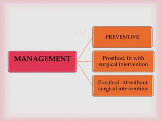
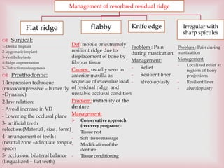
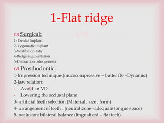
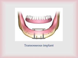
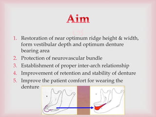
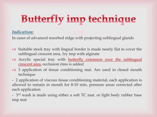
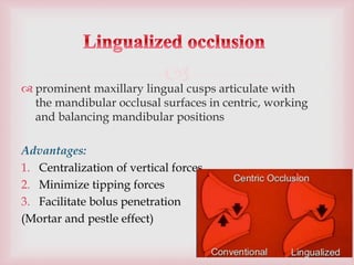
3.
Best treatment is to avoid total tooth extraction
…….. preservation of residual ridge
PREVENTION
4.
1. Support of the abutment teeth stresses of occlusion
spread over periodontal ligament mucosa of residual
ridge spared abuse
2. Maintenance of the alveolar bone
3. Sensory feedback and motor response
4. Minimal load threshold.
5. Tactile sensation (propioceptors)
6. Improved masticatory response
7. Reduction of psychological trauma
5.
Management of resorbred residual ridge
Flat ridge flabby Knife edge Irregular with
sharp spicules
Surgical:
1- Dental Implant
2- zygomatic implant
3-Vestibuloplasty
4-Ridge augmentation
5-Distraction osteogenesis
Prosthodontic:
1-Impression technique
(mucocompressive – butter fly
–Dynamic)
2-Jaw relation:
- Avoid increase in VD
- Lowering the occlusal plane
3- artificial teeth
selection:(Material , size , form)
4- arrangement of teeth :
(neutral zone –adequate tongue
space)
5- occlusion: bilateral balance
(lingualized – flat teeth)
Def: mobile or extremely
resilient ridge due to
displacement of bone by
fibrous tissue
Causes: usually seen in
anterior maxilla as
sequelae of excessive load
of residual ridge and
unstable occlusal condition
Problem: instability of the
denture
Management:
Conservative approach
(recovery programe):
- Tissue rest
- Soft tissue massage
- Modification of the
denture
- Tissue conditioning
Problem : Pain during
mastication
Management:
- Localized relief at
regions of bony
projections
- Resilient liner
- alveoloplasty
Problem : Pain
during mastication
Management:
- Relief
- Resilient liner
- alveoloplasty
6.
Management of resorbred residual ridge
Flat ridge flabby Knife edge Irregular with
sharp spicules
Prosthetic approach:
1-Impression:
selective tech , Sectional imp ( window tech),General selective
imp tech
2- Jaw relation:
Wax wafer method with easily displaceable recording material
3- Teeth selection , arrangement:
- Acc to Neutral zone
- Reduce BL width
- Crosslinked acrylic teeth
4- periodic check up
Surgical management:
1- Removal of fibrous tissue:
disadvantages:
- Trauma to underlying bone
- Decrease the sulcus depth
- Increase the bulk of denture material
2- Ridge augmentation by subperiosteal injection of hydroxyapatite
4-Waxing up
- Polished surface should
harmonize with the surrounding
musculature (by neutral zone
determination
- The lower denture should be
narrow at the premolar region
(modiolus)
5- Insertion:
- Lab, clinical remount: to clear
any occlusal prematurities
- Soft liner to decrease trauma
- Restricted instruction:
not to were denture at night to
give chance to the tissues to rest.
Type of food: (food doesn’t
require masticatory force exceed
tissue tolerance
6- recall maintenance
Unconventional denture:
Hollow denture,
Liquid denture
8.
4-Waxing up
- Polished surface should harmonize with the surrounding musculature (by neutral zone
determination
- The lower denture should be narrow at the premolar region (modiolus)
5- Insertion:
- Lab, clinical remount: to clear any occlusal prematurities
- Soft liner to decrease trauma
- Restricted instruction:
not to were denture at night to give chance to the tissues to rest.
Type of food: (food doesn’t require masticatory force exceed tissue tolerance
6- recall maintenance
Unconventional denture:
- Hollow denture
- Liquid denture
9.
Def: mobile or extremely resilient ridge due to
displacement of bone by fibrous tissue
Causes: usually seen in anterior maxilla as sequelae of
excessive load of residual ridge and unstable occlusal
condition
Problem: instability of the denture
Management:
Conservative approach (recovery programe):
- Tissue rest
- Soft tissue massage
- Modification of the denture
- Tissue conditioning
2- Flabby ridge
10.
Prosthetic approach:
1-Impression:
- selective tech
- Sectional imp ( window tech)
- General selective imp tech
2- Jaw relation:
Wax wafer method with easily displaceable recording material
3- Teeth selection , arrangement:
- Acc to Neutral zone
- Reduce BL width
- Crosslinked acrylic teeth
4- periodic check up
11.
Surgical management:
1- Removal of fibrous tissue:
disadvantages:
- Trauma to underlying bone
- Decrease the sulcus depth
- Increase the bulk of denture material
2- Ridge augmentation by subperiosteal injection of
hydroxyapatite
12.
Problem : Pain during mastication
Management:
- Relief
- Resilient liner
- alveolplasty
3- Knife edge ridge
13.
Problem : Pain during mastication
Management:
- Localized relief at regions of bony projections
- Resilient liner
- alveolplasty
4- Irregular ridge with sharp bony spicules
15.
Dental implants
Placement of two or more implants anteriorly in the area
between foramina can be of value in improving
horizontal stability and retention of the constructed
implant supported overdentures
16.
Maintenance of alveolar bone
Restoration& Maintenance of occlusal vertical dimension
Maintain facial esthetics improved phonetics
Improved occlusion
Improved psychological health
Regained propioception
Increased stability, retention
Improved masticatory performance
There is 20 fold decrease in the loss of structure with implants
when compared with resorption that occurs with removable
prosthesis
19.
Zygomatic implants:
When there is substantial amount of bone lost from the
upper jaw , and ordinary implants cannot be used
Longer than ordinary implants , engage bulk of
zygomatic bone
Can be used as an alternative to complex bone grafting
procedures
20.
Def:
Lowering muscle attachment, unattached mucosa into
deeper position into the sulcus.
Indication:
- Moderate bone resorption
- When vertical height of 12-15mm in mandible
22.
Removal of genial tubercles: This is done to provide
for an extension in the sublingual fold space.
Prominent mylohyoid ridge: It is some times
trimmed to allow proper extension of the lingual
flange of the mandibular denture
23.
DEF: Restore mandibular bulk by placing onlay bone graft
INDICATION
Severe bone resorption
Vertical height less than 1mm
TYPES OF BONE GRAFT:
1. Autogenous bone (illiac crest, ribs, cancellous bone,
sternum tibia and scapula)
2. Cartilage, fat and dermis
3. Preserved xenographs and homographs
4. Dimeneralized bone
5. Freez-dried grafts
6. Bone replacement materials e.g. hydroxyapatite crystals
25.
1. Restoration of near optimum ridge height & width,
form vestibular depth and optimum denture
bearing area
2. Protection of neurovascular bundle
3. Establishment of proper inter-arch relationship
4. Improvement of retention and stability of denture
5. Improve the patient comfort for wearing the
denture
29.
Advantages:
No need for donor site.
No limit for lengthening
Simultaneous lengthening of the surrounding soft
tissue such as skin, muscles, bl. Vessels and nerve
Disadvantages:
1. Long time
2. Danger of infection need for suitable distractors
30.
Aim : Preservation of remaining tissues
Factors to be considered:
1. Maximum tissue coverage
2. Correct vertical dimension
3. Occlusal balance
4. decrease occlusal table
5. Control amount & direction of force
6. Improve stability &retention of complete denture
31.
The cause of degenerative ridge will determine the type of
treatment
So a detailed examination must be performed
Complete mouth and panographic radiographs are essential
Dietary analysis: Their ability to chew will frequently dictate
their selection of food, and this will usually generate a diet high
in refined carbohydrates and lo in protein, vitamins and
minerals- just the opposite of what is needed to help stop bone
destruction
32.
Medical history
Record previous and existing systemic condition that may
contribute to mandibular atrophy
Comprehensive dental history:
1. Cause of extraction
2. Past denture experience, number of old dentures made
3. Frequency of denture rebasing should be appreciated to
estimate both the apparent rate of resorption & capability of
the individual to cope up with previous denture
4. Examination of the existing denture
33.
1. Saliva (quality, quantity,
viscosity)
2. Mucosa color
3. Health of ridge
,surrounding structure
4. Size ,shape of hard palate
5. Size, shape of the arch
6. Remaining root or bony
spicules
7. Interfering structure
(frenum)
8. Size position of
tongue
9. Sulcus depth
10. Lip position
11. Interarch space
12. Patient profile
Visual :
34.
1. Firmness of the ridge
2. Type of mucosa
3. Pattern of bone
resorption
4. Irregularities of the ridge
5. Bone exostosis
6. Tuberosities ,tori
s
7. External oblique
ridge
8. Undercut area
9. Lingual pouch
10. Painful area nature
of muscles
11. Buccal shelf
Digital
35.
Radiographic examination:
1. Assess degree of bone resorption, inadequate bone
thickness (risk of spontaneous fracture)
2. Position of the mental foramen, mandibular canal
dehiscence of the canal
Diagnostic models may also be taken to allow
case evaluation in the absence of the patient
36.
- Lack of ideal amount of supporting structures
decreases support.
- The encroachment of the surrounding mobile tissues
on to the denture border reduces both stability and
retention.
AIM:
1. Maximum extension without muscle impingement
2. Intimate contact with the tissue covered
3. Proper form of the border including the posterior border
of the maxillary denture
4. Proper relief of sensitive, hard area
38.
1. Adequate flow during impression to avoid uneven
pressure……….result in rebounding of compressed
tissues and /or sore spots
2. Should provide adequate reproduction of surface
detail
NB:
Accurate impression can only be recorded in their
healthy, full recovered state
Patients not allowed to wear their dentures for min
48 h before the impression
40.
I ry impression : compound
Acrylic special tray with occlusion rims on upper
,lower trays at acceptable VD.
Border molding using green stick compound
Final imp using ZOE while the patient is closing on
the occlusal rims
1896 by Greene
41.
Indication:
In cases of advanced resorbed ridge with projecting sublingual glands
Suitable stock tray with lingual border is made nearly flat to cover the
sublingual crescent area, 1ry imp with alginate
Acrylic special tray with butterfly extension over the sublingual
crescent area, occlusion rims is added
3 application of tissue conditioning mat. Are used in closed mouth
technique
- 2 application of viscous tissue conditioning material, each application is
allowed to remain in mouth for 8-10 min, pressure areas corrected after
each application
- 3rd wash is made using either a soft TC mat. or light body rubber base
imp mat
42.
Result : imp has tissue placing effect, very thick buccal border,
relatively thick lingual and sublingual crescent areas, covering the
max possible basal seat area with in functional limits of adjacent
tissues
43.
The impression material is shaped by the function of the
muscles and muscle attachments
Steps:
1. Special tray of acrylic resin
2. Three stops of impression compound are added to the fitting
surface ( one at the anterior,2 posteriorly in the 1 st molar
region) to allow space for 2mm for the imp material
3. Mandibular rests of imp compound are placed bilateral on the
occlusal surface of the tray in the molar region ( should be
concave to allow tongue movement) & compound tongue rest
is added in the anterior region to secure correct tongue
position during imp making
44.
1. Final impression using a thin mix of alginate impression.
2. The patient is asked to close slowly until the mandibular
rests firmly contact the maxillary arch , keep the tongue in
contact with the maxillary rest
3. The patient is instructed to swallow 3-5 times, forcefully
protrude the lips forward
The resulting impression covers the maximum possible basal
seat area and the borders are in harmony with adjacent
moving tissues
45.
J. F. McCord and K. W. Tyson, “A conservative prosthodontic option for the treatment of edentulous
patients with atrophic (flat) mandibular ridges,” British Dental Journal, vol. 182, no. 12, pp. 469–472, 1997.
50.
Tanvir H, et al. An innovative Wire Impression Technique of Highly
Resorbed Mandibular Ridge . Periodontics and Prosthodontics Vol.3 No.1:5
2017
51.
Determination of proper vertical dimension:
Avoid increasing the VD to avoid excessive forces on the ridge
Level of occlusal plane:
lowering the level of occl. plane toward the flatter ridge will decrease lateral
forces
Artificial teeth selection
- Material : acrylic preferred than porcelain ( cross linked acrylic teeth PTN)
- Size: decrease the No, width, length of teeth (BL,OG, MD)
- Form: non anatomical preferred than anatomical
Teeth arrangement:
Neutral zone ,,adequate tongue space
52.
Balanced occlusion :
Def:
Bilateral simultaneous anterior and posterior occlusal
contact of teeth in centric and eccentric position
Advantages:
To improve the stability of denture.
To reduce resorption of the residual ridge and
soreness.
To improve oral comfort & well being of the patient.
54.
prominent maxillary lingual cusps articulate with
the mandibular occlusal surfaces in centric, working
and balancing mandibular positions
Advantages:
1. Centralization of vertical forces
2. Minimize tipping forces
3. Facilitate bolus penetration
(Mortar and pestle effect)
55.
Flat teeth opposed by bladed teeth ,
Mandibular teeth are set to flat occlusal plane
No anterior interference in protrusive or lateral
movements
Provide consistent vertical seating force in both
centric and eccentric, hence the transverse vectors
area eliminated
56.
Balanced occlusion with cupless teeth can be achieved
by several ways:
inclination of the lower second molar
balancing ramps placed posterior to the most distal
molar.
steep compensatory curves
58.
Aim:
-record the range of muscle action , spaces into which the
denture can be extended without displacement,
- In this technique, complete utilization of the active and
passive tissues is obtained as the impression material is
being shaped by the function of the muscles and muscle
attachments allowing properly formed denture borders.
59.
The technique aims to construct a denture that is
shaped by muscle function and is in harmony with the
surrounding oral structures.
Advantages:
a) Improved stability and retention
b) Posterior teeth will be correctly positioned allowing
sufficient tongue space
c) Reduced food trapping adjacent
to the molar teeth
a) Good aesthetics due to
facial support.
63.
Polished surface of the denture
- Should harmonize with the surrounding musculature
(tongue , lip , cheeks) by neutral zone determination
- The lower denture should be narrow at the premolar
region because of the action od modiolus
Metal denture base is preferred to increase retention
by interfacial surface tension
Postdamming of retromolar area
64.
Lab, clinical remount: to clear any occlusal prematurities
Soft liner to decrease trauma
Restricted instruction:
• not to were denture at night to give chance to the tissues
to rest.
• Type of food: (food doesn’t require masticatory force
exceed tissue tolerance)
65.
Recall maintenance :
to ascertain amount of change in fitness & occlusion&
to evaluate and correct the osseous changes
67.
For more than 150 years, it was believed that the weight
of the lower denture contributes to both retention and
stability.
However, studies have shown that retention and
stability can be achieved by improving the fit of the
denture bases rather than addition of extra weight to
the dentures and also the weight of the lower
denture may not affect its retention and stability.
Hollowing the denture so as to reduce the weight of
the denture, thereby enhancing stability and
retention, reducing the further resorption of the jaws.
68.
In severe resorption of the edentulous ridges, Increased
inter-ridge space compounds this problem. To decrease the
leverage, reduction in the weight of the prosthesis was
recommended
Various weight reduction approaches have been achieved
using a solid three-dimensional spacer, including dental
stone, cellophane wrapped asbestos, silicone putty or
modeling clay during laboratory processing to exclude
denture base material from the planned hollow cavity of the
prosthesis.
69.
Aggarwal H et al, Lost salt technique for severely resorbed alveolar ridges: An innovative approach
71.
Advantages of Liquid-supported denture
are:
• preservation of residual ridge by
optimal distribution of forces,
• better retention, stability, support
• comfort due to close adaptation.
• shock absorbing effect
73.
Although challenging, the severely resorbed ridges
can be restored to a certain level of mastication with
the help of improved impression techniques , proper
selection of occlusion schemes, the use of specialized
dentures techniques and a regular follow up.
More recently, implant supported overdentures are
playing tremendous role in the treatment of the
severely resorbed ridges