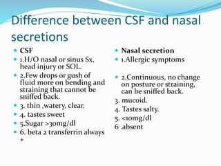
Download to read offline
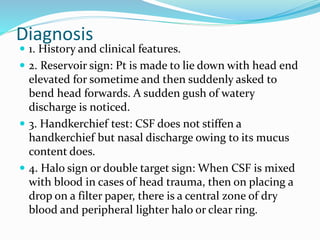
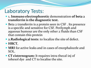


This document discusses cerebrospinal fluid (CSF) rhinorrhoea, or leakage of CSF into the nose. It defines CSF rhinorrhoea, describes the physiology of CSF production and absorption, and discusses the etiology, clinical features, diagnosis, and management of CSF leaks. The key causes are traumatic injuries or fractures that disrupt the barrier between the brain/spinal cord and nose. Diagnosis involves clinical exams and tests like the beta-2 transferrin immunoassay. Treatment focuses on conservative management initially but may require surgical repair using endoscopic or open approaches to graft the defect.


