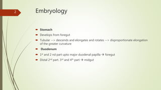
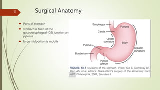
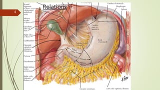
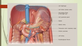
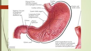
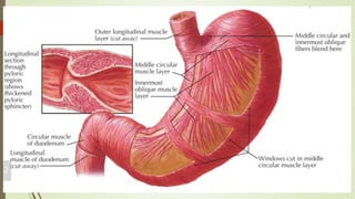
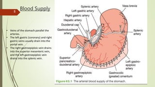
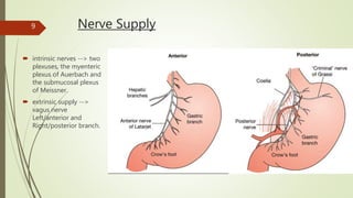
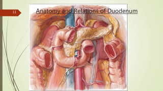
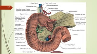
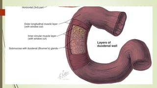
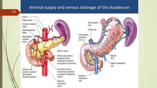
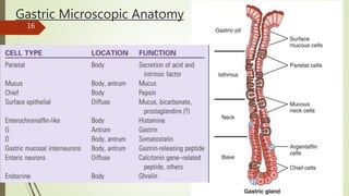
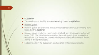
Stomach and Duodenum
Anatomy
Dr. Lala Robin. MS Gen. Surgery.
Senior Resident
CMC
1
Embryology
 Stomach
 Develops from foregut
 Tubular --> descends and elongates and rotates --> disproportionate elongation
of the greater curvature
 Duodenum
 1st and 2 nd part upto major duodenal papilla  foregut
 Distal 2nd part. 3rd and 4th part  midgut
2
Surgical Anatomy
 Parts of stomach
 stomach is fixed at the
gastroesophageal (GE) junction and
pylorus
 large midportion is mobile
3
Relations
4
5
6
7
Blood Supply
• Veins of the stomach parallel the
arteries.
• The left gastric (coronary) and right
gastric veins usually drain into the
portal vein.
• The right gastroepiploic vein drains
into the superior mesenteric vein,
and the left gastroepiploic vein
drains into the splenic vein.
8
Nerve Supply
 intrinsic nerves --> two
plexuses, the myenteric
plexus of Auerbach and
the submucosal plexus
of Meissner.
 extrinsic supply -->
vagus nerve
Left/anterior and
Right/posterior branch.
9
Lymphatic Drainage10
Anatomy and Relations of Duodenum11
12
13
14
PHYSIOLOGY OF THE STOMACH
AND DUODENUM15
Gastric Microscopic Anatomy
16
 Duodenum
 The duodenum is lined by a mucus-secreting columnar epithelium.
 Brunner glands
 Brunner glands are branched, tubuloalveolar glands with mucus-secreting acini
located in the duodenal
 Brunner glands produce a bicarbonate-rich fluid, also rich in epidermal growth
factor (EGF). The bicarbonate neutralises the acidic gastric juice entering the
duodenum. EGF inhibits HCl production of parietal cells and stimulates mitotic
activity in the epithelial cells.
 Endocrine cells in the duodenum produce cholecystokinin and secretin.
17
PHYSIOLOGY OF THE STOMACH AND
DUODENUM
 The stomach mechanically breaks up ingested food and, together with the actions
of acid and pepsin, forms chyme that passes into the duodenum.
 In contrast with the acidic environment of the stomach, the environment of the
duodenum is alkaline, due to the secretion of bicarbonate ions from both the
pancreas and the duodenum. This neutralises the acid chyme and adjusts the
luminal osmolarity to approximately that of plasma.
 Endocrine cells in the duodenum produce cholecystokinin, which stimulates the
pancreas to produce trypsin and the gall bladder to contract. Secretin is also
produced by the endocrine cells of the duodenum. This hormone inhibits gastric
acid secretion and promotes production of bicarbonate by the pancreas.
18
Gastric acid secretion
 Hydrogen ions are produced by the parietal cell by the proton pump.
 Histamine produced by ECL cells acts on H2 receptor in parietal cell in a paracrine
fashion.
 proton pump - final common pathway – hydrogen ion secretion.
 The ECL cell produces histamine in response to a number of stimuli that include the
vagus nerve and gastrin.
 Gastrin is released by the G cells in response to the presence of the food in the
stomach.
 The production of gastrin is inhibited by acid, creating a negative feedback loop.
19
 three phases of gastric secretion
 The cephalic phase is mediated by vagal activity, secondary to sensory arousal as
first demonstrated by Pavlov.
 The gastric phase is a response to food within the stomach, which is mediated
principally, but not exclusively, by gastrin.
 In the intestinal phase, the presence of chyme in the duodenum and small bowel
inhibits gastric emptying, and the acidification of the duodenum leads to the
production of secretin, which inhibits gastric acid secretion, along with numerous
other peptides originating from the gut.
 Somatostatin produced by D cells in stomach in response to a number of factors
including acidification, acts probably on the G cell, the ECL cell and the parietal
itself to inhibit the production of acid.
20
21
Gastric mucus and the gastric mucosal
barrier
 essential to the integrity of the gastric mucosa
 viscid layer of mucopolysaccharides produced by the mucus-producing cells of the
stomach and the pyloric glands
 buffering capacity is enhanced by the presence of bicarbonate ions within the
mucus
 bile, non-steroidal anti-inflammatory drugs (NSAIDs), alcohol, trauma and shock 
lead to breakdown.
 The stomach is the most sensitive to ischaemia following a hypovolaemic insult and
also the slowest to recover  stress ulceration
22
Gastroduodenal motor activity
 In the fasted state, and after food has cleared, in the small bowel there is a period
of quiescence lasting in the region of 40 minutes (phase I).
 There follows a series of waves of electrical and motor activity, also lasting for
about 40 minutes, propagated from the fundus of the stomach in a caudal
direction at a rate of about three per minute (phase II). These pass as far the
pylorus, but not beyond. Duodenal slow waves are generated in the duodenum at a
rate of about 10 per minute, which carry down the small bowel.
 The amplitude of these contractions increases to a maximum in phase III, which
lasts for about 10 minutes. This 90-minute cycle of activity is then repeated. From
the duodenum, the MMC moves distally at 5–10 cm/ min, reaching the terminal
ileum after 1.5 hours.
23
 Following a meal, the stomach exhibits receptive relaxation, which lasts for a few seconds.
Following this, adaptive relaxation occurs, which allows the proximal stomach to act as a
reservoir.
 Most of the peristaltic activity is found in the distal stomach (the antral mill) and the
proximal stomach demonstrates only tonic activity.
 The pylorus, which is most commonly open, contracts with the peristaltic wave and allows
only a few millilitres of chyme through at a time. The antral contraction against the closed
sphincter is important in the milling activity of the stomach.
 Although the duodenum is capable of generating 10 waves per minute, after a meal it only
contracts after an antral wave reaches the pylorus.
 The coordination of the motility of the antrum, pylorus and duodenum means that only
small quantities of food reach the small bowel at a time.
 This control of gastric emptying can be abolished after gastric surgery leading to significant
symptoms. Motility is influenced by numerous factors, including mechanical stimulation and
neuronal and endocrine influences
24

Stomach and duodenum anatomy and physiology

  • 1.
    Stomach and Duodenum Anatomy Dr.Lala Robin. MS Gen. Surgery. Senior Resident CMC 1
  • 2.
    Embryology  Stomach  Developsfrom foregut  Tubular --> descends and elongates and rotates --> disproportionate elongation of the greater curvature  Duodenum  1st and 2 nd part upto major duodenal papilla  foregut  Distal 2nd part. 3rd and 4th part  midgut 2
  • 3.
    Surgical Anatomy  Partsof stomach  stomach is fixed at the gastroesophageal (GE) junction and pylorus  large midportion is mobile 3
  • 4.
  • 5.
  • 6.
  • 7.
  • 8.
    Blood Supply • Veinsof the stomach parallel the arteries. • The left gastric (coronary) and right gastric veins usually drain into the portal vein. • The right gastroepiploic vein drains into the superior mesenteric vein, and the left gastroepiploic vein drains into the splenic vein. 8
  • 9.
    Nerve Supply  intrinsicnerves --> two plexuses, the myenteric plexus of Auerbach and the submucosal plexus of Meissner.  extrinsic supply --> vagus nerve Left/anterior and Right/posterior branch. 9
  • 10.
  • 11.
    Anatomy and Relationsof Duodenum11
  • 12.
  • 13.
  • 14.
  • 15.
    PHYSIOLOGY OF THESTOMACH AND DUODENUM15
  • 16.
  • 17.
     Duodenum  Theduodenum is lined by a mucus-secreting columnar epithelium.  Brunner glands  Brunner glands are branched, tubuloalveolar glands with mucus-secreting acini located in the duodenal  Brunner glands produce a bicarbonate-rich fluid, also rich in epidermal growth factor (EGF). The bicarbonate neutralises the acidic gastric juice entering the duodenum. EGF inhibits HCl production of parietal cells and stimulates mitotic activity in the epithelial cells.  Endocrine cells in the duodenum produce cholecystokinin and secretin. 17
  • 18.
    PHYSIOLOGY OF THESTOMACH AND DUODENUM  The stomach mechanically breaks up ingested food and, together with the actions of acid and pepsin, forms chyme that passes into the duodenum.  In contrast with the acidic environment of the stomach, the environment of the duodenum is alkaline, due to the secretion of bicarbonate ions from both the pancreas and the duodenum. This neutralises the acid chyme and adjusts the luminal osmolarity to approximately that of plasma.  Endocrine cells in the duodenum produce cholecystokinin, which stimulates the pancreas to produce trypsin and the gall bladder to contract. Secretin is also produced by the endocrine cells of the duodenum. This hormone inhibits gastric acid secretion and promotes production of bicarbonate by the pancreas. 18
  • 19.
    Gastric acid secretion Hydrogen ions are produced by the parietal cell by the proton pump.  Histamine produced by ECL cells acts on H2 receptor in parietal cell in a paracrine fashion.  proton pump - final common pathway – hydrogen ion secretion.  The ECL cell produces histamine in response to a number of stimuli that include the vagus nerve and gastrin.  Gastrin is released by the G cells in response to the presence of the food in the stomach.  The production of gastrin is inhibited by acid, creating a negative feedback loop. 19
  • 20.
     three phasesof gastric secretion  The cephalic phase is mediated by vagal activity, secondary to sensory arousal as first demonstrated by Pavlov.  The gastric phase is a response to food within the stomach, which is mediated principally, but not exclusively, by gastrin.  In the intestinal phase, the presence of chyme in the duodenum and small bowel inhibits gastric emptying, and the acidification of the duodenum leads to the production of secretin, which inhibits gastric acid secretion, along with numerous other peptides originating from the gut.  Somatostatin produced by D cells in stomach in response to a number of factors including acidification, acts probably on the G cell, the ECL cell and the parietal itself to inhibit the production of acid. 20
  • 21.
  • 22.
    Gastric mucus andthe gastric mucosal barrier  essential to the integrity of the gastric mucosa  viscid layer of mucopolysaccharides produced by the mucus-producing cells of the stomach and the pyloric glands  buffering capacity is enhanced by the presence of bicarbonate ions within the mucus  bile, non-steroidal anti-inflammatory drugs (NSAIDs), alcohol, trauma and shock  lead to breakdown.  The stomach is the most sensitive to ischaemia following a hypovolaemic insult and also the slowest to recover  stress ulceration 22
  • 23.
    Gastroduodenal motor activity In the fasted state, and after food has cleared, in the small bowel there is a period of quiescence lasting in the region of 40 minutes (phase I).  There follows a series of waves of electrical and motor activity, also lasting for about 40 minutes, propagated from the fundus of the stomach in a caudal direction at a rate of about three per minute (phase II). These pass as far the pylorus, but not beyond. Duodenal slow waves are generated in the duodenum at a rate of about 10 per minute, which carry down the small bowel.  The amplitude of these contractions increases to a maximum in phase III, which lasts for about 10 minutes. This 90-minute cycle of activity is then repeated. From the duodenum, the MMC moves distally at 5–10 cm/ min, reaching the terminal ileum after 1.5 hours. 23
  • 24.
     Following ameal, the stomach exhibits receptive relaxation, which lasts for a few seconds. Following this, adaptive relaxation occurs, which allows the proximal stomach to act as a reservoir.  Most of the peristaltic activity is found in the distal stomach (the antral mill) and the proximal stomach demonstrates only tonic activity.  The pylorus, which is most commonly open, contracts with the peristaltic wave and allows only a few millilitres of chyme through at a time. The antral contraction against the closed sphincter is important in the milling activity of the stomach.  Although the duodenum is capable of generating 10 waves per minute, after a meal it only contracts after an antral wave reaches the pylorus.  The coordination of the motility of the antrum, pylorus and duodenum means that only small quantities of food reach the small bowel at a time.  This control of gastric emptying can be abolished after gastric surgery leading to significant symptoms. Motility is influenced by numerous factors, including mechanical stimulation and neuronal and endocrine influences 24

Editor's Notes

  • #12 The duodenum is the most proximal section of the small intestine and is continuous proximally with the pylorus and distally with the jejunum. It forms a C-shaped loop around the head of the pancreas. The duodenum in adults is approximately 30 cm long (12 inches, hence its name duodenum ) and is subdivided into 4 sections (commonly termed the first, second, third, and fourth parts ), whose borders are delineated by angular course changes. Duodenum lies posterior to the peritoneum and thus is retroperitoneal.
  • #13 The first part of the duodenum is about 5 cm in length and courses rightward, upward, and backward from the pylorus. The proximal portion of the first part of the duodenum is also referred to as the duodenal bulb or cap . Loosely attached to the liver by the hepatoduodenal portion of the lesser omentum, the first part moves in response to movement by the pylorus. The gastroduodenal artery, bile duct, and the portal vein lie posterior, whereas the gallbladder lies anterior to the first part of the duodenum. The second part of the duodenum is 7 to 10 cm in length, coursing downward parallel and in front of the hilum of the right kidney and to the right in contact with the pancreatic head. Slightly inferior to the midpoint of the second part of the duodenum on the posteromedial wall, the nipple-like major duodenal papilla marks the location of the ampulla of Vater, through which the pancreaticobiliary ducts empty into the duodenum. On the same wall, 2 cm proximal to the major papilla, there may be a minor duodenal papilla that forms the opening for the accessory pancreatic duct. The third part of the duodenum is about 10 cm in length and courses transversely from right to left, crossing the midline anterior to the inferior vena cava, spine, and aorta. The superior mesenteric artery and vein course anterior to the third part of the duodenum generally to the right of midline. The fourth and final section of the duodenum is 5 cm long and courses upward to the left of the aorta to reach the inferior border of the pancreas. The junction between the duodenum and the jejunum (duodenojejunal flexure) is fixed posteriorly by the ligament of Treitz.
  • #14 The duodenal wall is composed of outer longitudinal and inner circular muscle layers. As is the case with the remainder of the small intestine, the luminal surface is lined with mucosa, forming circular folds known as theplicae circulares or val­vulae conniventes . An exception to this is the duodenal bulb, distinguished radiographically and endoscopically by its smooth, featureless mucosa.