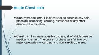
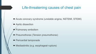
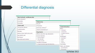
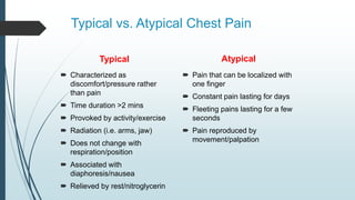
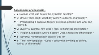
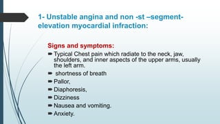
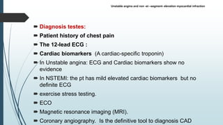
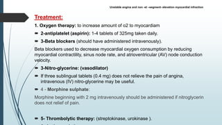
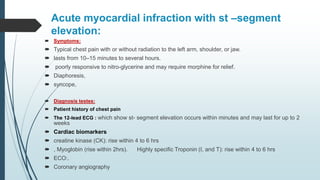
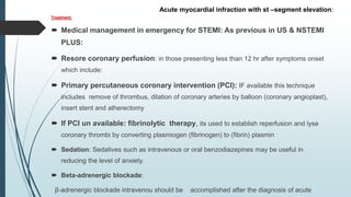
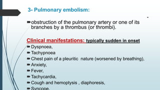
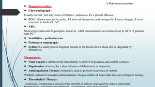
Acute chest pain
Acute Chest pain
is an imprecise term. It is often used to describe any pain,
pressure, squeezing, choking, numbness or any other
discomfort in the chest
Chest pain has many possible causes, all of which deserve
medical attention. The causes of chest pain fall into two
major categories — cardiac and non cardiac causes.
Life-threatening causes of chest pain
 Acute coronary syndrome (unstable angina, NSTEMI, STEMI)
 Aortic dissection
 Pulmonary embolism
 Pneumothorax (Tension pneumothorax)
 Pericardial tamponade
 Mediastinitis (e.g. esophageal rupture)
Differential diagnosis
UpToDate 2012
Typical vs. Atypical Chest Pain
Typical
 Characterized as
discomfort/pressure rather
than pain
 Time duration >2 mins
 Provoked by activity/exercise
 Radiation (i.e. arms, jaw)
 Does not change with
respiration/position
 Associated with
diaphoresis/nausea
 Relieved by rest/nitroglycerin
Atypical
 Pain that can be localized with
one finger
 Constant pain lasting for days
 Fleeting pains lasting for a few
seconds
 Pain reproduced by
movement/palpation
Assessment of chest pain .
 N Normal: what was before this symptom develop?
 O Onset : when start? What day &time? Suddenly or gradually?
 P Precipitating & palliative factors: as stress, position, and what can
relieve it?
 Q Quality & quantity: how does it feel? Is more/less than before?
 R Region & radiation: where it occur? Does it radiate to other region?
 S Severity: Numerical pain scale of 0 to 10.
 T Time: how long it last? Does it occur with anything as before,
during, or after meals?
1- Unstable angina and non -st –segment-
elevation myocardial infraction:
Signs and symptoms:
Typical Chest pain which radiate to the neck, jaw,
shoulders, and inner aspects of the upper arms, usually
the left arm.
 shortness of breath
Pallor,
Diaphoresis,
Dizziness
Nausea and vomiting.
Anxiety.
 Diagnosis testes:
 Patient history of chest pain
 The 12-lead ECG :
 Cardiac biomarkers (A cardiac-specific troponin)
 In Unstable angina: ECG and Cardiac biomarkers show no
evidence
 In NSTEMI: the pt has mild elevated cardiac biomarkers but no
definite ECG
 exercise stress testing.
 ECO
 Magnetic resonance imaging (MRI).
 Coronary angiography. Is the definitive tool to diagnosis CAD
Unstable angina and non -st –segment- elevation myocardial infraction
Treatment:
1. Oxygen therapy: to increase amount of o2 to myocardiam
 2-antiplatelet (aspirin): 1-4 tablets of 325mg taken daily.
 3-Beta blockers (should have administered intravenously).
Beta blockers used to decrease myocardial oxygen consumption by reducing
myocardial contractility, sinus node rate, and atrioventricular (AV) node conduction
velocity.
 3-Nitro-glycerine: (vasodilator)
 If three sublingual tablets (0.4 mg) does not relieve the pain of angina,
intravenous (IV) nitro-glycerine may be useful.
 4 - Morphine sulphate:
Morphine beginning with 2 mg intravenously should be administered if nitroglycerin
does not relief of pain.
 5- Thrombolytic therapy: (streptokinase, urokinase ).
Unstable angina and non -st –segment- elevation myocardial infraction
Acute myocardial infraction with st –segment
elevation:
 Symptoms:
 Typical chest pain with or without radiation to the left arm, shoulder, or jaw.
 lasts from 10–15 minutes to several hours.
 poorly responsive to nitro-glycerine and may require morphine for relief.
 Diaphoresis,
 syncope,
 Diagnosis testes:
 Patient history of chest pain
 The 12-lead ECG : which show st- segment elevation occurs within minutes and may last for up to 2
weeks
 Cardiac biomarkers
 creatine kinase (CK): rise within 4 to 6 hrs
 , Myoglobin (rise within 2hrs). Highly specific Troponin (I, and T): rise within 4 to 6 hrs
 ECO:.
 Coronary angiography
Treatment:
 Medical management in emergency for STEMI: As previous in US & NSTEMI
PLUS:
 Resore coronary perfusion: in those presenting less than 12 hr after symptoms onset
which include:
 Primary percutaneous coronary intervention (PCI): IF available this technique
includes remove of thrombus, dilation of coronary arteries by balloon (coronary angioplast),
insert stent and atherectomy
 If PCI un available: fibrinolytic therapy, its used to establish reperfusion and lyse
coronary thrombi by converting plasmiogen (fibrinogen) to (fibrin) plasmin
 Sedation: Sedatives such as intravenous or oral benzodiazepines may be useful in
reducing the level of anxiety.
 Beta-adrenergic blockade:
β-adrenergic blockade intravenou should be accomplished after the diagnosis of acute
Acute myocardial infraction with st –segment elevation:
3- Pulmonary embolism:

obstruction of the pulmonary artery or one of its
branches by a thrombus (or thrombi).
Clinical manifestations: typically sudden in onset
Dyspnoea,
Tachypnoea
Chest pain of a pleuritic nature (worsened by breathing),
Anxiety,
Fever,
Tachycardia,
Cough and hemoptysis , diaphoresis,
 Diagnostic studies:
 Chest radiograph :
Usually normal , but may shows infiltrates , atelectasis, Or a pleural effusion.
 ECG : Shows sinus tachycardia , PR interval depression ,and nonspecific T wave changes ,T wave
inversion in leads V1 , V2 .
 ABG:
Shows hypoxemia and hypocapnia, however, ABG measurements are normal in up to 20 % of patients
with PE .
 Ventilation – perfusion scan:
 Pulmonary angiography.
 D-dimer: a small protein fragment present in the blood after a blood clot is degraded by
fibrinolysis
Management:
 Nasal oxygen is administered immediately to relieve hypoxemia, and central cyanosis.
 Hypotension is treated by a slow infusion of dobutamine or dopamine.
 Anticoagulation Therapy: Heparin is used to prevent recurrence of emboli.
Warfarin sodium (Coumadin) administration is begun within 24 hours after the start of heparin therapy.
 Thrombolytic Therapy:
(urokinase, streptokinase,) resolves the thrombi or emboli more quickly, reduce pulmonary
3- Pulmonary embolism
4- Pericarditis:
 Pericarditis is inflammation of the pericardium. Acute pericarditis is pericarditis that lasts
no longer than 1 or 2 weeks. Inflammation often involves the adjoining diaphragm.
 Pericarditis can be a primary disease or occur secondarily as the result of some other
disorder, such as acute MI or renal failure
Causes
 Idiopathic or unknown (usually presumed to be viral)
 • Infectious OR • Bacterial
 • Tuberculosis
 • Autoimmune or inflammatory
 • Systemic lupus erythematosus
 • some Drugs and Vaccinations
 • Radiation therapy
 • Following device implantation, such as an implantable defibrillator
 • Acute myocardial infarction
 • Trauma to the chest wall or myocardium, including cardiopulmonary surgery
 Chronic renal failure requiring dialysis
Pericarditis Diagnosis:
 Medical history focuses on chest pain: Does the pain get worse when pt take a
deep breath? Is it worse when pt lie down? Does it get better after pt sit up and lean
forward?
 Physical examination focuses on careful listening through a stethoscope for
the scratchy sounds called a pericardial rub, which are produced by heart muscle
rubbing against the inflamed pericardium. These sounds strongly suggest the
diagnosis of pericarditis
 ECG.
 Note the diffuse upward concavity ST changes and the PR-segment depression.
Symptoms:
 Chest pain. The pain is felt below the sternum, and below the ribs on the left side of the
chest.
 Pericardial effusion: fluid accumulate between the pericardium and the heart
 Dyspnea.
 Distended veins in the neck
 Swollen ankles and feet.
Treatment:
 anti-inflammatory agents.
 Aspirin prescribed as initial therapy.
 nonsteroidal anti-inflammatory drugs (NSAIDs) agents are prescribed. (ibuprofen, and
naproxen) ,
 NSAIDs are usually quite effective in reducing inflammation and eliminating the pain
associated with pericarditis.
 Pericardiocentisis, a thin hollow needle is carefully inserted through the chest wall into
the area of accumulated fluid, and the fluid is drained through the needle.
4- Pericarditis
5- coronary spasm:
 Coronary artery spasm is a temporary, sudden narrowing of one of the coronary
arteries.
Causes:
 Alcohol withdrawal
 Emotional stress
 Exposure to cold
 Medications that cause narrowing of the blood vessels (vasoconstriction)
Symptomes:
 chest pain occurs at rest , Lasts from 5 to 30 minutes .
Exams and Tests:
 Tests to diagnose coronary artery spasm may include:
 Coronary angiography
 ECG
6-pleurisy:
 Inflammation of pleura.
Causes:
 Viral infection
 Bacterial infection
 Pneumonia
 Tuberculosis
 Pulmonary embolus
Symptoms:
 Pain in the muscles of the chest
 Persistent cough
 Fever
Diagnosis methods:
Physical examination - using a stethoscope, the doctor can hear the pleura
scraping against each other. Other breath sound abnormalities that suggest
pleurisy include rattling or crackling.
 Blood tests - to determine whether the cause is viral or bacterial.
 X-rays - of the chest, including CT scans or other imaging scans using
ultrasound.
 Thoracentesis - a small sample of pleural fluid is removed and examined.
 Bronchoscopy - a thin tube is inserted down the windpipe to examine the
airways.
Treatment:
 Treating the underlying cause - for example, treatment for tuberculosis
 Medications such as antibiotics and anti-inflammatory drugs
6-pleurisy
7- Costochondritis:
 Inflammation of the cartilages of the ribcage.
Symptoms:
 Pain: may have a sudden or gradual onset, and may be mild or severe,
 tenderness, swelling occur in one or more of the 4 upper ribs.
Diagnosis of Costochondritis:
 The medical history and physical examination are usually sufficient for
diagnosis.
Treatment:
 local heat, analgesics, anti-inflammatory drugs, or local steroid injections.
8-Acute cholecystitis:
 a sudden inflammation of the gallbladder that causes severe
abdominal pain and which can radiated to chest
Causes
 In 90% of cases, acute cholecystitis is caused by gallstones in
the gallbladder, and tumors of the gallbladder may also cause
cholecystitis.
Symptoms
 The main symptom is abdominal pain that is located on the upper
right side or upper middle of the abdomen.
 Abdominal fullness
 Fever
 Nausea and vomiting
Exams and Tests
 A physical exam will show that abdomen is tender to the touch.
 following blood tests are ordered :
 Amylase and lipase
 Bilirubin
 Complete blood count ( CBC) -- may show a higher than normal white blood cell count
 Liver function tests
 Imaging tests that can show gallstones or inflammation include:
 Abdominal ultrasound
 Abdominal CT scan
 Abdominal x-ray
 Oral cholecystogram
 Gallbladder radionuclide scan

 Treatment:
 Intravenous fluids, and antibiotics to fight infection.
Nursing roles for patients with chest pain
 Bed rest for a minimum of 12 to 24 hours
 Obtain 12-lead electrocardiogram (ECG) during chest pain to be read
within10 minutes.
 Start oxygen therapy
 Obtain laboratory blood specimens of cardiac biomarkers, including
Troponin.
 Minimize the number of times the patient’s skin is punctured and
 Avoid intramuscular injections
 Draw blood for laboratory tests when starting the IV line
 Start IV lines before thrombolytic therapy; designate one line to use for
blood draws
 Avoid continual use of noninvasive blood pressure cuff
 Monitor for acute Dysrhythmias and hypotension
 Monitor for reperfusion: resolution of angina or acute ST segment changes
Nursing roles for patients with chest pain
 Check for signs and symptoms of bleeding: decrease in hematocrit and
hemoglobin values, decrease in blood pressure, increase in heart rate, oozing or
bulging at invasive procedure sites, back pain, muscle weakness, changes in level
of consciousness, complaints of headache
 Treat major bleeding by discontinuing thrombolytic therapy and any anticoagulants;
apply direct pressure and notify the physician immediately, treat minor bleeding by
applying direct pressure if accessible and appropriate; continue to monitor
 Evaluate for indications for reperfusion therapy:
Acute_chest_pain.pptx educational systamic

Acute_chest_pain.pptx educational systamic

  • 1.
  • 2.
    Acute Chest pain isan imprecise term. It is often used to describe any pain, pressure, squeezing, choking, numbness or any other discomfort in the chest Chest pain has many possible causes, all of which deserve medical attention. The causes of chest pain fall into two major categories — cardiac and non cardiac causes.
  • 3.
    Life-threatening causes ofchest pain  Acute coronary syndrome (unstable angina, NSTEMI, STEMI)  Aortic dissection  Pulmonary embolism  Pneumothorax (Tension pneumothorax)  Pericardial tamponade  Mediastinitis (e.g. esophageal rupture)
  • 4.
  • 5.
    Typical vs. AtypicalChest Pain Typical  Characterized as discomfort/pressure rather than pain  Time duration >2 mins  Provoked by activity/exercise  Radiation (i.e. arms, jaw)  Does not change with respiration/position  Associated with diaphoresis/nausea  Relieved by rest/nitroglycerin Atypical  Pain that can be localized with one finger  Constant pain lasting for days  Fleeting pains lasting for a few seconds  Pain reproduced by movement/palpation
  • 6.
    Assessment of chestpain .  N Normal: what was before this symptom develop?  O Onset : when start? What day &time? Suddenly or gradually?  P Precipitating & palliative factors: as stress, position, and what can relieve it?  Q Quality & quantity: how does it feel? Is more/less than before?  R Region & radiation: where it occur? Does it radiate to other region?  S Severity: Numerical pain scale of 0 to 10.  T Time: how long it last? Does it occur with anything as before, during, or after meals?
  • 7.
    1- Unstable anginaand non -st –segment- elevation myocardial infraction: Signs and symptoms: Typical Chest pain which radiate to the neck, jaw, shoulders, and inner aspects of the upper arms, usually the left arm.  shortness of breath Pallor, Diaphoresis, Dizziness Nausea and vomiting. Anxiety.
  • 8.
     Diagnosis testes: Patient history of chest pain  The 12-lead ECG :  Cardiac biomarkers (A cardiac-specific troponin)  In Unstable angina: ECG and Cardiac biomarkers show no evidence  In NSTEMI: the pt has mild elevated cardiac biomarkers but no definite ECG  exercise stress testing.  ECO  Magnetic resonance imaging (MRI).  Coronary angiography. Is the definitive tool to diagnosis CAD Unstable angina and non -st –segment- elevation myocardial infraction
  • 9.
    Treatment: 1. Oxygen therapy:to increase amount of o2 to myocardiam  2-antiplatelet (aspirin): 1-4 tablets of 325mg taken daily.  3-Beta blockers (should have administered intravenously). Beta blockers used to decrease myocardial oxygen consumption by reducing myocardial contractility, sinus node rate, and atrioventricular (AV) node conduction velocity.  3-Nitro-glycerine: (vasodilator)  If three sublingual tablets (0.4 mg) does not relieve the pain of angina, intravenous (IV) nitro-glycerine may be useful.  4 - Morphine sulphate: Morphine beginning with 2 mg intravenously should be administered if nitroglycerin does not relief of pain.  5- Thrombolytic therapy: (streptokinase, urokinase ). Unstable angina and non -st –segment- elevation myocardial infraction
  • 10.
    Acute myocardial infractionwith st –segment elevation:  Symptoms:  Typical chest pain with or without radiation to the left arm, shoulder, or jaw.  lasts from 10–15 minutes to several hours.  poorly responsive to nitro-glycerine and may require morphine for relief.  Diaphoresis,  syncope,  Diagnosis testes:  Patient history of chest pain  The 12-lead ECG : which show st- segment elevation occurs within minutes and may last for up to 2 weeks  Cardiac biomarkers  creatine kinase (CK): rise within 4 to 6 hrs  , Myoglobin (rise within 2hrs). Highly specific Troponin (I, and T): rise within 4 to 6 hrs  ECO:.  Coronary angiography
  • 11.
    Treatment:  Medical managementin emergency for STEMI: As previous in US & NSTEMI PLUS:  Resore coronary perfusion: in those presenting less than 12 hr after symptoms onset which include:  Primary percutaneous coronary intervention (PCI): IF available this technique includes remove of thrombus, dilation of coronary arteries by balloon (coronary angioplast), insert stent and atherectomy  If PCI un available: fibrinolytic therapy, its used to establish reperfusion and lyse coronary thrombi by converting plasmiogen (fibrinogen) to (fibrin) plasmin  Sedation: Sedatives such as intravenous or oral benzodiazepines may be useful in reducing the level of anxiety.  Beta-adrenergic blockade: β-adrenergic blockade intravenou should be accomplished after the diagnosis of acute Acute myocardial infraction with st –segment elevation:
  • 12.
    3- Pulmonary embolism:  obstructionof the pulmonary artery or one of its branches by a thrombus (or thrombi). Clinical manifestations: typically sudden in onset Dyspnoea, Tachypnoea Chest pain of a pleuritic nature (worsened by breathing), Anxiety, Fever, Tachycardia, Cough and hemoptysis , diaphoresis,
  • 13.
     Diagnostic studies: Chest radiograph : Usually normal , but may shows infiltrates , atelectasis, Or a pleural effusion.  ECG : Shows sinus tachycardia , PR interval depression ,and nonspecific T wave changes ,T wave inversion in leads V1 , V2 .  ABG: Shows hypoxemia and hypocapnia, however, ABG measurements are normal in up to 20 % of patients with PE .  Ventilation – perfusion scan:  Pulmonary angiography.  D-dimer: a small protein fragment present in the blood after a blood clot is degraded by fibrinolysis Management:  Nasal oxygen is administered immediately to relieve hypoxemia, and central cyanosis.  Hypotension is treated by a slow infusion of dobutamine or dopamine.  Anticoagulation Therapy: Heparin is used to prevent recurrence of emboli. Warfarin sodium (Coumadin) administration is begun within 24 hours after the start of heparin therapy.  Thrombolytic Therapy: (urokinase, streptokinase,) resolves the thrombi or emboli more quickly, reduce pulmonary 3- Pulmonary embolism
  • 14.
    4- Pericarditis:  Pericarditisis inflammation of the pericardium. Acute pericarditis is pericarditis that lasts no longer than 1 or 2 weeks. Inflammation often involves the adjoining diaphragm.  Pericarditis can be a primary disease or occur secondarily as the result of some other disorder, such as acute MI or renal failure Causes  Idiopathic or unknown (usually presumed to be viral)  • Infectious OR • Bacterial  • Tuberculosis  • Autoimmune or inflammatory  • Systemic lupus erythematosus  • some Drugs and Vaccinations  • Radiation therapy  • Following device implantation, such as an implantable defibrillator  • Acute myocardial infarction  • Trauma to the chest wall or myocardium, including cardiopulmonary surgery  Chronic renal failure requiring dialysis
  • 15.
    Pericarditis Diagnosis:  Medicalhistory focuses on chest pain: Does the pain get worse when pt take a deep breath? Is it worse when pt lie down? Does it get better after pt sit up and lean forward?  Physical examination focuses on careful listening through a stethoscope for the scratchy sounds called a pericardial rub, which are produced by heart muscle rubbing against the inflamed pericardium. These sounds strongly suggest the diagnosis of pericarditis  ECG.  Note the diffuse upward concavity ST changes and the PR-segment depression.
  • 16.
    Symptoms:  Chest pain.The pain is felt below the sternum, and below the ribs on the left side of the chest.  Pericardial effusion: fluid accumulate between the pericardium and the heart  Dyspnea.  Distended veins in the neck  Swollen ankles and feet. Treatment:  anti-inflammatory agents.  Aspirin prescribed as initial therapy.  nonsteroidal anti-inflammatory drugs (NSAIDs) agents are prescribed. (ibuprofen, and naproxen) ,  NSAIDs are usually quite effective in reducing inflammation and eliminating the pain associated with pericarditis.  Pericardiocentisis, a thin hollow needle is carefully inserted through the chest wall into the area of accumulated fluid, and the fluid is drained through the needle. 4- Pericarditis
  • 17.
    5- coronary spasm: Coronary artery spasm is a temporary, sudden narrowing of one of the coronary arteries. Causes:  Alcohol withdrawal  Emotional stress  Exposure to cold  Medications that cause narrowing of the blood vessels (vasoconstriction) Symptomes:  chest pain occurs at rest , Lasts from 5 to 30 minutes . Exams and Tests:  Tests to diagnose coronary artery spasm may include:  Coronary angiography  ECG
  • 18.
    6-pleurisy:  Inflammation ofpleura. Causes:  Viral infection  Bacterial infection  Pneumonia  Tuberculosis  Pulmonary embolus Symptoms:  Pain in the muscles of the chest  Persistent cough  Fever
  • 19.
    Diagnosis methods: Physical examination- using a stethoscope, the doctor can hear the pleura scraping against each other. Other breath sound abnormalities that suggest pleurisy include rattling or crackling.  Blood tests - to determine whether the cause is viral or bacterial.  X-rays - of the chest, including CT scans or other imaging scans using ultrasound.  Thoracentesis - a small sample of pleural fluid is removed and examined.  Bronchoscopy - a thin tube is inserted down the windpipe to examine the airways. Treatment:  Treating the underlying cause - for example, treatment for tuberculosis  Medications such as antibiotics and anti-inflammatory drugs 6-pleurisy
  • 20.
    7- Costochondritis:  Inflammationof the cartilages of the ribcage. Symptoms:  Pain: may have a sudden or gradual onset, and may be mild or severe,  tenderness, swelling occur in one or more of the 4 upper ribs. Diagnosis of Costochondritis:  The medical history and physical examination are usually sufficient for diagnosis. Treatment:  local heat, analgesics, anti-inflammatory drugs, or local steroid injections.
  • 21.
    8-Acute cholecystitis:  asudden inflammation of the gallbladder that causes severe abdominal pain and which can radiated to chest Causes  In 90% of cases, acute cholecystitis is caused by gallstones in the gallbladder, and tumors of the gallbladder may also cause cholecystitis. Symptoms  The main symptom is abdominal pain that is located on the upper right side or upper middle of the abdomen.  Abdominal fullness  Fever  Nausea and vomiting
  • 22.
    Exams and Tests A physical exam will show that abdomen is tender to the touch.  following blood tests are ordered :  Amylase and lipase  Bilirubin  Complete blood count ( CBC) -- may show a higher than normal white blood cell count  Liver function tests  Imaging tests that can show gallstones or inflammation include:  Abdominal ultrasound  Abdominal CT scan  Abdominal x-ray  Oral cholecystogram  Gallbladder radionuclide scan   Treatment:  Intravenous fluids, and antibiotics to fight infection.
  • 23.
    Nursing roles forpatients with chest pain  Bed rest for a minimum of 12 to 24 hours  Obtain 12-lead electrocardiogram (ECG) during chest pain to be read within10 minutes.  Start oxygen therapy  Obtain laboratory blood specimens of cardiac biomarkers, including Troponin.  Minimize the number of times the patient’s skin is punctured and  Avoid intramuscular injections  Draw blood for laboratory tests when starting the IV line  Start IV lines before thrombolytic therapy; designate one line to use for blood draws  Avoid continual use of noninvasive blood pressure cuff  Monitor for acute Dysrhythmias and hypotension  Monitor for reperfusion: resolution of angina or acute ST segment changes
  • 24.
    Nursing roles forpatients with chest pain  Check for signs and symptoms of bleeding: decrease in hematocrit and hemoglobin values, decrease in blood pressure, increase in heart rate, oozing or bulging at invasive procedure sites, back pain, muscle weakness, changes in level of consciousness, complaints of headache  Treat major bleeding by discontinuing thrombolytic therapy and any anticoagulants; apply direct pressure and notify the physician immediately, treat minor bleeding by applying direct pressure if accessible and appropriate; continue to monitor  Evaluate for indications for reperfusion therapy: