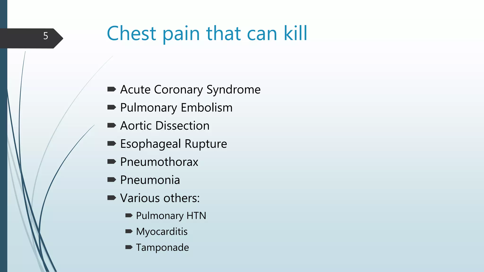
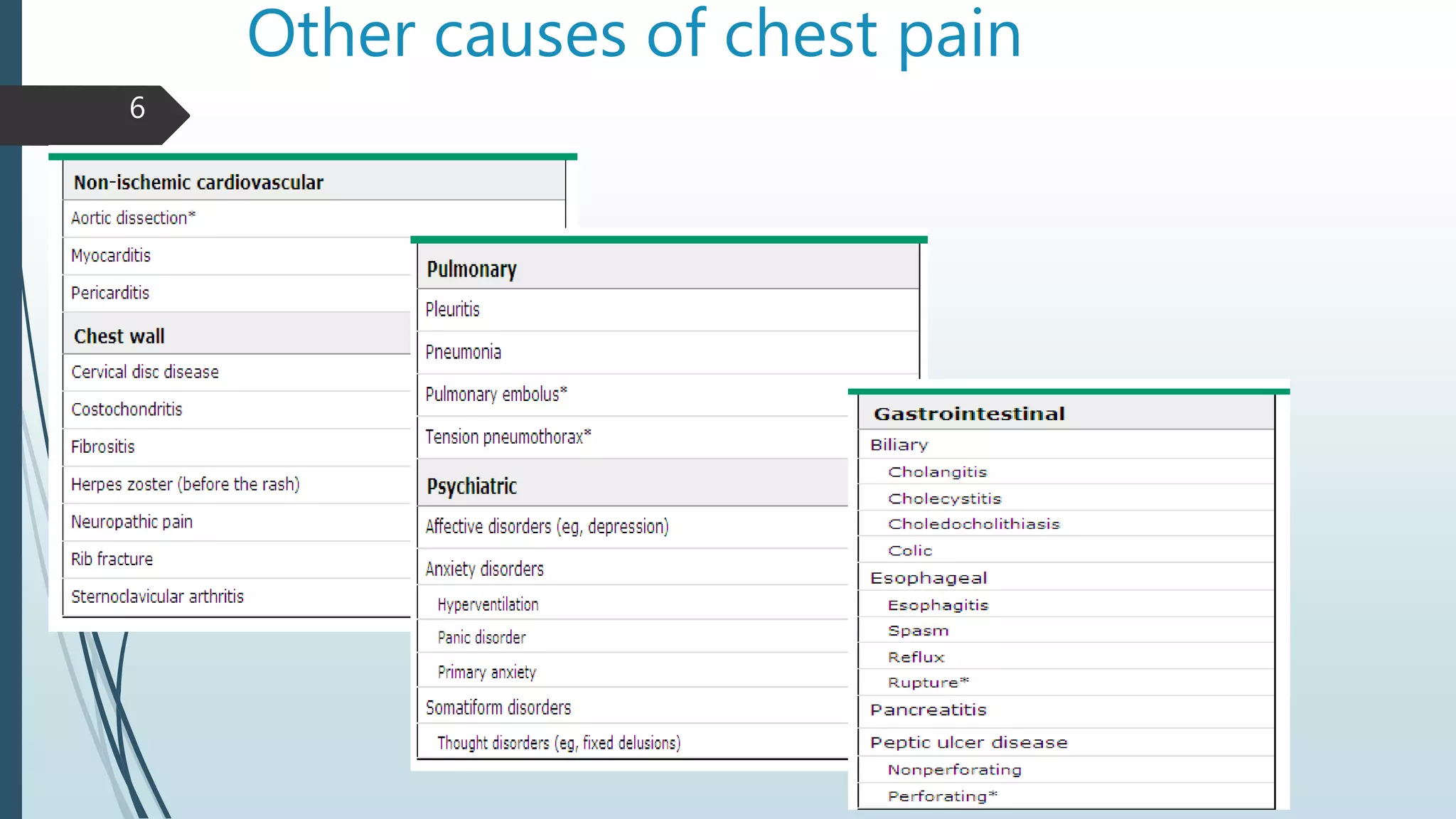
Chest Pain
Muqtasid
Ayesha Khan
Final year MBBS
Facilitated by: Dr. Afrasiab Altaf
Objectives
 Describe various etiologies for chest pain
 Typical vs Atypical chest pain
 Review approach to chest pain
 Focus on life threatening causes of chest pain
 Management of chest pain
 Review patient cases
2
Overview
 Chest pain accounts for 6 million annual visits to the Emergency
Departments in the United States
 Chest pain is the second most common Emergency Department
complaint after abdominal pain.
 Wide range of etiologies
 Cardiac, pulmonary, gastrointestinal, musculoskeletal
3
Causes of chest pain that
can kill
Chest pain that can kill
 Acute Coronary Syndrome
 Pulmonary Embolism
 Aortic Dissection
 Esophageal Rupture
 Pneumothorax
 Pneumonia
 Various others:
 Pulmonary HTN
 Myocarditis
 Tamponade
5
Other causes of chest pain
6
Do you know the types of
chest pain?
Types of chest pain
• Characterized as discomfort/pressure rather than pain
• Time duration >2 mins
• Provoked by activity/exercise
• Radiation (i.e. arms, jaw)
• Does not change with respiration/position
• Associated with diaphoresis/nausea
• Relieved by rest/nitroglycerin
Typical
• Pain that can be localized with one finger
• Constant pain lasting for days
• Fleeting pains lasting for a few seconds
• Pain reproduced by movement/palpation
Atypical
8
Approach to a patient with chest pain
 History
 Examination
 Stabilization
 Investigations
 Diagnosis
 Management
9
What are the key parts of HOPI in
chest pain patient?
What can you get out of patient in 4
minutes?
10
History matters
 Location: Central, left, or right
 Timing: Gradual or sudden onset
 Duration: since onset
 Character: crushing, stabbing, tearing, squeezing, sharp
 Associated symptoms: SOB, sweating, nausea
 Aggravating factors: What makes it worse or better?
 Quality: Visceral vs somatic
 Radiation: Back, neck, arm
 Severity: Scale of 1-10
11
Rest of the history
 Past medical and surgical: cardiovascular disease, pulmonary disease
 Medication: Nitroglycerin, ASA etc.
 Allergies: Always important!
 Social: Smoker, Alcoholic, Cocaine,
 Family: Sudden Death, Early MI, DVT, Pulmonary embolism
12
Key points in physical
examination
what can you examine in 2 minutes?
13
Physical examination
 General Appearance
 Vital Signs
 Heart (Muffled, Regular, Fast,)
 Lungs (Equal, Wet, Tympanitic,)
 Neck (JVD)
 Abdomen (Distention)
 Lower Extremities (Edema, calf tenderness,)
14
This man is brought to ED through
ambulance
What do you do next?
15
Approach in emergency department
First 60 seconds
 How does the patient look?
 What are the patient’s vital signs?
 Ambulance story?
16
Next 5 minutes
What are 2 bedside tests to consider?
What is an important and cheap medication you
should consider?
17
Next 5 minutes
 Brief History
 Brief Physical
 ABCs, IV line, O2, monitor vitals, pulse oximetry
 What are 2 bedside tests that can be done to help stratify the
patient?
 ECG
 Portable CXR
 What is an important and cheap medication you should consider?
 ASA (More on this later)
18
Next 10 minutes
 Patient already stabilized, initial data gathered, and
initial orders submitted
 Secondary survey: More detailed history and physical
exam
 Address patient’s pain
 Goal now is to categorize patient
1) Chest wall pain- Musculoskeletal
2) Pleuritic chest pain- Respiratory
3) Visceral chest pain- Cardiac
19
Case presentation
Case 1
 64 year old man presents with 5 hours of chest pain and chest pressure
associated with SOB, nausea and diaphoresis. Gradual onset while
shoveling the snow. Pain radiated towards left jaw. Improved with rest.
 Past medical history: HTN, DM
 General: Nontoxic appearing, apprehensive, mildly diaphoretic
 Vitals: 37.5ºC, RR16, HR 100, BP 160/95
 CVS: RRR, Normal S1, S2, no M/R/G
 Respiratory: CTAB, easy respirations
 Abdomen: Soft, NTND
 Extremities: No calf tenderness or swelling, no edema, strong distal pulses
21
Diagnosis?
What to do next?
22
Case 1 Acute Coronary Syndrome
ECG:
 This will differentiate what you must do now.
(Specific but not sensitive)
ST elevation in 2 contiguous leads: STEMI
New LBBB
Ischemia/strain: ST depressions, new T wave inversions, Q
waves
Nonspecific: T wave flattening/inversions or Q waves
without old EKG
23
Case 1 ACS
 CXR
 To look for failure and evaluate for other cause of chest pain
 Cardiac Enzymes
Marker Elevation Peak Duratio
CK-MB 3-12 h 18-24 h 2 days
Troponin-I 3-12 h 18 h 5-10 d
Troponin-T 3-12 h 12 h 5-14 d
24
What else can you do for this
ACS patient?
25
Treatment of ACS
 ABCs, IV line, O2, monitor vitals, pulse oximetry
 Morphine sulphate 5–10 mg or diamorphine 2.5–5 mg
 Aspirin 300mg plus clopedogril 600mg within 12 hours
 Aspirin continued-75–300 mg daily
 Heparin-fondaparinux 2.5 mg daily-8 days
 Sublingual glyceryl trinitrate (300–500 μg)or isosorbide dinitrate 1–2 mg/hour
 Atenolol 5–10 mg or metoprolol 5–15 mg given over 5 mins
 Fibrinolysis with streptokinase or Alteplase(tpA)
 Primary percutaneous coronary intervention (PCI)
26
Case 2
 30 years old male had an open reduction internal fixation of ankle
fracture 2 weeks ago, now presented with sudden onset of chest
pain.
27
Pulmonary Embolism
Signs and symptoms?
28
Pulmonary embolism
Symptoms
SOB or dyspnea- Present in 90%
Chest pain (pleuritic)- 66% of patients with PE
Cough
Sudden onset
Signs
Tachycardia > 100 beats per minute
Tachypnea > 20 breaths per minute
Hypoxia < 95% on RA (no other cause)
Lower extremity swelling
29
Pulmonary Embolus Risk Factors
 Hypercoaguability
 Malignancy, pregnancy, estrogen use, factor V Leiden, protein C/S
deficiency
 Venous stasis
 Bedrest > 48 hours, recent hospitalization, long distance travel
 Venous injury
 Recent trauma or surgery
30
Pulmonary embolism: diagnosis
 CT pulmonary angiography (first line diagnostic test)
 Electrocardiography
Rule out MI, pericarditis and other causes
 Arterial blood gases
reduced PaO2, normal or low PaCO2
 D-dimer
 Ventilation–perfusion scanning
31
Treatment of PE
 Oxygen, keep O2 saturation above 90%
 IV fluid to maintain blood pressure
 Heparin (Will limit propagation but does not dissolve clot)
Unfractionated: 80 u/kg bolus, 18 h/kg/hr
Fractionated (Lovenox): 1 mg/kg SC BID
 Fibrinolytics
Alteplase 50–100 mg infused over 2–6 hrs, (bolus in severe shock)
32
Case 3
 35 years old male with sudden
ripping pain radiating to back.
Diagnosis?
33
Aortic dissection
Blood violates aortic
intimal and adventitial
layers
False lumen is created
Dissection may extend
proximally, distally, or
in both directions
34
Classification; aortic dissection35
Etiology; Aortic Dissection
 Bimodal distribution
 Young: Connective tissue (Marfan) or pregnancy
 Older: Most commonly > 50 (mean age 63)
 Risk factors
 Male: 66% of patients
 Hypertension: 72% of patients
 Connective tissue disease
30% of Marfan’s patients get dissections
 Cocaine Use
 Syphilis
36
Clinical presentation
 Difficult clinical diagnosis
 85% have chest or back pain
 “Ripping” or “tearing” pain in 50%
 Neurologic symptoms(paraplegia) in 20%
 Hematuria
 Asymmetric pulses(brachial, carotid, femoral)
 Occlusion of aortic branches may cause
 MI (coronary)
 stroke (carotid)
 mesenteric infarction with an acute abdomen (coeliac and superior
mesenteric),
 renal failure (renal)
 acute limb (usually leg) ischaemia.
37
How do you confirm the
diagnosis of this disease?
38
Aortic dissection; diagnosis
 CXR-
 Widened mediastinum, abnormal aortic knob, pleural effusions
 Chest CT-
 Very sensitive and specific
 Quickly obtained
 Must think about kidney + contrast
 Angiography-
 Gold standard
 Most reliable anatomy of dissection
 Transoesophageal echocardiography
 Bedside U/S – evaluate aorta and look at heart to rule out tampanode.
39
Ct chest (aortic dissection) Angiography (aortic dissection)
40
Management
 2 large bore IV’s, monitor vitals, Type and crossmatch blood, ECG
 Ascending dissections will need aortic replacement surgery (type
A dissection)
 If dissection is only descending, management is medical
 Blood pressure control
 Maintain systolic BP at120-130 mmHg
 Beta blockers are first line (Labetalol and Esmolol)
 Can add vasodilators i.e. nitroprusside
 Percutaneous endoluminal repair
 Stent graft implantation
 Admission to ICU
41
Case 4
 18 year old healthy male was lifting weights when he had sudden onset of
sharp Chest Pain + Shortness Of Breath.
 HR 122, RR 34, BP 70/P, Sat 88%
 Decreased breath sounds on left side
42
What is your diagnosis?
Pneumothorax!
43
Diagnosis; pneumothorax
Chest XRay
Free air appears black
Collapse of lung may occur
Mediastinum shifted to
other side
44
Management
 Primary spontaneous pneumothorax
 If small
 Observe: resolves in 10 days
 Small chest tube with one way valve may help
 If larger
 100% O2 supply
 Analgesics
 Needle aspiration followed by chest tube placement
 Secondary spontaneous pneumothorax
 Chest tube drainage
 Surgical intervention to prevent recurrence
45
Tension pneumothorax
 Medical emergency
 Chest decompression with a large bore needle
 Placement of chest tube
46
Chest decompression
47
Placement of chest tube48
Case 5
40 years old man presented with chest pain since 3 weeks with heart burn specially
after meals. He has difficulty in swallowing with sour taste in mouth.
On examination he is anemic and has lost weight.
Diagnosis?
49
Gastroesophageal reflux disease
 SIGNS AND SYMPTOMS
 Heartburn (30-60min after meal)
 Spontaneous reflux of sour or bitter gastric contents into the mouth.
 Noncardiac chest pain
 Chronic cough
 Alarming features
 Troublesome dysphagia
 Odynophagia,
 Weight loss
 Iron deficiency anemia
50
Investigations
 Upper endoscopy
 Barium esophagography
 Esophageal pH or combined esophageal pH-impedance
 testing
51
Management
 Life style modification
 Eating smaller meals and elimination of acidic foods
 Weight loss and smoking cessation
 Avoid lying down within 3 hours after meals
 Initial therapy
 PPIs; omeprazole or rabeprazole, 20 mg taken 30min before breakfast-4-6
weeks
 Long term therapy
 PPIs therapy can be discontinued after 8–12 weeks
 H2-receptor antagonists (cimetidine 200 mg) may be used to control
symptoms
 Surgical fundoplication
52
Summary
 Chest pain is a very common complaint but has a broad differential
 Always try to rule out the life-threatening causes of chest pain
 It is important to remember that troponin elevation DOES NOT
always mean ACS
 Use the history, physical exam, labs, EKG and imaging to commit to
a diagnosis
 Whenever you are stuck, ask for help. Your seniors are here to help
you!
53
Thank you!
Questions?

Chest pain Case Presentation with management

  • 1.
    Chest Pain Muqtasid Ayesha Khan Finalyear MBBS Facilitated by: Dr. Afrasiab Altaf
  • 2.
    Objectives  Describe variousetiologies for chest pain  Typical vs Atypical chest pain  Review approach to chest pain  Focus on life threatening causes of chest pain  Management of chest pain  Review patient cases 2
  • 3.
    Overview  Chest painaccounts for 6 million annual visits to the Emergency Departments in the United States  Chest pain is the second most common Emergency Department complaint after abdominal pain.  Wide range of etiologies  Cardiac, pulmonary, gastrointestinal, musculoskeletal 3
  • 4.
    Causes of chestpain that can kill
  • 5.
    Chest pain thatcan kill  Acute Coronary Syndrome  Pulmonary Embolism  Aortic Dissection  Esophageal Rupture  Pneumothorax  Pneumonia  Various others:  Pulmonary HTN  Myocarditis  Tamponade 5
  • 6.
    Other causes ofchest pain 6
  • 7.
    Do you knowthe types of chest pain?
  • 8.
    Types of chestpain • Characterized as discomfort/pressure rather than pain • Time duration >2 mins • Provoked by activity/exercise • Radiation (i.e. arms, jaw) • Does not change with respiration/position • Associated with diaphoresis/nausea • Relieved by rest/nitroglycerin Typical • Pain that can be localized with one finger • Constant pain lasting for days • Fleeting pains lasting for a few seconds • Pain reproduced by movement/palpation Atypical 8
  • 9.
    Approach to apatient with chest pain  History  Examination  Stabilization  Investigations  Diagnosis  Management 9
  • 10.
    What are thekey parts of HOPI in chest pain patient? What can you get out of patient in 4 minutes? 10
  • 11.
    History matters  Location:Central, left, or right  Timing: Gradual or sudden onset  Duration: since onset  Character: crushing, stabbing, tearing, squeezing, sharp  Associated symptoms: SOB, sweating, nausea  Aggravating factors: What makes it worse or better?  Quality: Visceral vs somatic  Radiation: Back, neck, arm  Severity: Scale of 1-10 11
  • 12.
    Rest of thehistory  Past medical and surgical: cardiovascular disease, pulmonary disease  Medication: Nitroglycerin, ASA etc.  Allergies: Always important!  Social: Smoker, Alcoholic, Cocaine,  Family: Sudden Death, Early MI, DVT, Pulmonary embolism 12
  • 13.
    Key points inphysical examination what can you examine in 2 minutes? 13
  • 14.
    Physical examination  GeneralAppearance  Vital Signs  Heart (Muffled, Regular, Fast,)  Lungs (Equal, Wet, Tympanitic,)  Neck (JVD)  Abdomen (Distention)  Lower Extremities (Edema, calf tenderness,) 14
  • 15.
    This man isbrought to ED through ambulance What do you do next? 15
  • 16.
    Approach in emergencydepartment First 60 seconds  How does the patient look?  What are the patient’s vital signs?  Ambulance story? 16
  • 17.
    Next 5 minutes Whatare 2 bedside tests to consider? What is an important and cheap medication you should consider? 17
  • 18.
    Next 5 minutes Brief History  Brief Physical  ABCs, IV line, O2, monitor vitals, pulse oximetry  What are 2 bedside tests that can be done to help stratify the patient?  ECG  Portable CXR  What is an important and cheap medication you should consider?  ASA (More on this later) 18
  • 19.
    Next 10 minutes Patient already stabilized, initial data gathered, and initial orders submitted  Secondary survey: More detailed history and physical exam  Address patient’s pain  Goal now is to categorize patient 1) Chest wall pain- Musculoskeletal 2) Pleuritic chest pain- Respiratory 3) Visceral chest pain- Cardiac 19
  • 20.
  • 21.
    Case 1  64year old man presents with 5 hours of chest pain and chest pressure associated with SOB, nausea and diaphoresis. Gradual onset while shoveling the snow. Pain radiated towards left jaw. Improved with rest.  Past medical history: HTN, DM  General: Nontoxic appearing, apprehensive, mildly diaphoretic  Vitals: 37.5ºC, RR16, HR 100, BP 160/95  CVS: RRR, Normal S1, S2, no M/R/G  Respiratory: CTAB, easy respirations  Abdomen: Soft, NTND  Extremities: No calf tenderness or swelling, no edema, strong distal pulses 21
  • 22.
  • 23.
    Case 1 AcuteCoronary Syndrome ECG:  This will differentiate what you must do now. (Specific but not sensitive) ST elevation in 2 contiguous leads: STEMI New LBBB Ischemia/strain: ST depressions, new T wave inversions, Q waves Nonspecific: T wave flattening/inversions or Q waves without old EKG 23
  • 24.
    Case 1 ACS CXR  To look for failure and evaluate for other cause of chest pain  Cardiac Enzymes Marker Elevation Peak Duratio CK-MB 3-12 h 18-24 h 2 days Troponin-I 3-12 h 18 h 5-10 d Troponin-T 3-12 h 12 h 5-14 d 24
  • 25.
    What else canyou do for this ACS patient? 25
  • 26.
    Treatment of ACS ABCs, IV line, O2, monitor vitals, pulse oximetry  Morphine sulphate 5–10 mg or diamorphine 2.5–5 mg  Aspirin 300mg plus clopedogril 600mg within 12 hours  Aspirin continued-75–300 mg daily  Heparin-fondaparinux 2.5 mg daily-8 days  Sublingual glyceryl trinitrate (300–500 μg)or isosorbide dinitrate 1–2 mg/hour  Atenolol 5–10 mg or metoprolol 5–15 mg given over 5 mins  Fibrinolysis with streptokinase or Alteplase(tpA)  Primary percutaneous coronary intervention (PCI) 26
  • 27.
    Case 2  30years old male had an open reduction internal fixation of ankle fracture 2 weeks ago, now presented with sudden onset of chest pain. 27
  • 28.
  • 29.
    Pulmonary embolism Symptoms SOB ordyspnea- Present in 90% Chest pain (pleuritic)- 66% of patients with PE Cough Sudden onset Signs Tachycardia > 100 beats per minute Tachypnea > 20 breaths per minute Hypoxia < 95% on RA (no other cause) Lower extremity swelling 29
  • 30.
    Pulmonary Embolus RiskFactors  Hypercoaguability  Malignancy, pregnancy, estrogen use, factor V Leiden, protein C/S deficiency  Venous stasis  Bedrest > 48 hours, recent hospitalization, long distance travel  Venous injury  Recent trauma or surgery 30
  • 31.
    Pulmonary embolism: diagnosis CT pulmonary angiography (first line diagnostic test)  Electrocardiography Rule out MI, pericarditis and other causes  Arterial blood gases reduced PaO2, normal or low PaCO2  D-dimer  Ventilation–perfusion scanning 31
  • 32.
    Treatment of PE Oxygen, keep O2 saturation above 90%  IV fluid to maintain blood pressure  Heparin (Will limit propagation but does not dissolve clot) Unfractionated: 80 u/kg bolus, 18 h/kg/hr Fractionated (Lovenox): 1 mg/kg SC BID  Fibrinolytics Alteplase 50–100 mg infused over 2–6 hrs, (bolus in severe shock) 32
  • 33.
    Case 3  35years old male with sudden ripping pain radiating to back. Diagnosis? 33
  • 34.
    Aortic dissection Blood violatesaortic intimal and adventitial layers False lumen is created Dissection may extend proximally, distally, or in both directions 34
  • 35.
  • 36.
    Etiology; Aortic Dissection Bimodal distribution  Young: Connective tissue (Marfan) or pregnancy  Older: Most commonly > 50 (mean age 63)  Risk factors  Male: 66% of patients  Hypertension: 72% of patients  Connective tissue disease 30% of Marfan’s patients get dissections  Cocaine Use  Syphilis 36
  • 37.
    Clinical presentation  Difficultclinical diagnosis  85% have chest or back pain  “Ripping” or “tearing” pain in 50%  Neurologic symptoms(paraplegia) in 20%  Hematuria  Asymmetric pulses(brachial, carotid, femoral)  Occlusion of aortic branches may cause  MI (coronary)  stroke (carotid)  mesenteric infarction with an acute abdomen (coeliac and superior mesenteric),  renal failure (renal)  acute limb (usually leg) ischaemia. 37
  • 38.
    How do youconfirm the diagnosis of this disease? 38
  • 39.
    Aortic dissection; diagnosis CXR-  Widened mediastinum, abnormal aortic knob, pleural effusions  Chest CT-  Very sensitive and specific  Quickly obtained  Must think about kidney + contrast  Angiography-  Gold standard  Most reliable anatomy of dissection  Transoesophageal echocardiography  Bedside U/S – evaluate aorta and look at heart to rule out tampanode. 39
  • 40.
    Ct chest (aorticdissection) Angiography (aortic dissection) 40
  • 41.
    Management  2 largebore IV’s, monitor vitals, Type and crossmatch blood, ECG  Ascending dissections will need aortic replacement surgery (type A dissection)  If dissection is only descending, management is medical  Blood pressure control  Maintain systolic BP at120-130 mmHg  Beta blockers are first line (Labetalol and Esmolol)  Can add vasodilators i.e. nitroprusside  Percutaneous endoluminal repair  Stent graft implantation  Admission to ICU 41
  • 42.
    Case 4  18year old healthy male was lifting weights when he had sudden onset of sharp Chest Pain + Shortness Of Breath.  HR 122, RR 34, BP 70/P, Sat 88%  Decreased breath sounds on left side 42
  • 43.
    What is yourdiagnosis? Pneumothorax! 43
  • 44.
    Diagnosis; pneumothorax Chest XRay Freeair appears black Collapse of lung may occur Mediastinum shifted to other side 44
  • 45.
    Management  Primary spontaneouspneumothorax  If small  Observe: resolves in 10 days  Small chest tube with one way valve may help  If larger  100% O2 supply  Analgesics  Needle aspiration followed by chest tube placement  Secondary spontaneous pneumothorax  Chest tube drainage  Surgical intervention to prevent recurrence 45
  • 46.
    Tension pneumothorax  Medicalemergency  Chest decompression with a large bore needle  Placement of chest tube 46
  • 47.
  • 48.
  • 49.
    Case 5 40 yearsold man presented with chest pain since 3 weeks with heart burn specially after meals. He has difficulty in swallowing with sour taste in mouth. On examination he is anemic and has lost weight. Diagnosis? 49
  • 50.
    Gastroesophageal reflux disease SIGNS AND SYMPTOMS  Heartburn (30-60min after meal)  Spontaneous reflux of sour or bitter gastric contents into the mouth.  Noncardiac chest pain  Chronic cough  Alarming features  Troublesome dysphagia  Odynophagia,  Weight loss  Iron deficiency anemia 50
  • 51.
    Investigations  Upper endoscopy Barium esophagography  Esophageal pH or combined esophageal pH-impedance  testing 51
  • 52.
    Management  Life stylemodification  Eating smaller meals and elimination of acidic foods  Weight loss and smoking cessation  Avoid lying down within 3 hours after meals  Initial therapy  PPIs; omeprazole or rabeprazole, 20 mg taken 30min before breakfast-4-6 weeks  Long term therapy  PPIs therapy can be discontinued after 8–12 weeks  H2-receptor antagonists (cimetidine 200 mg) may be used to control symptoms  Surgical fundoplication 52
  • 53.
    Summary  Chest painis a very common complaint but has a broad differential  Always try to rule out the life-threatening causes of chest pain  It is important to remember that troponin elevation DOES NOT always mean ACS  Use the history, physical exam, labs, EKG and imaging to commit to a diagnosis  Whenever you are stuck, ask for help. Your seniors are here to help you! 53
  • 54.