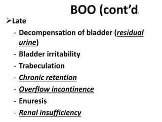
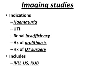
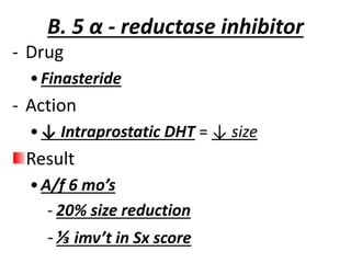
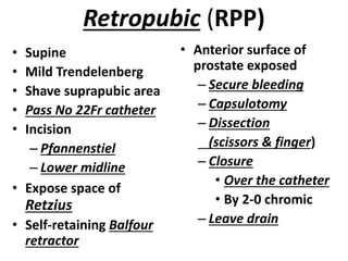
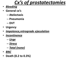
Benign Prostatic
Hyperplasia
(BPH)
pathology
• BPH affects
– Glandular epith (sub mucosal glands)
– Stromal cells
– ↑ed growth
 Glands of peripheral zone compressed to
form
1. Lateral lobes
2. False capsule
 Growth of central zone →”middle” lobe.
 Growth can be “global” “local”
Pathogenesis and etiology
Etiology
- Uncertain
- Theories
a) Hormonal
• Androgens
• Estrogens
• Stromal – epithelial interactions
• GF’S
• Neurotransmitters
b) Neoplastic
• Benign neoplasm (fibromyoadenoma)
pathogenesis
(Gland enlargement)
• Disrupted equilibrium b/n
–Cell death
–Cell proliferation
• Androgen
–Required during
Development
Puberty
Aging
No androgen = no BPH
Patho (cont’d)
• Androgen receptors
- Pst responds to androgen through out life
- ↑ in age
↑ in estrogen
↓
↑ expression of AR in aging prostate
↓
↑ in pst size despite low serum [ ] of
androgen
↓
maintained androgen dependent cell growth
symptomatology
• Prostatism →LUTS
• Classified into
 Obstructive
- Hesitancy
- Poor flow
- Intermittency
- Dribbling
- Sensation of poor
bladder emptying
- Episodes of near
retention
 Irritative
- Frequency
- Nocturia
- Urgency
- Urge
incontinence
- Nocturnal
incontinence
(enuresis)
Symptomatology (cont’d)
• Scoring systems
 IPSS (international prostate sx score)
- Used for assessment of sx severity
- According to IPSS
- Mild (0 to 7)
- Moderate ( 8 to 19)
- Severe ( 20 to 35)
 Sx score is 1o determinant for
- Rx response
- Ds progression (“ watchful waiting”)
IPSS can not be used to establish the Dx of BPH
Effects of BPH
1. LUTS & BOO
• No Sx, No BOO
• No Sx but urodynamic evidence of BOO
• Sx of prostatism with BOO
2. Retention
3. Haematuria
4. Urinary infection
5. Stone formation
6. Trabeculation
7. Lengthening of urethra
8. RF
BOO
• Urodynamic concept
– Low flow rate
– High voiding pressure
• Dx’ed only by pressure
flow studies
• DDx
– BPH
– BNC
– BNH
– Prostatic ca
– Urethral stricture
– Functional obstruction
• Effects of BOO
 Early
- Flow rate
Low = < 10ml/sec
- Voiding pressure
High = > 80 cm H2 o
- Detrusor instability
- AUR
 May be 1st Sx of BOO
 Precipitated by
- Postponement
- Over indulgence
- Operation
- confinement
BOO (cont’d)
Late
- Decompensation of bladder (residual
urine)
- Bladder irritability
- Trabeculation
- Chronic retention
- Overflow incontinence
- Enuresis
- Renal insufficiency
Evaluation of pt’s with LUTS
• Hx
 Which Sx’s are
predominant
Irritative
Obstructive
 Where does the
pt lies by IPSS
- Mild
- Moderate
- Severe
 Ask specifically
Haematuria
UTI
Diabetes
Nervous sys. Ds
Urinary retention
Surgery of LUT
Physical examination
• General assessment
 Chest
 CVS
 Anemia
 DHN
Abdominal exam
 Suprapubic
- Fullness
- Dullness
- Tenderness
PE (cont’d)
 DRE
- Size
• not for deciding active Rx
• poorly correlates with
- Severity of Sx’s
- Degree urodynamic obstruction
- Rx outcomes
- Prostate
• Large , smooth, convex, elastic, firm
• mucosa moves over the prostate
 NS exam ( R/O caudaequina lesions)
Urinalysis (U/A)
• Deep stick
• Spun sediment
• Used to R/O
–Prostatic ca
–UTI
• Cytology
–Severe irritable Sx’s
Serum creatinine
• To R/O renal insufficiency 2o to BPH
(~13%)
• RI increases risk of post-op cx
–25% risk with RI
–17% risk without RI
• “silent prostatism”
–Occult & progressive renal damage
with minimal or no urinary complaint
–Identified by creatinine measurement
Serum PSA
• Prostatic Ca can coexist with BPH
& gives Sx’s ~ to BPH
• Should be done if Ca identification
alters BPH Mx.
• Value overlaps b/n BPH & Ca
• Value > 4nmol/lit
–Trans rectal us & multiple bx
• Will not done if DRE is NL
uroflowmetry
• Electronic recording of the urine flow
rate
• Non invasive urodynamic test
• Quantifies strength of urine stream
• 2 to 3 voids , with voided vlm 150 to
200ml in flow rate clinic
• Typical hx + FR <10ml/sec for voided
vlm >200ml =
Recommend Rx
Post void residual urine (PVR)
• Vlm of urine immediately after
micturation
• NL value
–<5ml = 78% of men
–<12ml = 100% of men
• Measured by trans abdominal
u/s
Pressure flow studies
• Done to distinguish b/n low PFR 20 to
–BOO
–Decompensated or neurogenic
bladder
• Reliable
• If Boo is not dx’ed by
–Initial evaluation
–Flow rate
–PVR
Filling cystometry (cystometrography)
• Invasive UD study
• Gives information about
–Bladder capacity
–Bladder compliance
• Indications
–In pt’s with suspected 10 bladder or
neurologic lesions who can not urinate
Urethroscopy
• Indications
–Haematuria
–Urethral stricture
–Bladder ca
–Prior LUT surgery
• Benefits
–Prostate enlargement
–BN – obstruction (flexible)
–Trabeculation
–Diverticula
Imaging studies
• Indications
–Haematuria
–UTI
–Renal insufficiency
–Hx of urolithiasis
–Hx of UT surgery
• Includes
–IVU, US, KUB
Goal of Rx
• Relieving LUTS
• Decreasing BOO
• Improving bladder emptying
• Ameliorating detrusor instability
• Reversing renal insufficiency
• Preventing
–Future episodes of gross haematuria
–UTI
–Urinary retention
Over view
• Dramatic expansion of Rx
option in the last 2 decades
with
1. Medical therapy
2. Minimally invasive therapy
3. Operative therapy
“watchful waiting”
• Patient with BPH
–Mild Sx’s (AUA-Sx < 8)
–PFR > 10ml/sec
–PVR < 100ml/sec?
• Annual base line evaluation
• General advice
–Limit fluid intake in evening
–Never indulge alcohol
Medical therapy
A. α-adrenergic receptor blockers
- Drugs
• Doxazosin
• Trazosin
• Tamsulosin
- Action
• Relax prostatic smooth muscles & relieve
“dynamic” component of obstruction
- SE
• Asthenia
• Headache
• Dizziness
B. 5 α - reductase inhibitor
- Drug
•Finasteride
- Action
•↓ Intraprostatic DHT = ↓ size
Result
•A/f 6 mo’s
- 20% size reduction
-⅓ imv’t in Sx score
prophylaxis
• Potential role of medical Rx for prevention of
– Development
– Progression
• Should be given before 50 yrs
• Limitations
– Long term drug exposure
– Adverse effects
– Costs
– Effective Rx’py when BPH occur
prostatectomies
• Types
– TURP
– RPP
– TVP
• Success rate
– AUR & CUR = 100%
– Severe Sx & urodynamically proven BOO = 90%
– Mild SX = 65%
– Unobstructed detrusor instability = do not respond
well.
Pre-op preparation
• Prepare 2 units of bld
• Counseling
–Obtain consent
–Inform benefit & risk
- Retrograde ejaculation (65%)
- Erectile dysfunction (5%)
- Urinary incontinence
- UTI
- Urethral stricture
TURP
• 1920 & 30’s
• Gold standard
• >90% of prostatectomies
• Indications
– AUR
– Recurrent infection
– Recurrent haematuria
↓
Gland size < 40gm
• Anesthesia
–GA (SA)
–LA + sedation
• Prophylaxis
–Indwelling
catheter
–Recent UTI
–Chronic retention
–Prosthetic heart
valves
–Cephalosporins
Complications of TURP
• Short term
–TUR syndrome
• 2% of pt’s
• Confusion
• Nausea
• Vomiting
• HPN
• Bradycardia
– Bleeding (5%)
– perforation
• Long term cx’s
–Incontinence
(1%)
–Impotence (5 to
10%)
–Retrograde
ejaculation (78 to
80%)
Retropubic (RPP)
• Supine
• Mild Trendelenberg
• Shave suprapubic area
• Pass No 22Fr catheter
• Incision
– Pfannenstiel
– Lower midline
• Expose space of
Retzius
• Self-retaining Balfour
retractor
• Anterior surface of
prostate exposed
– Secure bleeding
– Capsulotomy
– Dissection
(scissors & finger)
– Closure
• Over the catheter
• By 2-0 chromic
– Leave drain
TVP
• Position, scrubbing
& draping ~ to RPP
• Insert No 22Fr
catheter
–Drain residual
urine
–Instill 250ml of
saline
–Clamp catheter
• Incision ~ to RPP
• Exposure
– Peritoneum swept
• Cephalad
– Place self-retaining
Balfour retractor
– Stay stitch
• 3cm apart
• Just below
peritoneal reflection
Post-op Mx
• In put out put
• Bladder irrigation
• Effective pain Mx
• 1st POD
–Fluid diet
– Ambulation
– Deflate balloon(30ml)
– Irrigate residual clot
• 2nd POD
–Regular diet
• 3rd POD
– Remove retropubic
• 4th POD
– Discharge with
catheter
• 5th to 7th POD
– Remove catheter
Cx’s of prostatectomies
• Bleeding
• General cx’s
– Atelectasis
– Pneumonia
– DVT
• Urgency
• Impotence,retrograde ejaculation
• Incontinence
– Urge
– Stress
– Total (none)
• BNC
• Death (0.2 to 0.3%)
URINARY LITHIASIS
• Stones are found in the
kidneys & Ureter
Bladder & Urethra
Very Common in Ethiopia
~40% of urologic problem
Stone disease is treatable not curable.
Rapid advance in intervention in the last 25
years.
URINARY LITHIASIS
• The origin of surgery is the ancient
operation for bladder stones.
• Incidence varies in different
populations, it is uncommon in many
parts of Africa.
• The incidence is about 4%.
• Dehydration and over consumption of
protein predispose to stone formation.
Even though there has been a rapid
advance in treatment, our
understanding of the pathogenesis lags
behind
URINARY LITHIASIS
Types of stones
• Calcium Oxalate (~60-70%)
• Phosphate (~20-30%)
• Urate (~10%)
• Cystine(~1%)
Most stones are mixed and contain organic
matrix
URINARY LITHIASIS
Calcium Oxalate (CaC2O4.H2O)
oxalate waste product of COOH
Metabolism l
Oxalate
COOH
(Glycine and Ascorbic acid)
15% originates from diet
Ca Oxalate stone forms in Acidic urine and it is
radiopaque
Phosphate stones
Calcium phosphate & Magnesium ammonium phosphate
• Ca5 (PO4) 3OH (Triple phosphate)
H2PO4
- = HPO4
-- + H HPO4
-- = PO4
-- + H
• MgNH4PO4.H2O (Struvite)
Forms in Alkaline urine
Assume shape of pelvis & calyces (Stag horn)
Associated with UTI (Urea splitting bacteria)
URINARY LITHIASIS
Urate and Cystine
• Uric Acid is the by product of purine metabolism
Uric Acid == H+ + Urate
Solubility at PH 5 60mg / liter
Solubility at PH 7 1600mg / liter
Therefore forms in Acidic urine
Pure Urate stones are radiolucent
• Cystine stones
Occurs in patients with cystinuria
URINARY LITHIASIS
Formation of urinary stones
• Stones form as a result of precipitation of
stone forming particles (Salts) in urine
Concentration
U.Volume
excretion
Inhibitors
(Citrate)
Ph
Stasis
Infection
URINARY LITHIASIS
Causes of Urolithiasis
• Increased Excretion in urine
1. Calcium
Hyperparathyroidism (Ca Phosphate stones)
Idiopathic hypercalciuria ( Ca Oxalate)
Hypervitaminoses D , Neoplastic disease
2. Oxalate
Dietary, Enzymatic defect, Bowel resection
3.Uric acid
Dietary, Enzymatic defect
• Decreased Excretion (Citrate)
• Renal tubular disorders (Acidosis)
• Secondary Infections splitting urea (Proteus), obstructions
*High protein diet and low urine output states predispose to stone*
No obvious cause in most patients.
URINARY LITHIASIS
CLINICAL FEATURES
Depends on location, size & Complications
• Kidney stones
– Asymptomatic ( No distension or obstruction)
– Pain ( Loin pain, Renal colic)
– Hematuria
• (Gross Vs Microscopic)
– Infection
• Pyelonephritis, pyonephrosis, perinephric abscess leading
to septicemia
-Bilateral obstruction (anuria or R Failure)
URINARY LITHIASIS
CLINICAL FEATURES
• Stones in the ureter
Present with colic (agonizing pain)
If hydronephrosis progress pain disappear
May present with pyonephrosis– septicemia
• Stones in the bladder
– Pain in the suprapubic or genital area
– Hematuria and interruption of micturition
URINARY LITHIASIS
Diagnosis and Investigation
• Urinalysis, urine culture & RFT are
essential
• Ultrasonography
– Detects stone in kidney and
bladder
– Detects obstructive changes
– Asses parenchyma loss
Plain abdominal x-ray detects
90% of stones
IVU is the best investigation for
this purpose
URINARY LITHIASIS
(Plain Film of Abdomen)
URINARY LITHIASIS
(Intravenous urogram (IVU)
URINARY LITHIASIS
(Intravenous urogram (IVU)
URINARY LITHIASIS
(Intravenous urogram (IVU)
URINARY LITHIASIS
(Intravenous urogram (IVU)
URINARY LITHIASIS
(Intravenous urogram (IVU)
URINARY LITHIASIS
(Intravenous urogram (IVU)
URINARY LITHIASIS
Management
Expectant treatment.
– FOR STONES <5mm
– For ureteric stones advancing down (4-6 weeks)
– Hydronephrosis need urgent intervention
• Extracorporeal shock wave lithotripsy (ESWL)
• Ureteroscopy
• Percutaneous nephrolithotomy (PCNL)
• Open surgery rarely required (<5%)
URINARY LITHIASIS
(Treatment Options)
• 1. ESWL 2. URETEROSCOPY 3.PCNL
4. Surgery
ESWL
ESWL
URETEROSCOPY
URETEROSCOPY
• RIGID FLEXIBLE
PCNL
Open Surgery
Nephrolitotomy, Pyelolithotomy,
Ureterolithotomy &cysolithotomy (Nephrectomy
may be required)
Anatrophic Nephrolithotomy
TREATMENT OF UROLITHIASIS
• KIDNEY
– STAG HORN
• PCNL+ESWL/ OPEN SURGERY
– PELVIS
• ESWL(<2CM)
• PCNL(>2.5CM)
– CALYCEAL
• ESWL/PCNL
• URETER
– UPPER (ESWL/URETEROSCOPY)
– MID (URETEROSCOPY/ESWL)
– DISTAL (URETEROSCOPY/ESWL)
• BLADDER
– OPEN SURGERY/LITHOPLAXY/ESWL
URINARY LITHIASIS
Drug treatment and prevention
• Recurrence is a major problem (20-
75%)
Measures to prevent recurrence
1. Identify the cause
 Stone analysis
 Blood chemistry Calcium, Po4, Uric acid…..
 24 hours urine calcium, oxalate, citrate , uric acid…
2. General measures
High fluid intake (urine>2 liters)
Reduce protein and salt intake
URINARY LITHIASIS
Drug treatment and prevention
3. Specific treatment
1. Uric acid
 Alkalinization of urine (K-Citrate)
 Haloperidol
 Dietary
2. Calcium stones (depends on lab finding)
 Idiopathic hypercalciuria (thiazides)
 Dietary
3. Secondary stones
 Treat Infection and obstruction
URINARY LITHIASIS
Drug treatment and prevention
There is no one time cure for stone disease.
Needs a continuous care like diabetes.
LOWER ABDOMINAL PAIN (PID)
• PID - Ascending infection of the uterus,
fallopian tubes, ovaries, &peritoneum
• Sexually transmitted
• Causes include N.gonorrhea, C.trachomatis &
Anaerobes i.e (poly microbial)
• Bilateral lower abdominal pain & vaginal
discharge support diagnosis
70
PID contd
• Other surgical and gynecological causes of lower
abdominal pain such as
– ectopic pregnancy
– appendicitis
– bowel disease
– Bowel obstruction
– Perforation
– Diverticulitis
– Bleeding ulcer
– Gall bladder disease
– Pancreatitis
– Ischaemia
– Inflammatory
71
Contd....
should be ruled out
• Women with HIV may have severe PID
 Diabetic acidosis
 Pelvic inflammatory disease
 Renal stones
 Acute urinary retention
 Kidney infections
 Labour pains
 Aortic aneurysm
Lead poisoning
Sickle cell crisis
72
Diagnosis of PID
• Diagnosis is often difficult & inconsistent
clinical presentations are common
• History should include
- Erratic bleeding
- Missed period
- Recent delivery
- Miscarriage
73
Diagnosis contd
• Physical examination
- Check temperature
- Palpate abdomen for tenderness, guarding & mass
- Check for vaginal bleeding &abnormal discharge
74
Diagnosis contd
• Empiric treatment should be initiated in
sexually active young women and other
women at risk for STls if the following
minimum criteria are present and no other
cause(s) for the illness can be identified:
- uterine/adnexal tenderness or
- cervical motion tenderness
75
Complications of PID
• Peritonitis and intra-abdominal abscess
• Adhesion and intestinal obstruction
• Ectopic pregnancy
• Infertility
• Chronic pelvic pain
76
Indications for hospitalizations in PID
• Uncertain diagnosis
• Acute abdomen can not be excluded
• Pelvic abscess is suspected
• Severe illness precludes management on an
outpatient basis
• Pregnancy
• The patient is unable to follow or tolerate an
outpatient regimen
• The patient has failed to respond to outpatient
therapy
77
cervical excitation,
tenderness or
lower abdominal
tenderness & VD
complains of lower abdominal pain
Take history & examine (abdominal & vaginal)
•Missed/ overdue period,
•Pregnancy
•Recent delivery/ abortion
/Miscarriage
• Abdominal guarding/
rebound tenderness
• Vaginal bleeding
•Abdominal mass
Yes
Yes
• Refer patient for surgical or
gynaecological assessment
• set up IV line
• Resuscitate if required
No
Treat for PID
& review in 3 days
No
improved? No Refer
patient for
admission
Yes
• Continue treatment
• Educate on RR
• Offer HTC
• Condom use
•partner(s)treatment
•Recording and reporting
Any other
illness?
Yes
Manage
appropriately
78
Recommended treatment for PID
Out patient In patent
Ceftriaxone 250 mg IM stat/
Spectinomycin 2 gm IM stat
Plus
Azithromycin 1gm po stat/Doxycycline
tablet 100 mg po bid for 14 days
Plus
Metronidazole 500 mg bid for 14 days
Admit if there is no improvement within
72 hours
The prefered regimen is Ceftriaxone
250mg IM stat plus Azithromycin
1gm po stat plus
Metronidazole 500 mg bid for 7
days
Ceftriaxone 250 mg IV/IM daily
/Spectinomycin 2 gm im bid
Plus
Doxycycline 100 mg bid for 14 days
Plus
Metronidazole 500 mg bid for 14 days
or chloramphenicol 500 mg IV qid.
79
• For inpatient PID, ceftriaxone or
spectinomycin should continue for 24hrs after
the patient remain clinically improved, after
which doxycycline and metronidazole should
continue for a total of 14 days
80
Chap.VII.pptx
Chap.VII.pptx
Chap.VII.pptx
Chap.VII.pptx
Chap.VII.pptx
Chap.VII.pptx
Chap.VII.pptx
Chap.VII.pptx
Chap.VII.pptx
Chap.VII.pptx
Chap.VII.pptx
Chap.VII.pptx
Chap.VII.pptx
Chap.VII.pptx
Chap.VII.pptx

Chap.VII.pptx

  • 3.
  • 4.
    pathology • BPH affects –Glandular epith (sub mucosal glands) – Stromal cells – ↑ed growth  Glands of peripheral zone compressed to form 1. Lateral lobes 2. False capsule  Growth of central zone →”middle” lobe.  Growth can be “global” “local”
  • 5.
    Pathogenesis and etiology Etiology -Uncertain - Theories a) Hormonal • Androgens • Estrogens • Stromal – epithelial interactions • GF’S • Neurotransmitters b) Neoplastic • Benign neoplasm (fibromyoadenoma)
  • 6.
    pathogenesis (Gland enlargement) • Disruptedequilibrium b/n –Cell death –Cell proliferation • Androgen –Required during Development Puberty Aging No androgen = no BPH
  • 7.
    Patho (cont’d) • Androgenreceptors - Pst responds to androgen through out life - ↑ in age ↑ in estrogen ↓ ↑ expression of AR in aging prostate ↓ ↑ in pst size despite low serum [ ] of androgen ↓ maintained androgen dependent cell growth
  • 8.
    symptomatology • Prostatism →LUTS •Classified into  Obstructive - Hesitancy - Poor flow - Intermittency - Dribbling - Sensation of poor bladder emptying - Episodes of near retention  Irritative - Frequency - Nocturia - Urgency - Urge incontinence - Nocturnal incontinence (enuresis)
  • 9.
    Symptomatology (cont’d) • Scoringsystems  IPSS (international prostate sx score) - Used for assessment of sx severity - According to IPSS - Mild (0 to 7) - Moderate ( 8 to 19) - Severe ( 20 to 35)  Sx score is 1o determinant for - Rx response - Ds progression (“ watchful waiting”) IPSS can not be used to establish the Dx of BPH
  • 10.
    Effects of BPH 1.LUTS & BOO • No Sx, No BOO • No Sx but urodynamic evidence of BOO • Sx of prostatism with BOO 2. Retention 3. Haematuria 4. Urinary infection 5. Stone formation 6. Trabeculation 7. Lengthening of urethra 8. RF
  • 11.
    BOO • Urodynamic concept –Low flow rate – High voiding pressure • Dx’ed only by pressure flow studies • DDx – BPH – BNC – BNH – Prostatic ca – Urethral stricture – Functional obstruction • Effects of BOO  Early - Flow rate Low = < 10ml/sec - Voiding pressure High = > 80 cm H2 o - Detrusor instability - AUR  May be 1st Sx of BOO  Precipitated by - Postponement - Over indulgence - Operation - confinement
  • 12.
    BOO (cont’d) Late - Decompensationof bladder (residual urine) - Bladder irritability - Trabeculation - Chronic retention - Overflow incontinence - Enuresis - Renal insufficiency
  • 13.
    Evaluation of pt’swith LUTS • Hx  Which Sx’s are predominant Irritative Obstructive  Where does the pt lies by IPSS - Mild - Moderate - Severe  Ask specifically Haematuria UTI Diabetes Nervous sys. Ds Urinary retention Surgery of LUT
  • 14.
    Physical examination • Generalassessment  Chest  CVS  Anemia  DHN Abdominal exam  Suprapubic - Fullness - Dullness - Tenderness
  • 15.
    PE (cont’d)  DRE -Size • not for deciding active Rx • poorly correlates with - Severity of Sx’s - Degree urodynamic obstruction - Rx outcomes - Prostate • Large , smooth, convex, elastic, firm • mucosa moves over the prostate  NS exam ( R/O caudaequina lesions)
  • 16.
    Urinalysis (U/A) • Deepstick • Spun sediment • Used to R/O –Prostatic ca –UTI • Cytology –Severe irritable Sx’s
  • 17.
    Serum creatinine • ToR/O renal insufficiency 2o to BPH (~13%) • RI increases risk of post-op cx –25% risk with RI –17% risk without RI • “silent prostatism” –Occult & progressive renal damage with minimal or no urinary complaint –Identified by creatinine measurement
  • 18.
    Serum PSA • ProstaticCa can coexist with BPH & gives Sx’s ~ to BPH • Should be done if Ca identification alters BPH Mx. • Value overlaps b/n BPH & Ca • Value > 4nmol/lit –Trans rectal us & multiple bx • Will not done if DRE is NL
  • 19.
    uroflowmetry • Electronic recordingof the urine flow rate • Non invasive urodynamic test • Quantifies strength of urine stream • 2 to 3 voids , with voided vlm 150 to 200ml in flow rate clinic • Typical hx + FR <10ml/sec for voided vlm >200ml = Recommend Rx
  • 20.
    Post void residualurine (PVR) • Vlm of urine immediately after micturation • NL value –<5ml = 78% of men –<12ml = 100% of men • Measured by trans abdominal u/s
  • 21.
    Pressure flow studies •Done to distinguish b/n low PFR 20 to –BOO –Decompensated or neurogenic bladder • Reliable • If Boo is not dx’ed by –Initial evaluation –Flow rate –PVR
  • 22.
    Filling cystometry (cystometrography) •Invasive UD study • Gives information about –Bladder capacity –Bladder compliance • Indications –In pt’s with suspected 10 bladder or neurologic lesions who can not urinate
  • 23.
    Urethroscopy • Indications –Haematuria –Urethral stricture –Bladderca –Prior LUT surgery • Benefits –Prostate enlargement –BN – obstruction (flexible) –Trabeculation –Diverticula
  • 24.
    Imaging studies • Indications –Haematuria –UTI –Renalinsufficiency –Hx of urolithiasis –Hx of UT surgery • Includes –IVU, US, KUB
  • 25.
    Goal of Rx •Relieving LUTS • Decreasing BOO • Improving bladder emptying • Ameliorating detrusor instability • Reversing renal insufficiency • Preventing –Future episodes of gross haematuria –UTI –Urinary retention
  • 26.
    Over view • Dramaticexpansion of Rx option in the last 2 decades with 1. Medical therapy 2. Minimally invasive therapy 3. Operative therapy
  • 27.
    “watchful waiting” • Patientwith BPH –Mild Sx’s (AUA-Sx < 8) –PFR > 10ml/sec –PVR < 100ml/sec? • Annual base line evaluation • General advice –Limit fluid intake in evening –Never indulge alcohol
  • 28.
    Medical therapy A. α-adrenergicreceptor blockers - Drugs • Doxazosin • Trazosin • Tamsulosin - Action • Relax prostatic smooth muscles & relieve “dynamic” component of obstruction - SE • Asthenia • Headache • Dizziness
  • 29.
    B. 5 α- reductase inhibitor - Drug •Finasteride - Action •↓ Intraprostatic DHT = ↓ size Result •A/f 6 mo’s - 20% size reduction -⅓ imv’t in Sx score
  • 30.
    prophylaxis • Potential roleof medical Rx for prevention of – Development – Progression • Should be given before 50 yrs • Limitations – Long term drug exposure – Adverse effects – Costs – Effective Rx’py when BPH occur
  • 31.
    prostatectomies • Types – TURP –RPP – TVP • Success rate – AUR & CUR = 100% – Severe Sx & urodynamically proven BOO = 90% – Mild SX = 65% – Unobstructed detrusor instability = do not respond well.
  • 32.
    Pre-op preparation • Prepare2 units of bld • Counseling –Obtain consent –Inform benefit & risk - Retrograde ejaculation (65%) - Erectile dysfunction (5%) - Urinary incontinence - UTI - Urethral stricture
  • 33.
    TURP • 1920 &30’s • Gold standard • >90% of prostatectomies • Indications – AUR – Recurrent infection – Recurrent haematuria ↓ Gland size < 40gm • Anesthesia –GA (SA) –LA + sedation • Prophylaxis –Indwelling catheter –Recent UTI –Chronic retention –Prosthetic heart valves –Cephalosporins
  • 34.
    Complications of TURP •Short term –TUR syndrome • 2% of pt’s • Confusion • Nausea • Vomiting • HPN • Bradycardia – Bleeding (5%) – perforation • Long term cx’s –Incontinence (1%) –Impotence (5 to 10%) –Retrograde ejaculation (78 to 80%)
  • 35.
    Retropubic (RPP) • Supine •Mild Trendelenberg • Shave suprapubic area • Pass No 22Fr catheter • Incision – Pfannenstiel – Lower midline • Expose space of Retzius • Self-retaining Balfour retractor • Anterior surface of prostate exposed – Secure bleeding – Capsulotomy – Dissection (scissors & finger) – Closure • Over the catheter • By 2-0 chromic – Leave drain
  • 36.
    TVP • Position, scrubbing &draping ~ to RPP • Insert No 22Fr catheter –Drain residual urine –Instill 250ml of saline –Clamp catheter • Incision ~ to RPP • Exposure – Peritoneum swept • Cephalad – Place self-retaining Balfour retractor – Stay stitch • 3cm apart • Just below peritoneal reflection
  • 37.
    Post-op Mx • Input out put • Bladder irrigation • Effective pain Mx • 1st POD –Fluid diet – Ambulation – Deflate balloon(30ml) – Irrigate residual clot • 2nd POD –Regular diet • 3rd POD – Remove retropubic • 4th POD – Discharge with catheter • 5th to 7th POD – Remove catheter
  • 38.
    Cx’s of prostatectomies •Bleeding • General cx’s – Atelectasis – Pneumonia – DVT • Urgency • Impotence,retrograde ejaculation • Incontinence – Urge – Stress – Total (none) • BNC • Death (0.2 to 0.3%)
  • 39.
    URINARY LITHIASIS • Stonesare found in the kidneys & Ureter Bladder & Urethra Very Common in Ethiopia ~40% of urologic problem Stone disease is treatable not curable. Rapid advance in intervention in the last 25 years.
  • 40.
    URINARY LITHIASIS • Theorigin of surgery is the ancient operation for bladder stones. • Incidence varies in different populations, it is uncommon in many parts of Africa. • The incidence is about 4%. • Dehydration and over consumption of protein predispose to stone formation. Even though there has been a rapid advance in treatment, our understanding of the pathogenesis lags behind
  • 41.
    URINARY LITHIASIS Types ofstones • Calcium Oxalate (~60-70%) • Phosphate (~20-30%) • Urate (~10%) • Cystine(~1%) Most stones are mixed and contain organic matrix
  • 42.
    URINARY LITHIASIS Calcium Oxalate(CaC2O4.H2O) oxalate waste product of COOH Metabolism l Oxalate COOH (Glycine and Ascorbic acid) 15% originates from diet Ca Oxalate stone forms in Acidic urine and it is radiopaque
  • 43.
    Phosphate stones Calcium phosphate& Magnesium ammonium phosphate • Ca5 (PO4) 3OH (Triple phosphate) H2PO4 - = HPO4 -- + H HPO4 -- = PO4 -- + H • MgNH4PO4.H2O (Struvite) Forms in Alkaline urine Assume shape of pelvis & calyces (Stag horn) Associated with UTI (Urea splitting bacteria)
  • 44.
    URINARY LITHIASIS Urate andCystine • Uric Acid is the by product of purine metabolism Uric Acid == H+ + Urate Solubility at PH 5 60mg / liter Solubility at PH 7 1600mg / liter Therefore forms in Acidic urine Pure Urate stones are radiolucent • Cystine stones Occurs in patients with cystinuria
  • 45.
    URINARY LITHIASIS Formation ofurinary stones • Stones form as a result of precipitation of stone forming particles (Salts) in urine Concentration U.Volume excretion Inhibitors (Citrate) Ph Stasis Infection
  • 46.
    URINARY LITHIASIS Causes ofUrolithiasis • Increased Excretion in urine 1. Calcium Hyperparathyroidism (Ca Phosphate stones) Idiopathic hypercalciuria ( Ca Oxalate) Hypervitaminoses D , Neoplastic disease 2. Oxalate Dietary, Enzymatic defect, Bowel resection 3.Uric acid Dietary, Enzymatic defect • Decreased Excretion (Citrate) • Renal tubular disorders (Acidosis) • Secondary Infections splitting urea (Proteus), obstructions *High protein diet and low urine output states predispose to stone* No obvious cause in most patients.
  • 47.
    URINARY LITHIASIS CLINICAL FEATURES Dependson location, size & Complications • Kidney stones – Asymptomatic ( No distension or obstruction) – Pain ( Loin pain, Renal colic) – Hematuria • (Gross Vs Microscopic) – Infection • Pyelonephritis, pyonephrosis, perinephric abscess leading to septicemia -Bilateral obstruction (anuria or R Failure)
  • 48.
    URINARY LITHIASIS CLINICAL FEATURES •Stones in the ureter Present with colic (agonizing pain) If hydronephrosis progress pain disappear May present with pyonephrosis– septicemia • Stones in the bladder – Pain in the suprapubic or genital area – Hematuria and interruption of micturition
  • 49.
    URINARY LITHIASIS Diagnosis andInvestigation • Urinalysis, urine culture & RFT are essential • Ultrasonography – Detects stone in kidney and bladder – Detects obstructive changes – Asses parenchyma loss Plain abdominal x-ray detects 90% of stones IVU is the best investigation for this purpose
  • 50.
  • 51.
  • 52.
  • 53.
  • 54.
  • 55.
  • 56.
  • 57.
    URINARY LITHIASIS Management Expectant treatment. –FOR STONES <5mm – For ureteric stones advancing down (4-6 weeks) – Hydronephrosis need urgent intervention • Extracorporeal shock wave lithotripsy (ESWL) • Ureteroscopy • Percutaneous nephrolithotomy (PCNL) • Open surgery rarely required (<5%)
  • 58.
    URINARY LITHIASIS (Treatment Options) •1. ESWL 2. URETEROSCOPY 3.PCNL 4. Surgery
  • 59.
  • 60.
  • 61.
  • 62.
  • 63.
  • 64.
    Open Surgery Nephrolitotomy, Pyelolithotomy, Ureterolithotomy&cysolithotomy (Nephrectomy may be required) Anatrophic Nephrolithotomy
  • 65.
    TREATMENT OF UROLITHIASIS •KIDNEY – STAG HORN • PCNL+ESWL/ OPEN SURGERY – PELVIS • ESWL(<2CM) • PCNL(>2.5CM) – CALYCEAL • ESWL/PCNL • URETER – UPPER (ESWL/URETEROSCOPY) – MID (URETEROSCOPY/ESWL) – DISTAL (URETEROSCOPY/ESWL) • BLADDER – OPEN SURGERY/LITHOPLAXY/ESWL
  • 66.
    URINARY LITHIASIS Drug treatmentand prevention • Recurrence is a major problem (20- 75%) Measures to prevent recurrence 1. Identify the cause  Stone analysis  Blood chemistry Calcium, Po4, Uric acid…..  24 hours urine calcium, oxalate, citrate , uric acid… 2. General measures High fluid intake (urine>2 liters) Reduce protein and salt intake
  • 67.
    URINARY LITHIASIS Drug treatmentand prevention 3. Specific treatment 1. Uric acid  Alkalinization of urine (K-Citrate)  Haloperidol  Dietary 2. Calcium stones (depends on lab finding)  Idiopathic hypercalciuria (thiazides)  Dietary 3. Secondary stones  Treat Infection and obstruction
  • 68.
    URINARY LITHIASIS Drug treatmentand prevention There is no one time cure for stone disease. Needs a continuous care like diabetes.
  • 70.
    LOWER ABDOMINAL PAIN(PID) • PID - Ascending infection of the uterus, fallopian tubes, ovaries, &peritoneum • Sexually transmitted • Causes include N.gonorrhea, C.trachomatis & Anaerobes i.e (poly microbial) • Bilateral lower abdominal pain & vaginal discharge support diagnosis 70
  • 71.
    PID contd • Othersurgical and gynecological causes of lower abdominal pain such as – ectopic pregnancy – appendicitis – bowel disease – Bowel obstruction – Perforation – Diverticulitis – Bleeding ulcer – Gall bladder disease – Pancreatitis – Ischaemia – Inflammatory 71
  • 72.
    Contd.... should be ruledout • Women with HIV may have severe PID  Diabetic acidosis  Pelvic inflammatory disease  Renal stones  Acute urinary retention  Kidney infections  Labour pains  Aortic aneurysm Lead poisoning Sickle cell crisis 72
  • 73.
    Diagnosis of PID •Diagnosis is often difficult & inconsistent clinical presentations are common • History should include - Erratic bleeding - Missed period - Recent delivery - Miscarriage 73
  • 74.
    Diagnosis contd • Physicalexamination - Check temperature - Palpate abdomen for tenderness, guarding & mass - Check for vaginal bleeding &abnormal discharge 74
  • 75.
    Diagnosis contd • Empirictreatment should be initiated in sexually active young women and other women at risk for STls if the following minimum criteria are present and no other cause(s) for the illness can be identified: - uterine/adnexal tenderness or - cervical motion tenderness 75
  • 76.
    Complications of PID •Peritonitis and intra-abdominal abscess • Adhesion and intestinal obstruction • Ectopic pregnancy • Infertility • Chronic pelvic pain 76
  • 77.
    Indications for hospitalizationsin PID • Uncertain diagnosis • Acute abdomen can not be excluded • Pelvic abscess is suspected • Severe illness precludes management on an outpatient basis • Pregnancy • The patient is unable to follow or tolerate an outpatient regimen • The patient has failed to respond to outpatient therapy 77
  • 78.
    cervical excitation, tenderness or lowerabdominal tenderness & VD complains of lower abdominal pain Take history & examine (abdominal & vaginal) •Missed/ overdue period, •Pregnancy •Recent delivery/ abortion /Miscarriage • Abdominal guarding/ rebound tenderness • Vaginal bleeding •Abdominal mass Yes Yes • Refer patient for surgical or gynaecological assessment • set up IV line • Resuscitate if required No Treat for PID & review in 3 days No improved? No Refer patient for admission Yes • Continue treatment • Educate on RR • Offer HTC • Condom use •partner(s)treatment •Recording and reporting Any other illness? Yes Manage appropriately 78
  • 79.
    Recommended treatment forPID Out patient In patent Ceftriaxone 250 mg IM stat/ Spectinomycin 2 gm IM stat Plus Azithromycin 1gm po stat/Doxycycline tablet 100 mg po bid for 14 days Plus Metronidazole 500 mg bid for 14 days Admit if there is no improvement within 72 hours The prefered regimen is Ceftriaxone 250mg IM stat plus Azithromycin 1gm po stat plus Metronidazole 500 mg bid for 7 days Ceftriaxone 250 mg IV/IM daily /Spectinomycin 2 gm im bid Plus Doxycycline 100 mg bid for 14 days Plus Metronidazole 500 mg bid for 14 days or chloramphenicol 500 mg IV qid. 79
  • 80.
    • For inpatientPID, ceftriaxone or spectinomycin should continue for 24hrs after the patient remain clinically improved, after which doxycycline and metronidazole should continue for a total of 14 days 80