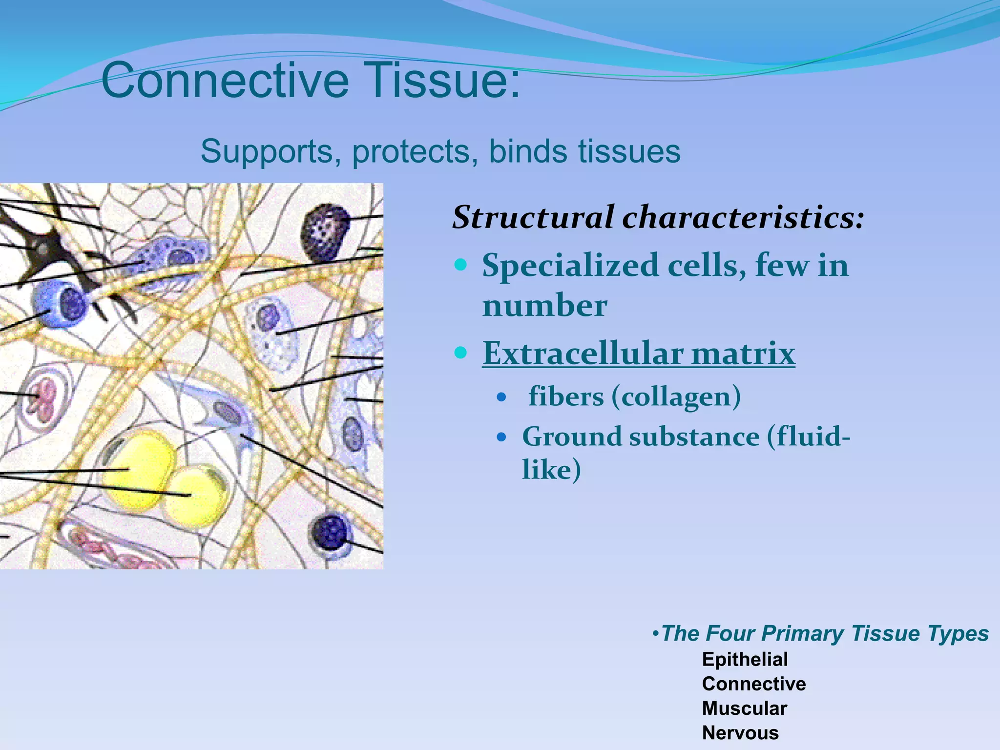
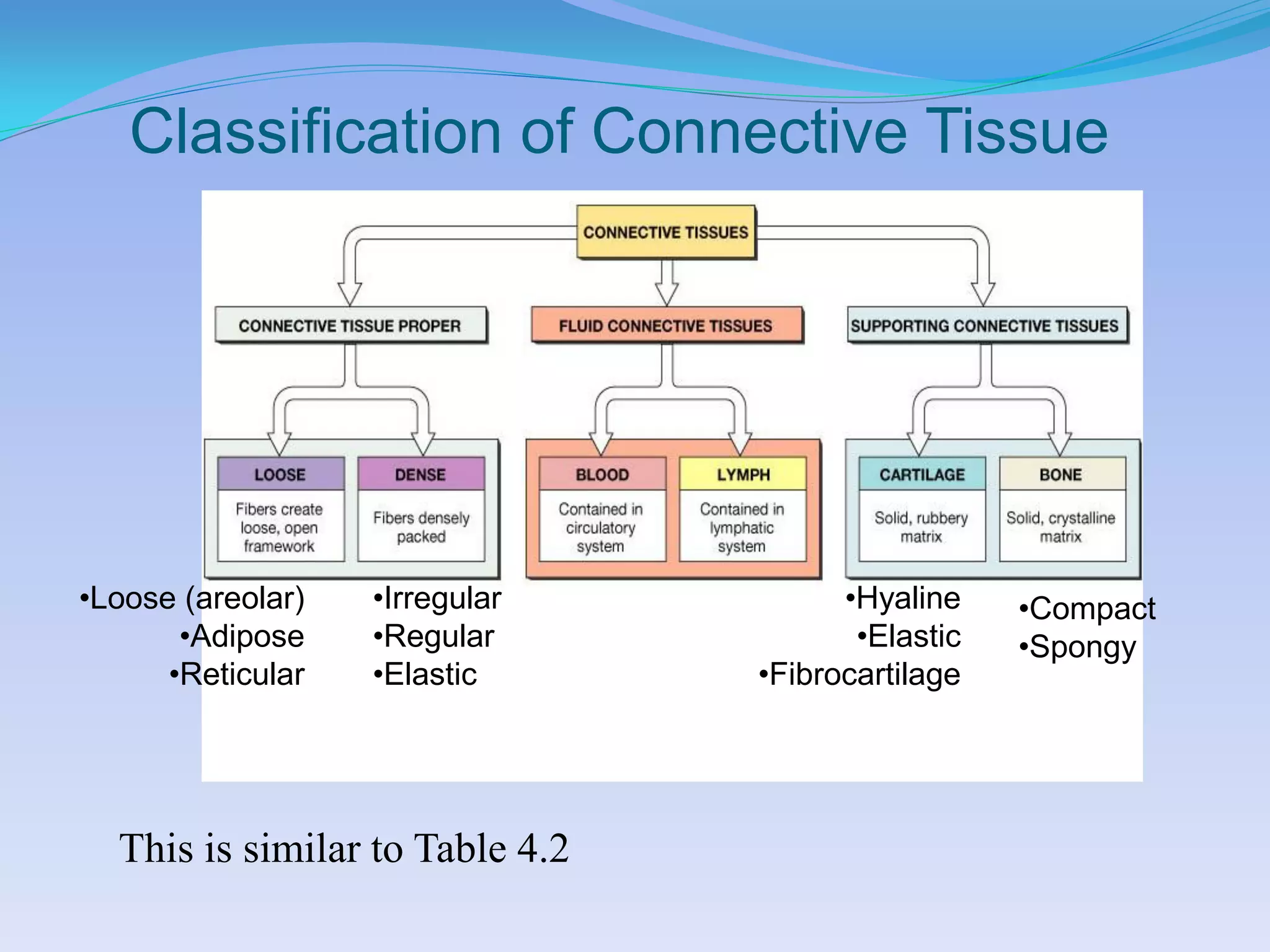
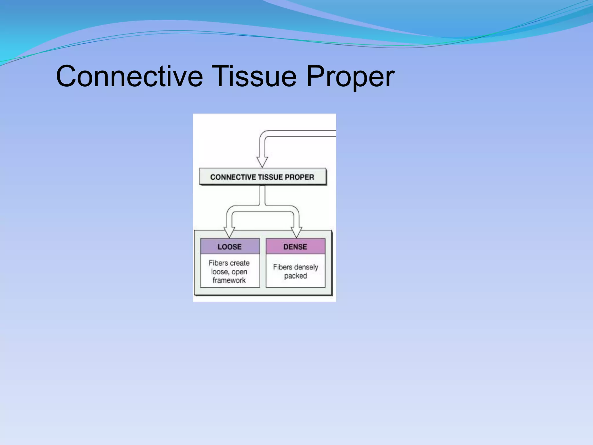
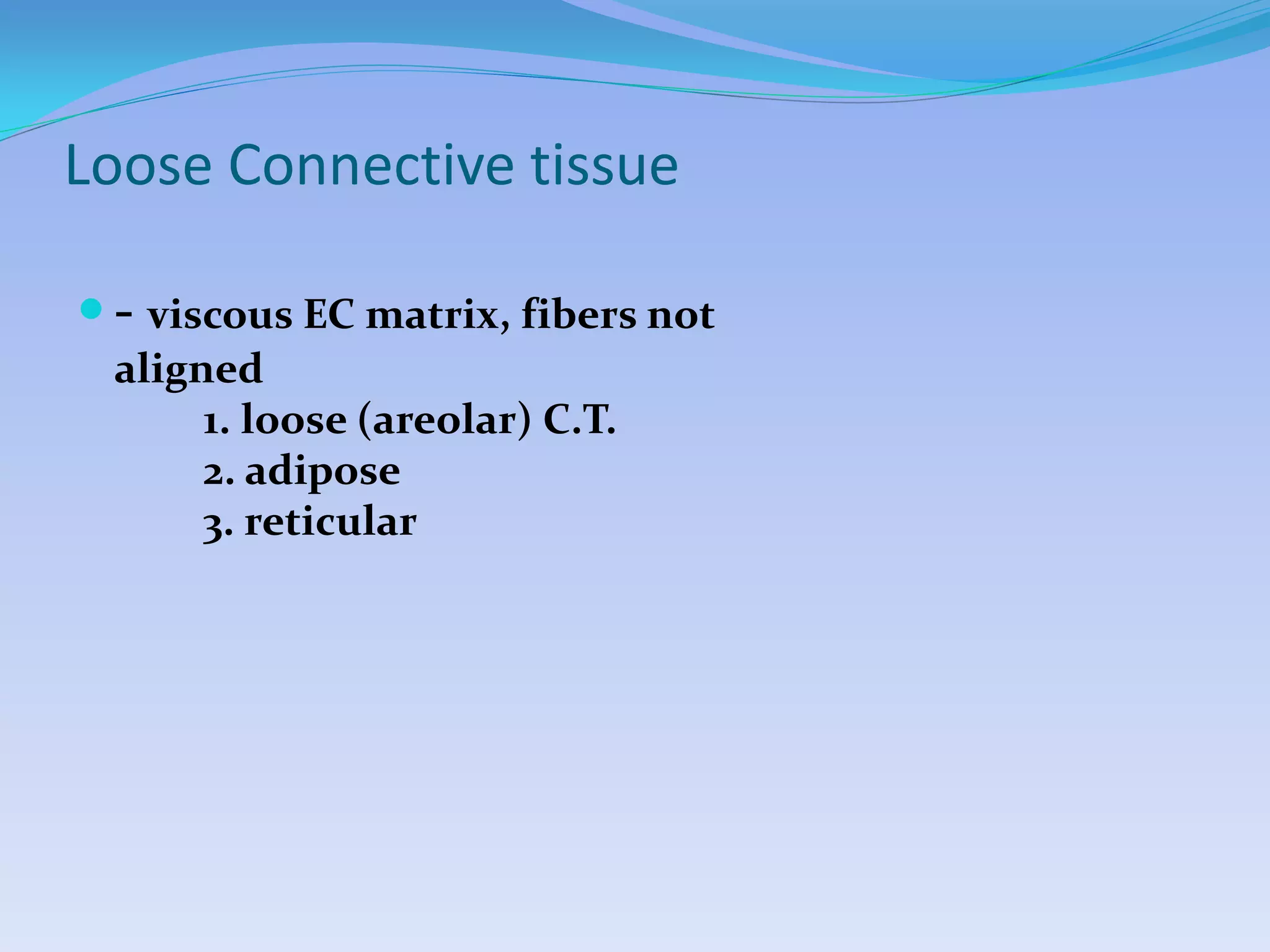
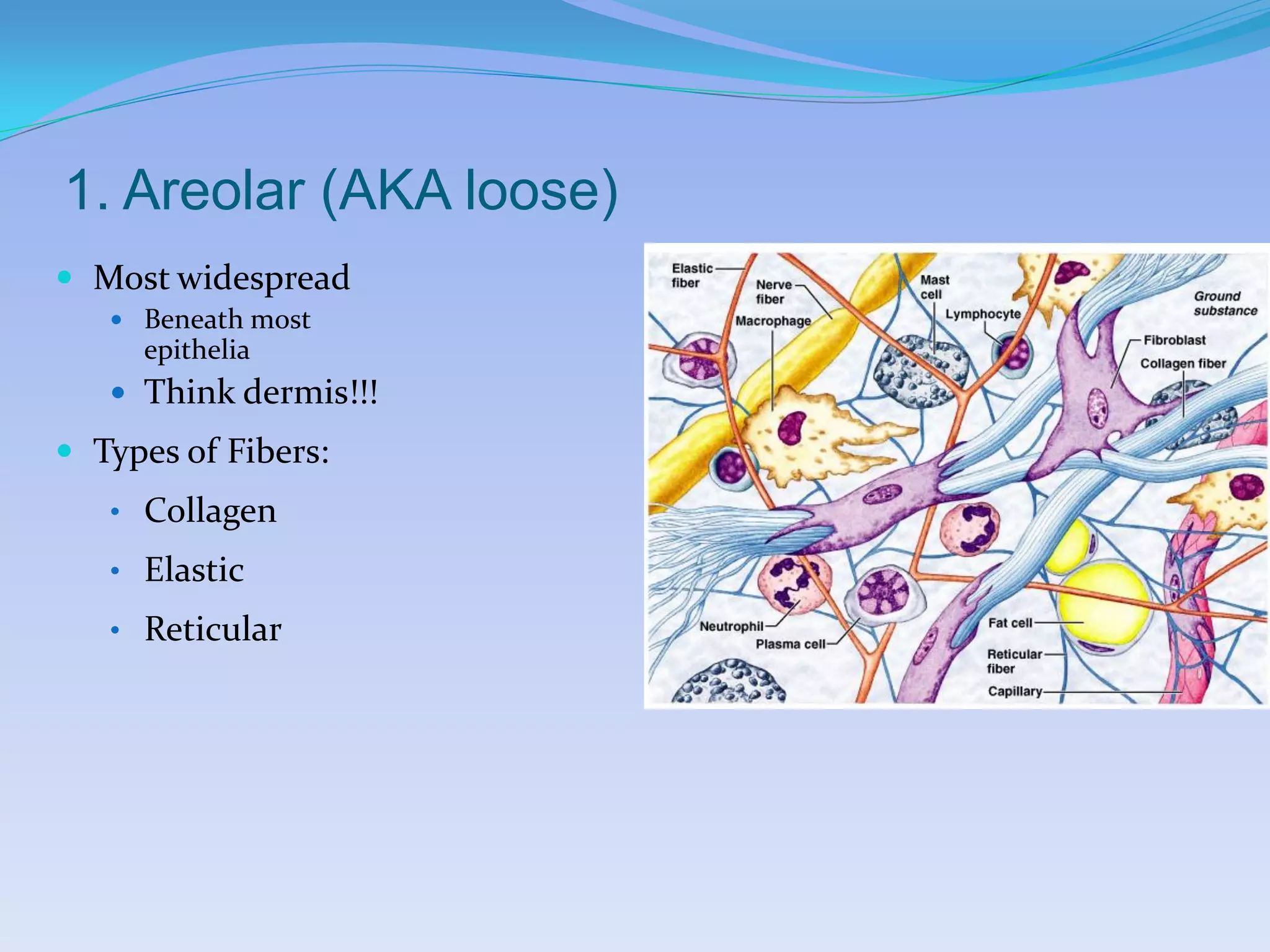
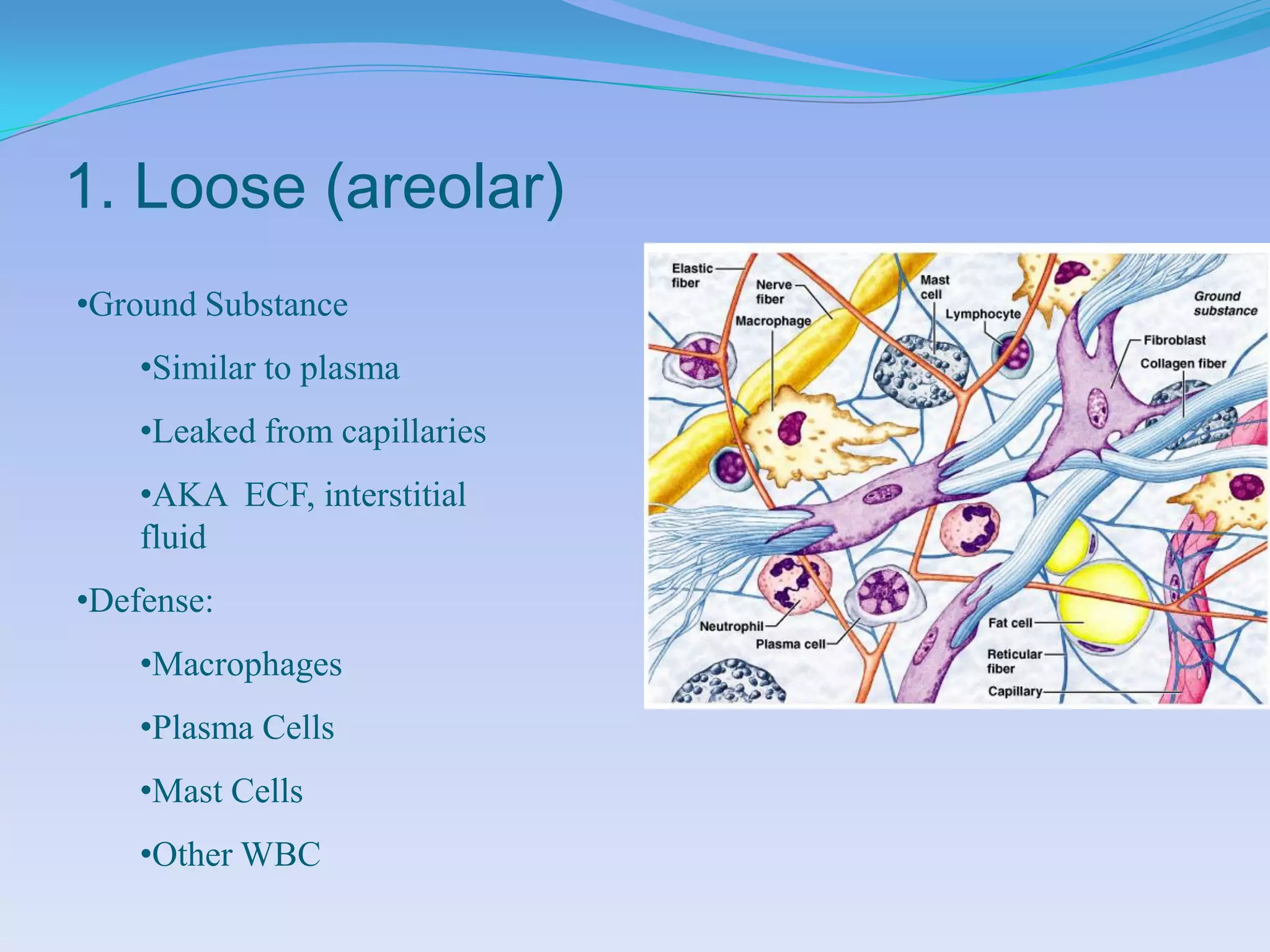
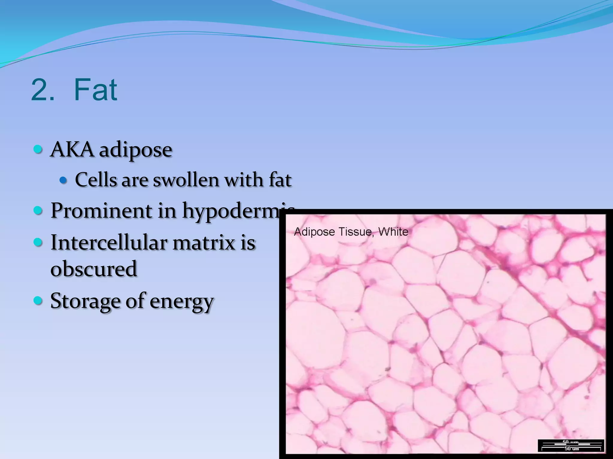
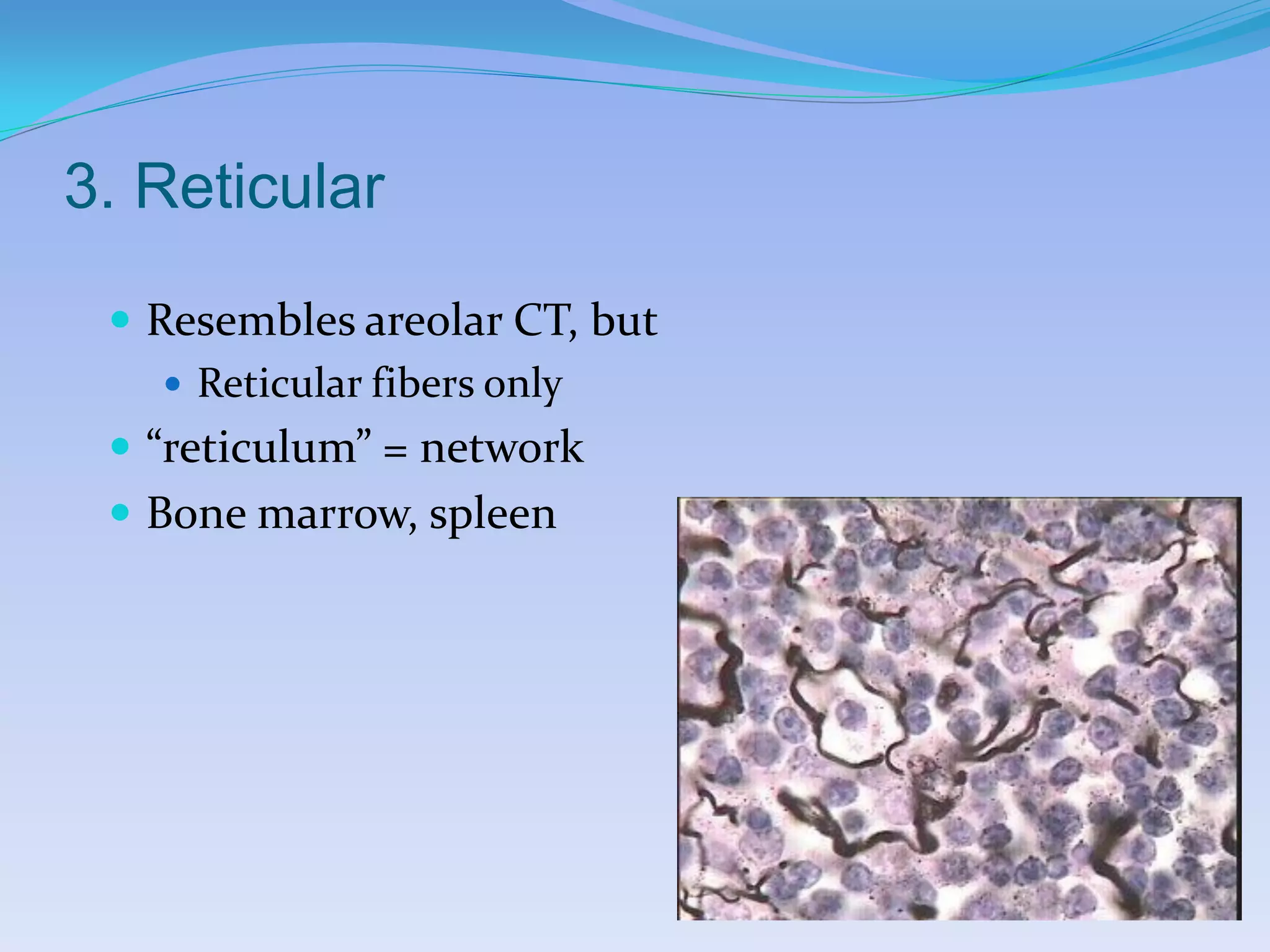
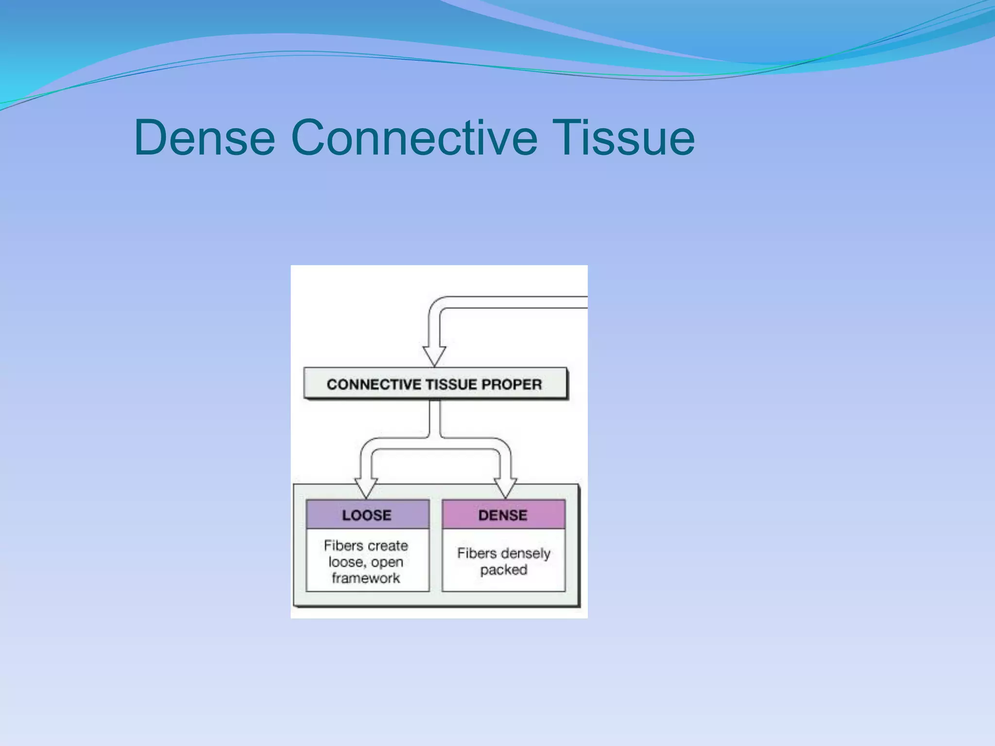
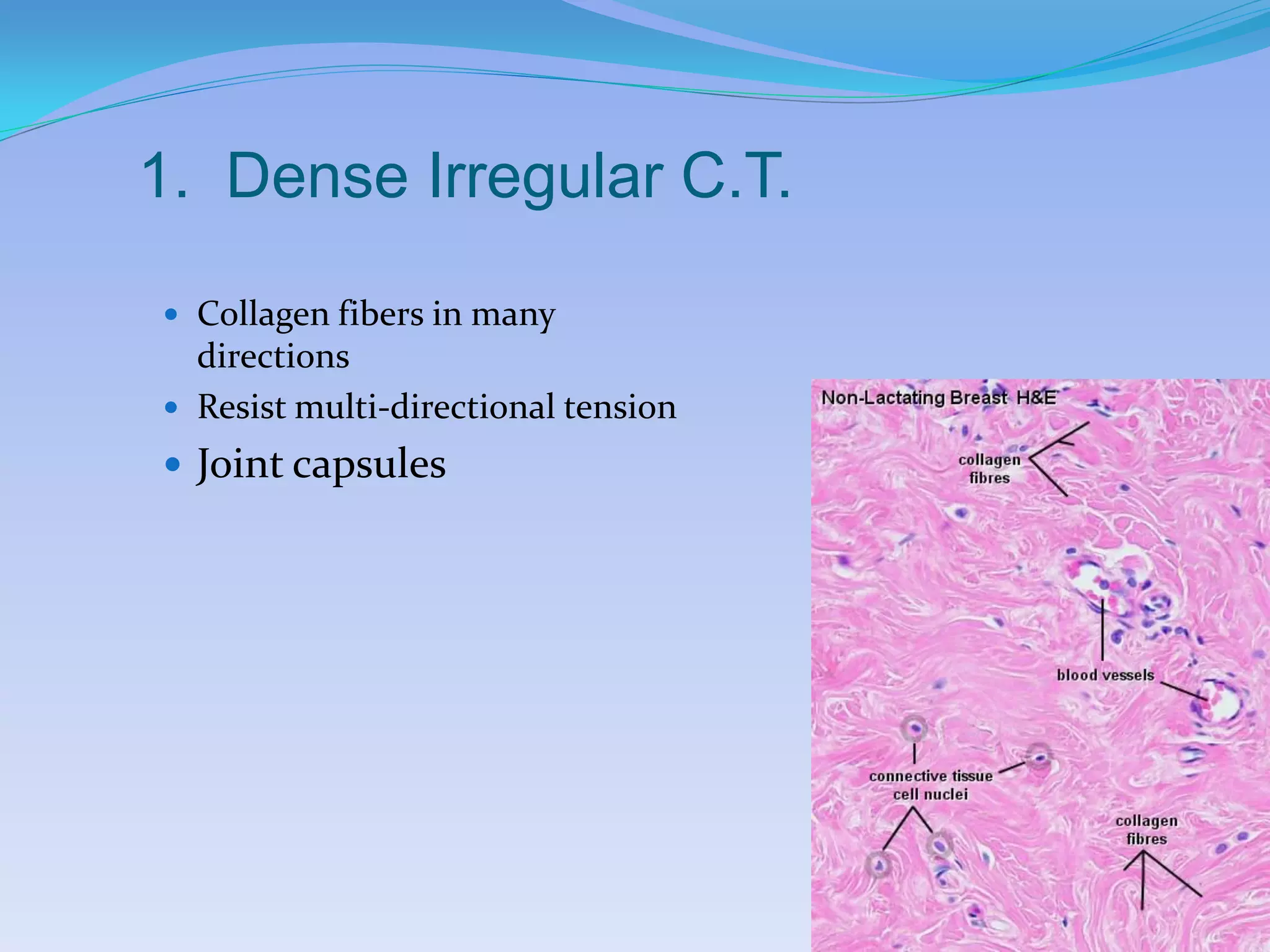
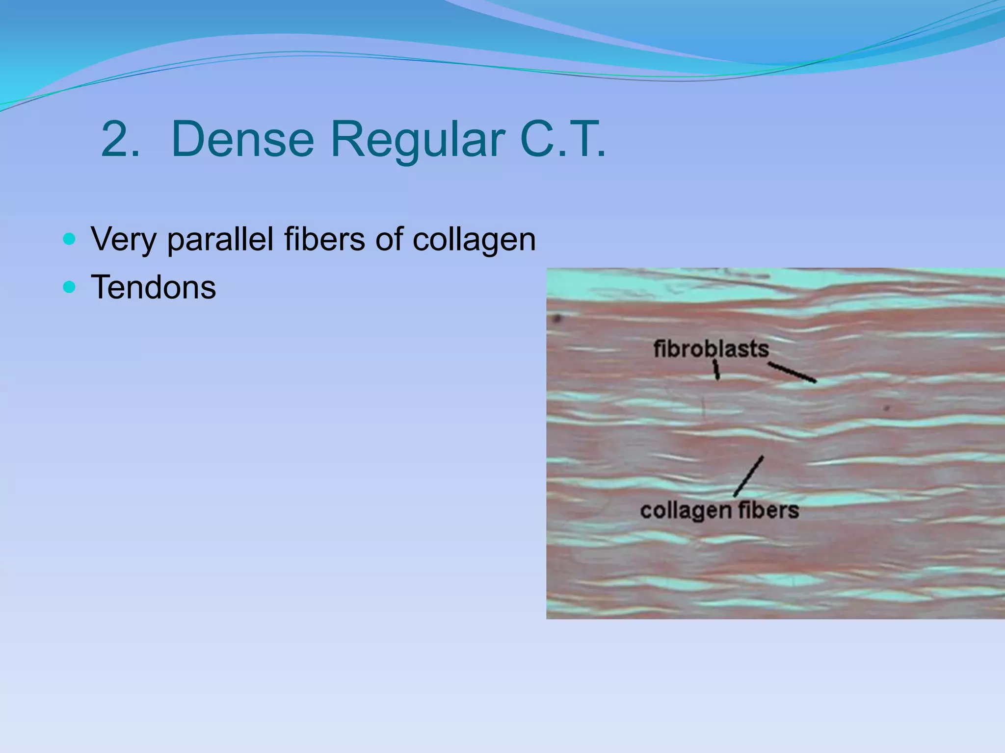
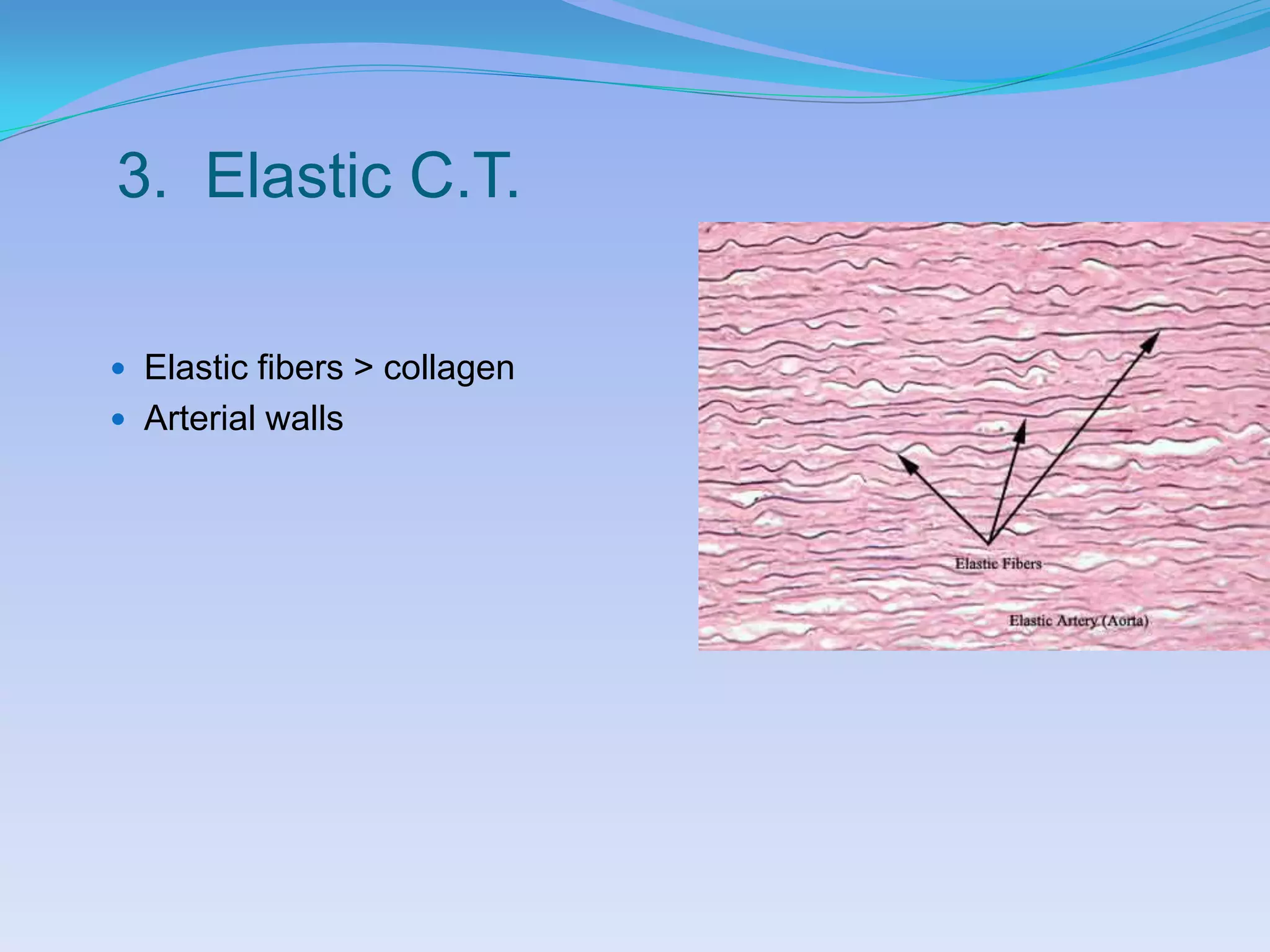
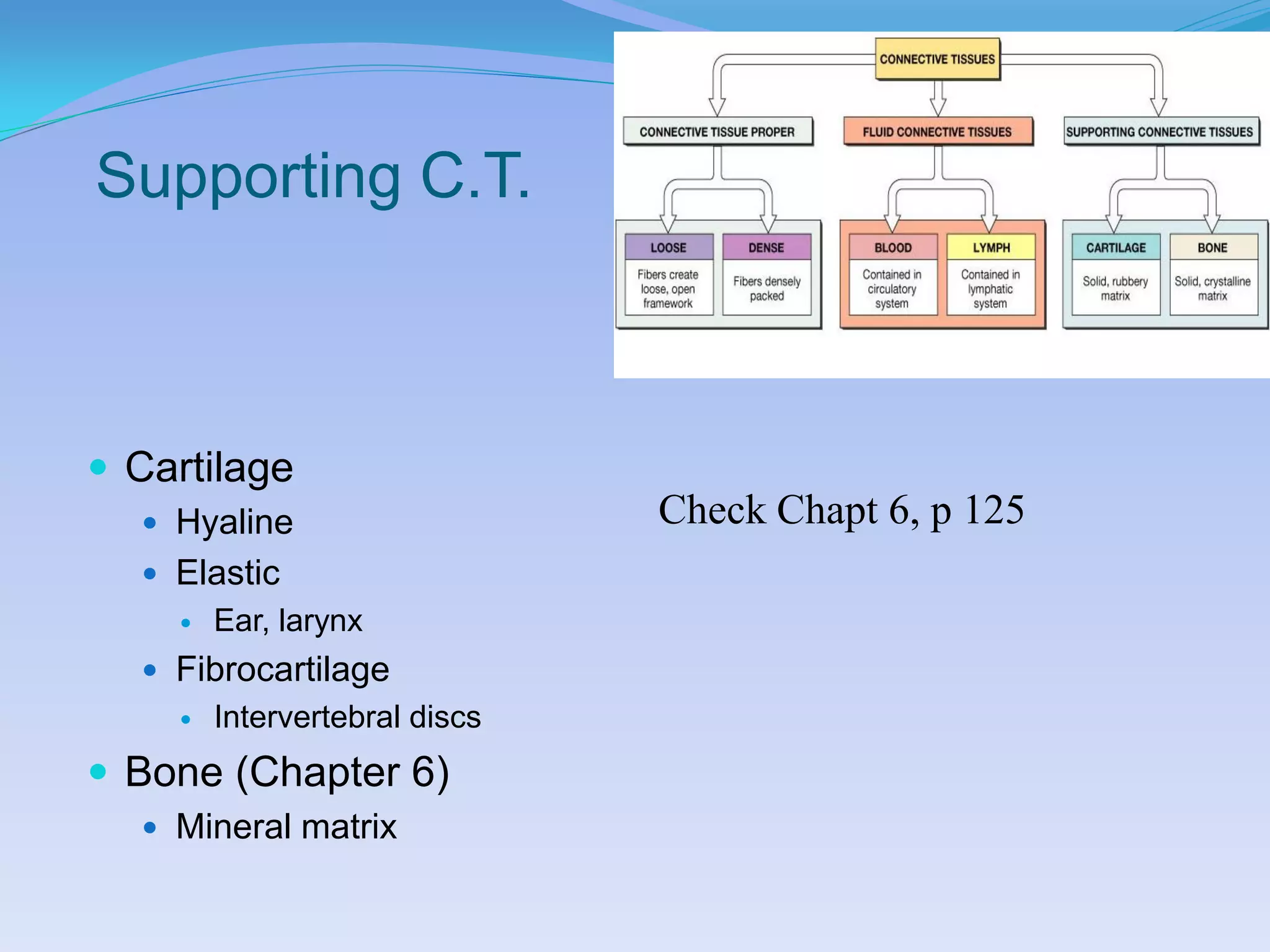
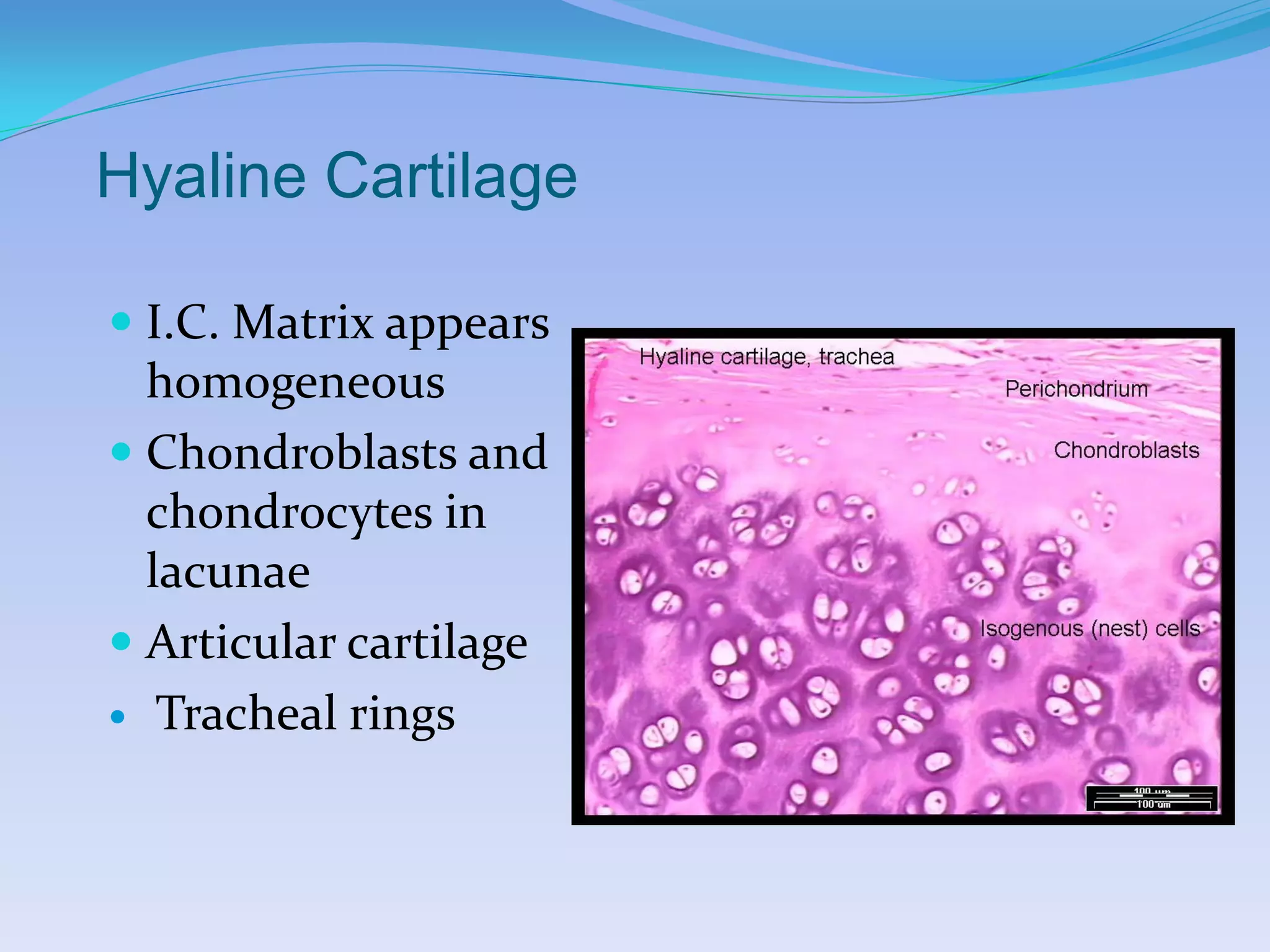
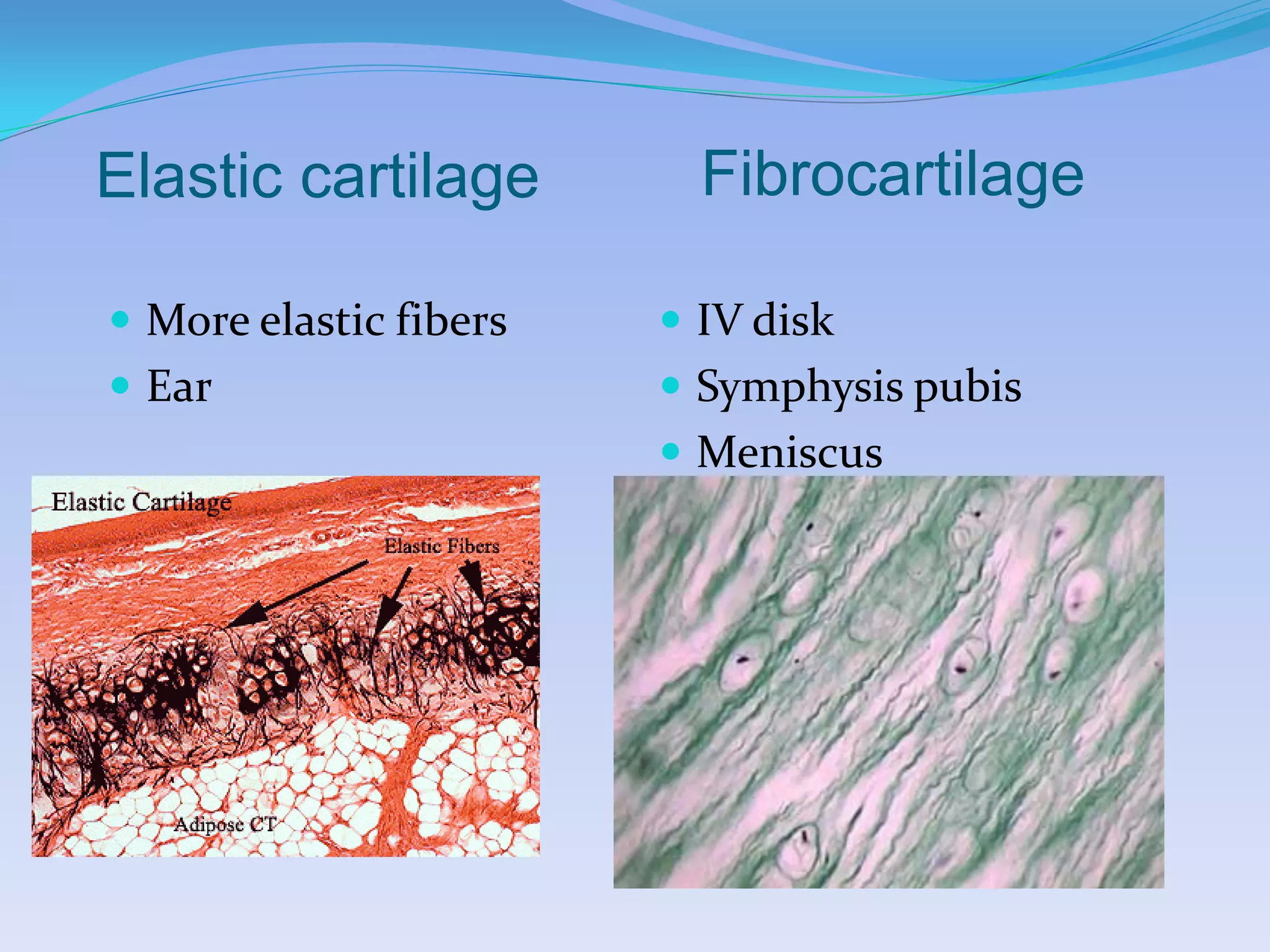
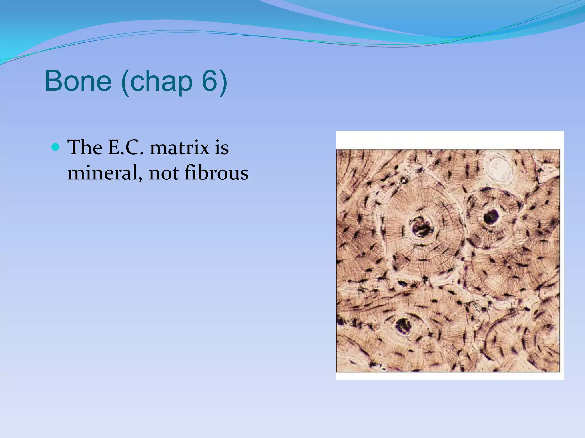
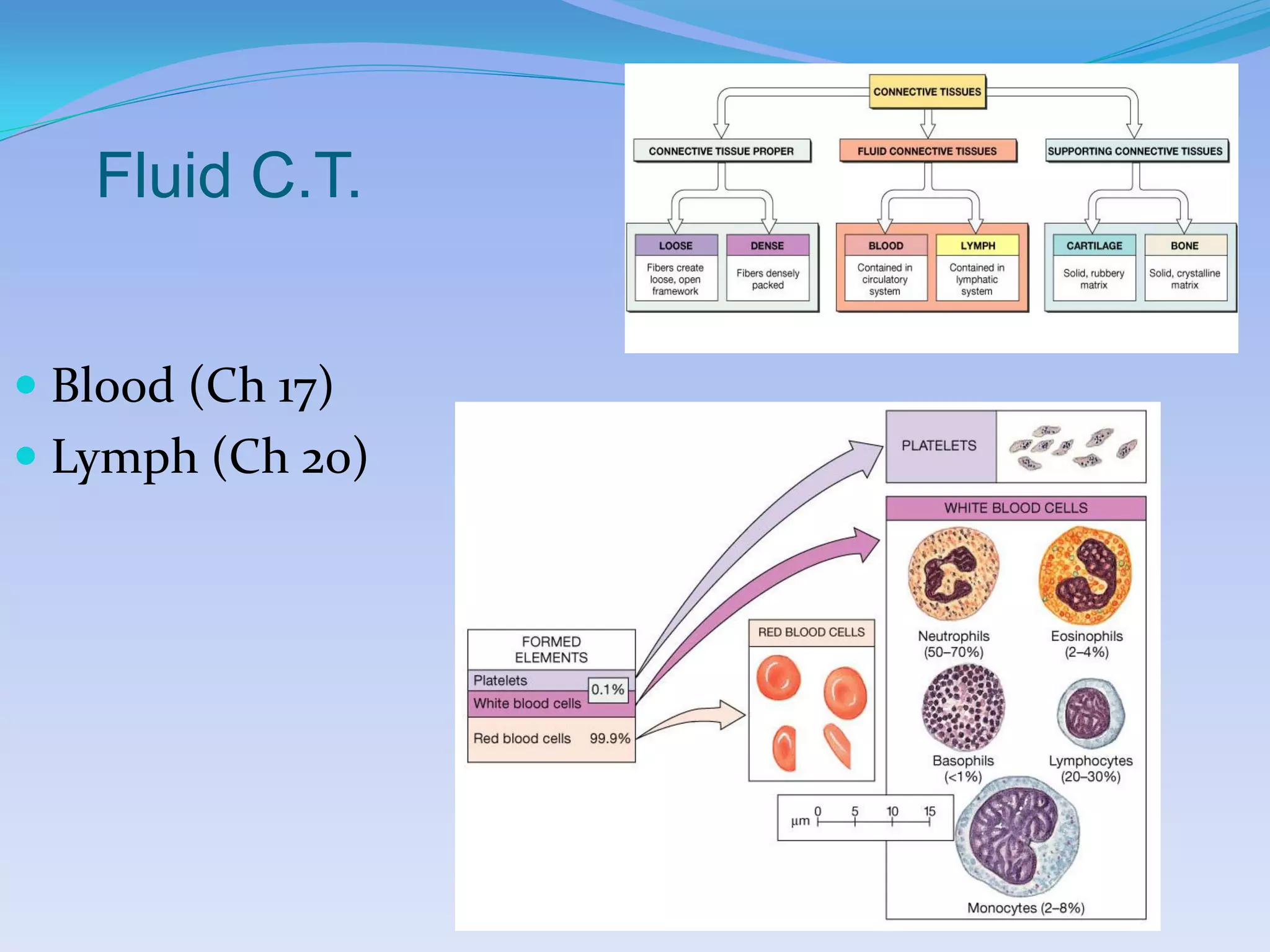
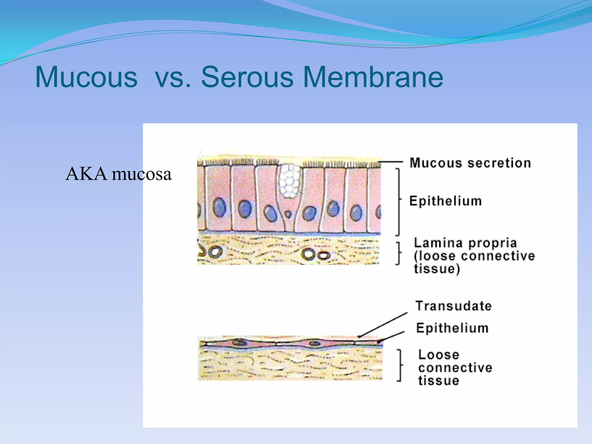
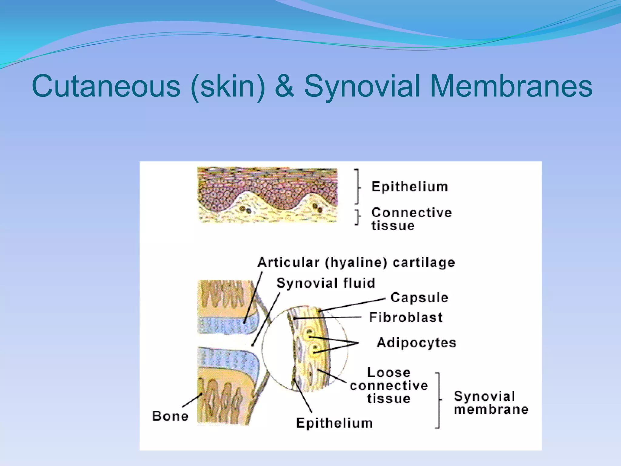
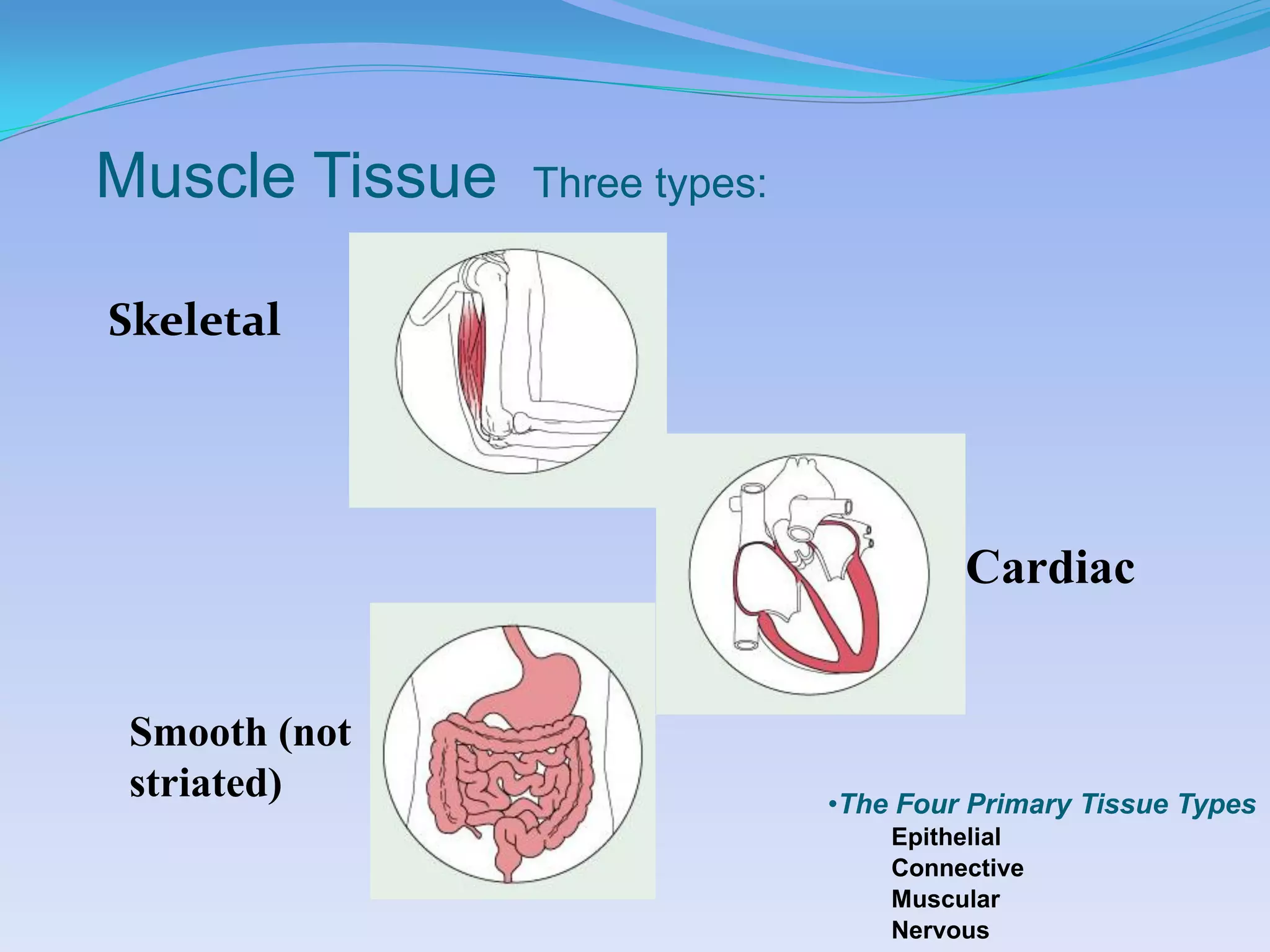
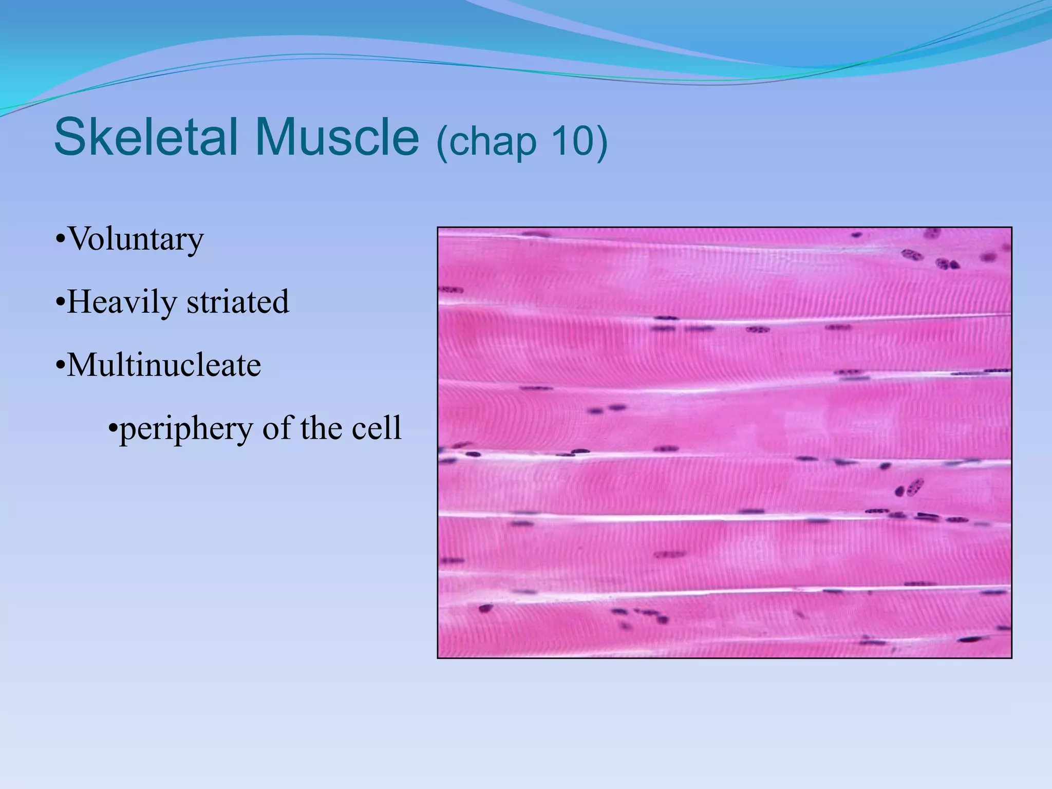
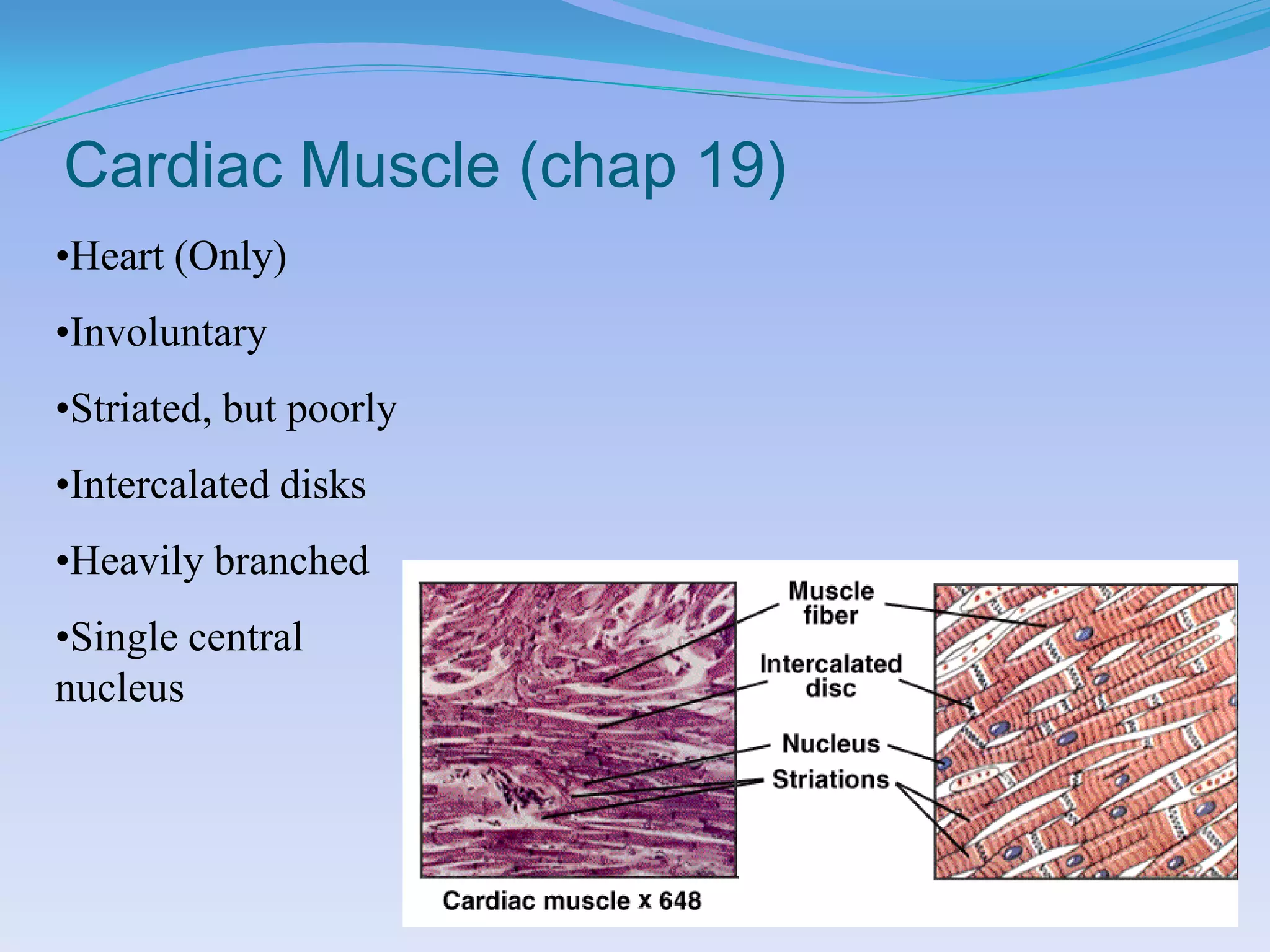
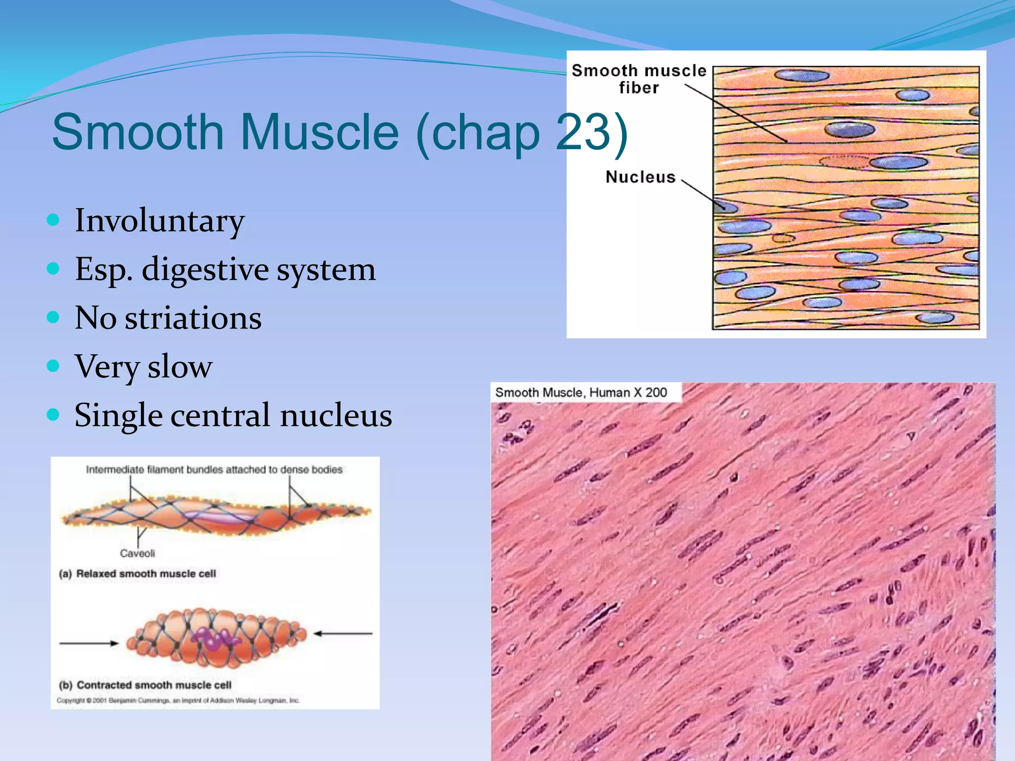
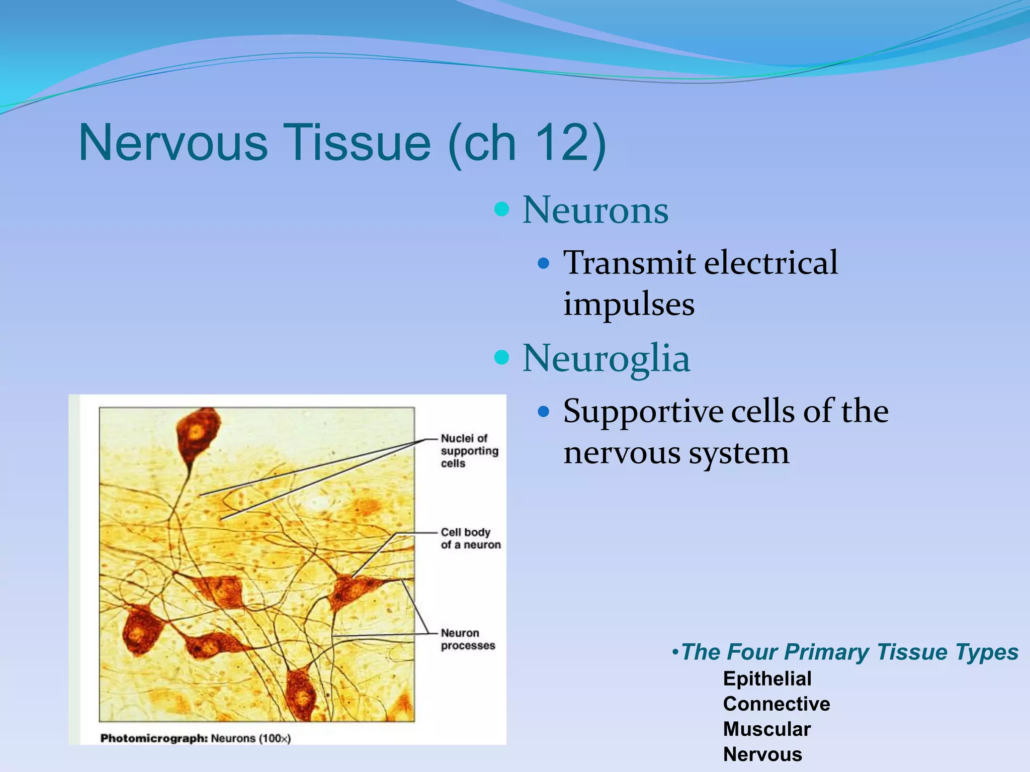
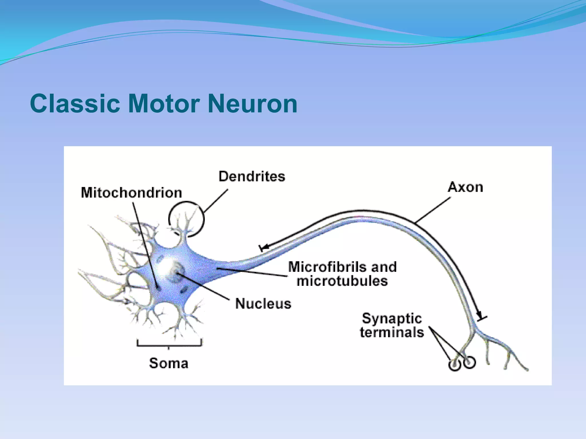
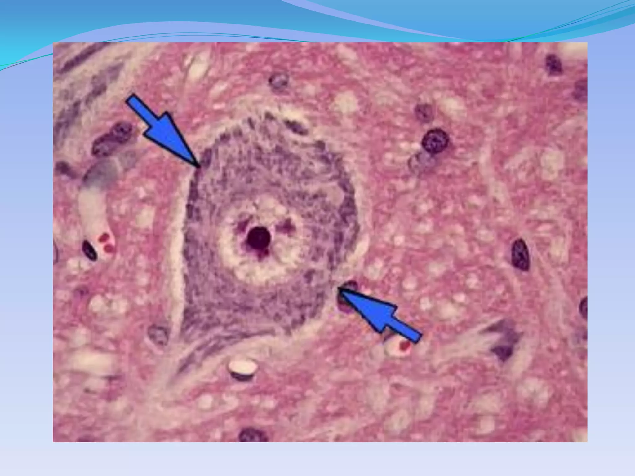
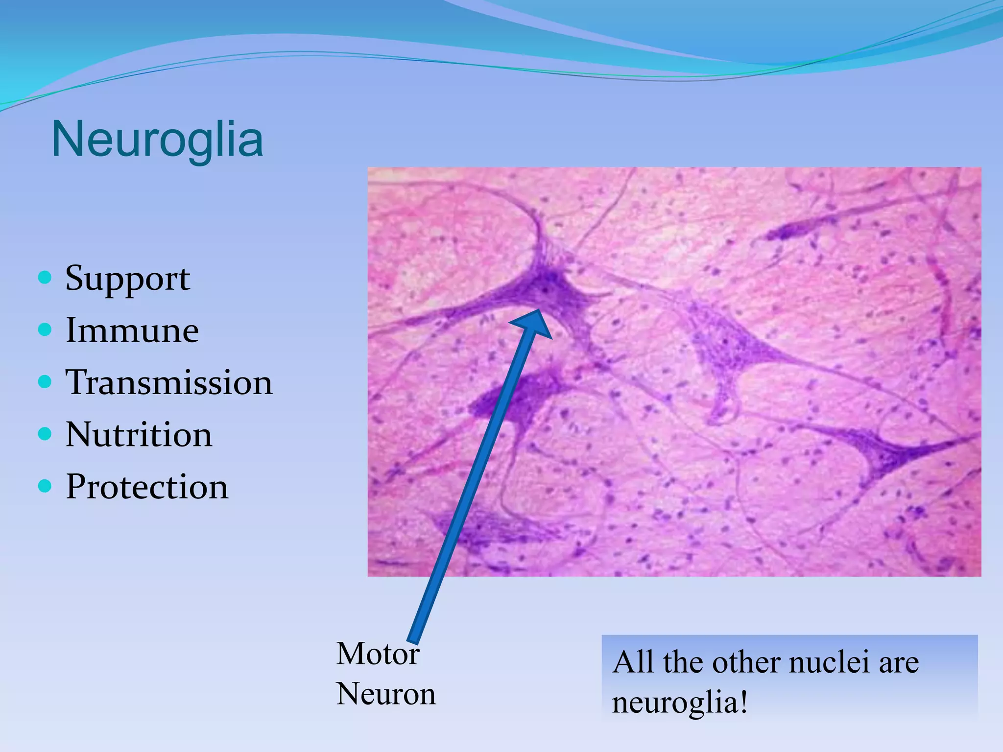
Chapter 4, Tissues, Part 2

•Histology = The study of tissues
•Tissue = A collection of cells that perform related
functions, and are similar in structure

•The Four Primary Tissue Types:
   •Epithelial
   •Connective
   •Muscular
   •Nervous


                                                Developed by
                                    John Gallagher, MS, DVM
Connective Tissue:
    Supports, protects, binds tissues
                     Structural characteristics:
                      Specialized cells, few in
                       number
                      Extracellular matrix
                         fibers (collagen)
                         Ground substance (fluid-
                          like)


                                    •The Four Primary Tissue Types
                                        Epithelial
                                        Connective
                                        Muscular
                                        Nervous
Functions of C.T.
 Structural framework for body
 Transport of fluids and dissolved
  materials
 Support and protection for organs
 Energy storage (fat)
 Defense


                               •The Four Primary Tissue Types
                                   Epithelial
                                   Connective
                                   Muscular
                                   Nervous
Classification of Connective Tissue


•Loose (areolar)   •Irregular          •Hyaline    •Compact
       •Adipose    •Regular             •Elastic   •Spongy
      •Reticular   •Elastic      •Fibrocartilage


  This is similar to Table 4.2
Connective Tissue Proper
Loose Connective tissue

 - viscous EC matrix, fibers not
 aligned
      1. loose (areolar) C.T.
      2. adipose
      3. reticular
1. Areolar (AKA loose)
 Most widespread
    Beneath most
     epithelia
    Think dermis!!!
 Types of Fibers:
   • Collagen
   • Elastic
   • Reticular
1. Loose (areolar)
•Ground Substance
    •Similar to plasma
    •Leaked from capillaries
    •AKA ECF, interstitial
    fluid
•Defense:
    •Macrophages
    •Plasma Cells
    •Mast Cells
    •Other WBC
2. Fat
 AKA adipose
    Cells are swollen with fat
 Prominent in hypodermis
 Intercellular matrix is
  obscured
 Storage of energy
3. Reticular

  Resembles areolar CT, but
     Reticular fibers only
  “reticulum” = network
  Bone marrow, spleen
Dense Connective Tissue
1. Dense Irregular C.T.

 Collagen fibers in many
  directions
 Resist multi-directional tension
 Joint capsules
2. Dense Regular C.T.
 Very parallel fibers of collagen
 Tendons
3. Elastic C.T.

 Elastic fibers > collagen
 Arterial walls
Supporting C.T.


 Cartilage
   Hyaline                    Check Chapt 6, p 125
   Elastic
       Ear, larynx
   Fibrocartilage
       Intervertebral discs
 Bone (Chapter 6)
   Mineral matrix
Hyaline Cartilage

 I.C. Matrix appears
  homogeneous
 Chondroblasts and
  chondrocytes in
  lacunae
 Articular cartilage
   Tracheal rings
Elastic cartilage         Fibrocartilage

 More elastic fibers    IV disk
 Ear                    Symphysis pubis
                         Meniscus
Bone (chap 6)

 The E.C. matrix is
 mineral, not fibrous
Fluid C.T.


 Blood (Ch 17)
 Lymph (Ch 20)
Membranes = Combination of
Epithelia & C.T. (p 88)
Mucous membranes,
   Lining of cavities that communicate with exterior


 Serous membranes,
   Lining of sealed, internal cavities
                                                  Know special names of
 Cutaneous membranes,                            serous membranes
   Skin                                          depending on location
 Synovial membranes,                             Also know difference
   Joints                                        between “parietal” and
                                                  “visceral”
Mucous vs. Serous Membrane


 AKA mucosa
Cutaneous (skin) & Synovial Membranes
Muscle Tissue   Three types:


Skeletal


                                        Cardiac


 Smooth (not
 striated)                     •The Four Primary Tissue Types
                                   Epithelial
                                   Connective
                                   Muscular
                                   Nervous
Skeletal Muscle (chap 10)
•Voluntary
•Heavily striated
•Multinucleate
   •periphery of the cell
Cardiac Muscle (chap 19)
•Heart (Only)
•Involuntary
•Striated, but poorly
•Intercalated disks
•Heavily branched
•Single central
nucleus
Smooth Muscle (chap 23)
 Involuntary
 Esp. digestive system
 No striations
 Very slow
 Single central nucleus
Nervous Tissue (ch 12)
                  Neurons
                    Transmit electrical
                    impulses
                  Neuroglia
                    Supportive cells of the
                    nervous system


                             •The Four Primary Tissue Types
                                 Epithelial
                                 Connective
                                 Muscular
                                 Nervous
Classic Motor Neuron
Neuroglia

 Support
 Immune
 Transmission
 Nutrition
 Protection


                 Motor    All the other nuclei are
                 Neuron   neuroglia!
Muir Pass, Sierra Nevada

Chapter4 tissuesmariebpart2

  • 1.
    Chapter 4, Tissues,Part 2 •Histology = The study of tissues •Tissue = A collection of cells that perform related functions, and are similar in structure •The Four Primary Tissue Types: •Epithelial •Connective •Muscular •Nervous Developed by John Gallagher, MS, DVM
  • 2.
    Connective Tissue: Supports, protects, binds tissues Structural characteristics:  Specialized cells, few in number  Extracellular matrix  fibers (collagen)  Ground substance (fluid- like) •The Four Primary Tissue Types Epithelial Connective Muscular Nervous
  • 3.
    Functions of C.T. Structural framework for body  Transport of fluids and dissolved materials  Support and protection for organs  Energy storage (fat)  Defense •The Four Primary Tissue Types Epithelial Connective Muscular Nervous
  • 4.
    Classification of ConnectiveTissue •Loose (areolar) •Irregular •Hyaline •Compact •Adipose •Regular •Elastic •Spongy •Reticular •Elastic •Fibrocartilage This is similar to Table 4.2
  • 5.
  • 6.
    Loose Connective tissue - viscous EC matrix, fibers not aligned 1. loose (areolar) C.T. 2. adipose 3. reticular
  • 7.
    1. Areolar (AKAloose)  Most widespread  Beneath most epithelia  Think dermis!!!  Types of Fibers: • Collagen • Elastic • Reticular
  • 8.
    1. Loose (areolar) •GroundSubstance •Similar to plasma •Leaked from capillaries •AKA ECF, interstitial fluid •Defense: •Macrophages •Plasma Cells •Mast Cells •Other WBC
  • 9.
    2. Fat  AKAadipose  Cells are swollen with fat  Prominent in hypodermis  Intercellular matrix is obscured  Storage of energy
  • 10.
    3. Reticular Resembles areolar CT, but  Reticular fibers only  “reticulum” = network  Bone marrow, spleen
  • 11.
  • 12.
    1. Dense IrregularC.T.  Collagen fibers in many directions  Resist multi-directional tension  Joint capsules
  • 13.
    2. Dense RegularC.T.  Very parallel fibers of collagen  Tendons
  • 14.
    3. Elastic C.T. Elastic fibers > collagen  Arterial walls
  • 15.
    Supporting C.T.  Cartilage  Hyaline Check Chapt 6, p 125  Elastic  Ear, larynx  Fibrocartilage  Intervertebral discs  Bone (Chapter 6)  Mineral matrix
  • 16.
    Hyaline Cartilage  I.C.Matrix appears homogeneous  Chondroblasts and chondrocytes in lacunae  Articular cartilage  Tracheal rings
  • 17.
    Elastic cartilage Fibrocartilage  More elastic fibers  IV disk  Ear  Symphysis pubis  Meniscus
  • 18.
    Bone (chap 6) The E.C. matrix is mineral, not fibrous
  • 19.
    Fluid C.T.  Blood(Ch 17)  Lymph (Ch 20)
  • 20.
    Membranes = Combinationof Epithelia & C.T. (p 88) Mucous membranes,  Lining of cavities that communicate with exterior  Serous membranes,  Lining of sealed, internal cavities Know special names of  Cutaneous membranes, serous membranes  Skin depending on location  Synovial membranes, Also know difference  Joints between “parietal” and “visceral”
  • 21.
    Mucous vs. SerousMembrane AKA mucosa
  • 22.
    Cutaneous (skin) &Synovial Membranes
  • 23.
    Muscle Tissue Three types: Skeletal Cardiac Smooth (not striated) •The Four Primary Tissue Types Epithelial Connective Muscular Nervous
  • 24.
    Skeletal Muscle (chap10) •Voluntary •Heavily striated •Multinucleate •periphery of the cell
  • 25.
    Cardiac Muscle (chap19) •Heart (Only) •Involuntary •Striated, but poorly •Intercalated disks •Heavily branched •Single central nucleus
  • 26.
    Smooth Muscle (chap23)  Involuntary  Esp. digestive system  No striations  Very slow  Single central nucleus
  • 27.
    Nervous Tissue (ch12)  Neurons  Transmit electrical impulses  Neuroglia  Supportive cells of the nervous system •The Four Primary Tissue Types Epithelial Connective Muscular Nervous
  • 28.
  • 30.
    Neuroglia  Support  Immune Transmission  Nutrition  Protection Motor All the other nuclei are Neuron neuroglia!
  • 31.