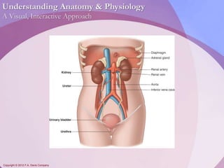
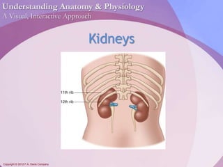
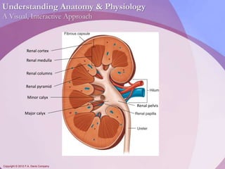
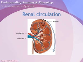
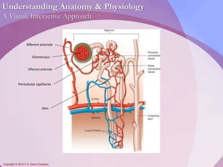
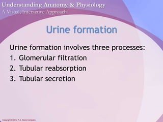
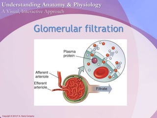
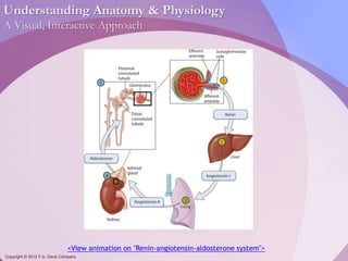
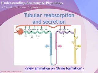
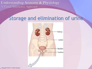
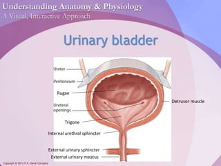
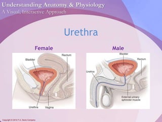
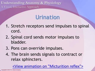
Understanding Anatomy & Physiology 
A Visual, Interactive Approach 
Copyright © 2012 F.A. Davis Company 
Chapter 18 
The Urinary System
Understanding Anatomy & Physiology 
A Visual, Interactive Approach 
Copyright © 2012 F.A. Davis Company
Understanding Anatomy & Physiology 
A Visual, Interactive Approach 
Copyright © 2012 F.A. Davis Company 
Kidneys
Understanding Anatomy & Physiology 
A Visual, Interactive Approach 
Renal cortex 
Renal medulla 
Renal columns 
Renal pyramid 
Minor calyx 
Major calyx 
Copyright © 2012 F.A. Davis Company 
Renal pelvis
Understanding Anatomy & Physiology 
A Visual, Interactive Approach 
Blood vessels, nerves, and the ureter enter 
and leave the kidney from the: 
A. renal pelvis. 
B. hilum. 
C. calyx. 
D. renal pyramid. 
Copyright © 2012 F.A. Davis Company
Understanding Anatomy & Physiology 
A Visual, Interactive Approach 
Correct answer: B 
Rationale: 
 The renal pelvis, formed by the convergence 
of the major calyces, channels urine out of 
the kidney. 
 The calyx collects urine leaving the papilla. 
 Renal pyramids are the cone-shaped sections 
in the kidney. 
Copyright © 2012 F.A. Davis Company
Understanding Anatomy & Physiology 
A Visual, Interactive Approach 
Copyright © 2012 F.A. Davis Company 
Renal circulation 
Renal artery 
Renal vein 
Nephron
Understanding Anatomy & Physiology 
A Visual, Interactive Approach 
Afferent arteriole 
Glomerulus 
Efferent arteriole 
Peritubular capillaries 
Vein 
Copyright © 2012 F.A. Davis Company
Understanding Anatomy & Physiology 
A Visual, Interactive Approach 
Copyright © 2012 F.A. Davis Company 
Renal corpuscle 
Bowman’s 
capsule
Understanding Anatomy & Physiology 
A Visual, Interactive Approach 
Copyright © 2012 F.A. Davis Company 
Proximal convoluted tubule 
Loop of Henle 
Distal convoluted tubule 
Collecting duct
Understanding Anatomy & Physiology 
A Visual, Interactive Approach 
What are the filtration units of the 
kidney? 
A. Renal corpuscle 
B. Glomerulus 
C. Nephron 
D. Renal pelvis 
Copyright © 2012 F.A. Davis Company
Understanding Anatomy & Physiology 
A Visual, Interactive Approach 
Correct answer: C 
Rationale: 
 The renal corpuscle is part of the nephron. 
 The renal corpuscle contains the glomerulus. 
 The renal pelvis funnels urine out of the 
kidney. 
Copyright © 2012 F.A. Davis Company
Understanding Anatomy & Physiology 
A Visual, Interactive Approach 
Copyright © 2012 F.A. Davis Company 
Urine formation 
Urine formation involves three processes: 
1. Glomerular filtration 
2. Tubular reabsorption 
3. Tubular secretion
Understanding Anatomy & Physiology 
A Visual, Interactive Approach 
Glomerular filtration 
Copyright © 2012 F.A. Davis Company
Understanding Anatomy & Physiology 
A Visual, Interactive Approach 
Copyright © 2012 F.A. Davis Company 
Regulation of GFR 
 Glomerular filtration rate [GFR] should be 
constant, despite changes in blood 
pressure. 
 If flow is too high, the body will lose 
excessive water and nutrients. 
 If flow is too low, tubules may reabsorb 
toxins.
Understanding Anatomy & Physiology 
A Visual, Interactive Approach 
<View animation on "Renin-angiotensin-aldosterone system"> 
Copyright © 2012 F.A. Davis Company
Understanding Anatomy & Physiology 
A Visual, Interactive Approach 
Copyright © 2012 F.A. Davis Company 
Tubular reabsorption 
and secretion 
<View animation on "Urine formation">
Understanding Anatomy & Physiology 
A Visual, Interactive Approach 
Most sodium is reabsorbed from the 
glomerular filtrate in the: 
A. proximal convoluted tubule. 
B. distal convoluted tubule. 
C. collection duct. 
D. glomerulus. 
Copyright © 2012 F.A. Davis Company
Understanding Anatomy & Physiology 
A Visual, Interactive Approach 
Correct answer: A 
Rationale: 
 The distal convoluted tubule and collection 
ducts reabsorb variable amounts of water 
and salt. 
 In the glomerulus, water and small solutes 
filter out of the blood and into Bowman’s 
capsule. 
Copyright © 2012 F.A. Davis Company
Understanding Anatomy & Physiology 
A Visual, Interactive Approach 
Hormones that affect the 
Copyright © 2012 F.A. Davis Company 
urinary system 
HORMONE EFFECT ON RENAL TUBULE 
Aldosterone Reabsorbs: NaCl; H2O 
Excretes: K+ 
Atrial natriuretic peptide (ANP) Excretes: NaCl; H2O 
Antidiuretic hormone (ADH) Excretes: H2O 
Parathyroid hormone (PTH) Reabsorbs: Calcium 
Excretes: Phosphate
Understanding Anatomy & Physiology 
A Visual, Interactive Approach 
Composition of urine 
 Consists of 95% water; 5% dissolved 
substances 
 Reveals information about health of 
kidneys and other organs 
Copyright © 2012 F.A. Davis Company
Understanding Anatomy & Physiology 
A Visual, Interactive Approach 
Storage and elimination of urine 
Copyright © 2012 F.A. Davis Company 
Ureter
Understanding Anatomy & Physiology 
A Visual, Interactive Approach 
Copyright © 2012 F.A. Davis Company 
Urinary bladder 
Rugae 
Trigone 
Internal urethral sphincter 
External urinary sphincter 
External urinary meatus 
Detrusor muscle
Understanding Anatomy & Physiology 
A Visual, Interactive Approach 
Copyright © 2012 F.A. Davis Company 
Urethra 
Female Male
Understanding Anatomy & Physiology 
A Visual, Interactive Approach 
Copyright © 2012 F.A. Davis Company 
Urination 
1. Stretch receptors send impulses to spinal 
cord. 
2. Spinal cord sends motor impulses to 
bladder. 
3. Pons can override impulses. 
4. The brain sends signals to contract or 
relax sphincters. 
<View animation on "Micturition reflex">
Understanding Anatomy & Physiology 
A Visual, Interactive Approach 
The triangular-shaped smooth area on 
the floor of the bladder is called the: 
A. rugae. 
B. papilla. 
C. cortex. 
D. trigone. 
Copyright © 2012 F.A. Davis Company
Understanding Anatomy & Physiology 
A Visual, Interactive Approach 
Correct answer: D 
Rationale: 
 Rugae are folds of tissue on the inside of the 
bladder. 
 Papillae are the blunt points of the renal 
pyramids. 
 The cortex is the outer zone of the kidney. 
Copyright © 2012 F.A. Davis Company

Chapter18 - Urinary System

  • 1.
    Understanding Anatomy &Physiology A Visual, Interactive Approach Copyright © 2012 F.A. Davis Company Chapter 18 The Urinary System
  • 2.
    Understanding Anatomy &Physiology A Visual, Interactive Approach Copyright © 2012 F.A. Davis Company
  • 3.
    Understanding Anatomy &Physiology A Visual, Interactive Approach Copyright © 2012 F.A. Davis Company Kidneys
  • 4.
    Understanding Anatomy &Physiology A Visual, Interactive Approach Renal cortex Renal medulla Renal columns Renal pyramid Minor calyx Major calyx Copyright © 2012 F.A. Davis Company Renal pelvis
  • 5.
    Understanding Anatomy &Physiology A Visual, Interactive Approach Blood vessels, nerves, and the ureter enter and leave the kidney from the: A. renal pelvis. B. hilum. C. calyx. D. renal pyramid. Copyright © 2012 F.A. Davis Company
  • 6.
    Understanding Anatomy &Physiology A Visual, Interactive Approach Correct answer: B Rationale:  The renal pelvis, formed by the convergence of the major calyces, channels urine out of the kidney.  The calyx collects urine leaving the papilla.  Renal pyramids are the cone-shaped sections in the kidney. Copyright © 2012 F.A. Davis Company
  • 7.
    Understanding Anatomy &Physiology A Visual, Interactive Approach Copyright © 2012 F.A. Davis Company Renal circulation Renal artery Renal vein Nephron
  • 8.
    Understanding Anatomy &Physiology A Visual, Interactive Approach Afferent arteriole Glomerulus Efferent arteriole Peritubular capillaries Vein Copyright © 2012 F.A. Davis Company
  • 9.
    Understanding Anatomy &Physiology A Visual, Interactive Approach Copyright © 2012 F.A. Davis Company Renal corpuscle Bowman’s capsule
  • 10.
    Understanding Anatomy &Physiology A Visual, Interactive Approach Copyright © 2012 F.A. Davis Company Proximal convoluted tubule Loop of Henle Distal convoluted tubule Collecting duct
  • 11.
    Understanding Anatomy &Physiology A Visual, Interactive Approach What are the filtration units of the kidney? A. Renal corpuscle B. Glomerulus C. Nephron D. Renal pelvis Copyright © 2012 F.A. Davis Company
  • 12.
    Understanding Anatomy &Physiology A Visual, Interactive Approach Correct answer: C Rationale:  The renal corpuscle is part of the nephron.  The renal corpuscle contains the glomerulus.  The renal pelvis funnels urine out of the kidney. Copyright © 2012 F.A. Davis Company
  • 13.
    Understanding Anatomy &Physiology A Visual, Interactive Approach Copyright © 2012 F.A. Davis Company Urine formation Urine formation involves three processes: 1. Glomerular filtration 2. Tubular reabsorption 3. Tubular secretion
  • 14.
    Understanding Anatomy &Physiology A Visual, Interactive Approach Glomerular filtration Copyright © 2012 F.A. Davis Company
  • 15.
    Understanding Anatomy &Physiology A Visual, Interactive Approach Copyright © 2012 F.A. Davis Company Regulation of GFR  Glomerular filtration rate [GFR] should be constant, despite changes in blood pressure.  If flow is too high, the body will lose excessive water and nutrients.  If flow is too low, tubules may reabsorb toxins.
  • 16.
    Understanding Anatomy &Physiology A Visual, Interactive Approach <View animation on "Renin-angiotensin-aldosterone system"> Copyright © 2012 F.A. Davis Company
  • 17.
    Understanding Anatomy &Physiology A Visual, Interactive Approach Copyright © 2012 F.A. Davis Company Tubular reabsorption and secretion <View animation on "Urine formation">
  • 18.
    Understanding Anatomy &Physiology A Visual, Interactive Approach Most sodium is reabsorbed from the glomerular filtrate in the: A. proximal convoluted tubule. B. distal convoluted tubule. C. collection duct. D. glomerulus. Copyright © 2012 F.A. Davis Company
  • 19.
    Understanding Anatomy &Physiology A Visual, Interactive Approach Correct answer: A Rationale:  The distal convoluted tubule and collection ducts reabsorb variable amounts of water and salt.  In the glomerulus, water and small solutes filter out of the blood and into Bowman’s capsule. Copyright © 2012 F.A. Davis Company
  • 20.
    Understanding Anatomy &Physiology A Visual, Interactive Approach Hormones that affect the Copyright © 2012 F.A. Davis Company urinary system HORMONE EFFECT ON RENAL TUBULE Aldosterone Reabsorbs: NaCl; H2O Excretes: K+ Atrial natriuretic peptide (ANP) Excretes: NaCl; H2O Antidiuretic hormone (ADH) Excretes: H2O Parathyroid hormone (PTH) Reabsorbs: Calcium Excretes: Phosphate
  • 21.
    Understanding Anatomy &Physiology A Visual, Interactive Approach Composition of urine  Consists of 95% water; 5% dissolved substances  Reveals information about health of kidneys and other organs Copyright © 2012 F.A. Davis Company
  • 22.
    Understanding Anatomy &Physiology A Visual, Interactive Approach Storage and elimination of urine Copyright © 2012 F.A. Davis Company Ureter
  • 23.
    Understanding Anatomy &Physiology A Visual, Interactive Approach Copyright © 2012 F.A. Davis Company Urinary bladder Rugae Trigone Internal urethral sphincter External urinary sphincter External urinary meatus Detrusor muscle
  • 24.
    Understanding Anatomy &Physiology A Visual, Interactive Approach Copyright © 2012 F.A. Davis Company Urethra Female Male
  • 25.
    Understanding Anatomy &Physiology A Visual, Interactive Approach Copyright © 2012 F.A. Davis Company Urination 1. Stretch receptors send impulses to spinal cord. 2. Spinal cord sends motor impulses to bladder. 3. Pons can override impulses. 4. The brain sends signals to contract or relax sphincters. <View animation on "Micturition reflex">
  • 26.
    Understanding Anatomy &Physiology A Visual, Interactive Approach The triangular-shaped smooth area on the floor of the bladder is called the: A. rugae. B. papilla. C. cortex. D. trigone. Copyright © 2012 F.A. Davis Company
  • 27.
    Understanding Anatomy &Physiology A Visual, Interactive Approach Correct answer: D Rationale:  Rugae are folds of tissue on the inside of the bladder.  Papillae are the blunt points of the renal pyramids.  The cortex is the outer zone of the kidney. Copyright © 2012 F.A. Davis Company

Editor's Notes

  • #3 The urinary system consists of kidneys, ureters, urinary bladder, and urethra.
  • #4 The kidneys lie against the posterior abdominal wall, underneath the 12th rib. They are retroperitoneal (posterior to the parietal peritoneum). The ribs help protect the kidneys, as does a heavy cushion of fat encasing each organ. Each kidney measures about 4 inches (10 cm) long, 2 inches (5 cm) wide, and 1 inch (2.5 cm) thick; they extend from the level of the T12 vertebra to the L3 vertebra. The right kidney sits a little lower than the left because of the liver, which occupies the space above it. Structures (such as blood vessels, the ureters, and nerves) enter and leave the kidney through a slit called the hilum—located in a concave notch on the medial side.
  • #5 A tough, fibrous capsule surrounds each kidney. The interior consists of the renal cortex and renal medulla; the renal cortex forms the outer region and the renal medulla forms the inner region. Extensions from the renal cortex, called renal columns, divide the interior into cone-shaped sections called renal pyramids. The base of each pyramid faces outward toward the cortex; the point of the pyramid, called the renal papilla, faces the hilum. The renal papilla extends into a cup called a minor calyx. The calyx collects urine leaving the papilla. Two or three minor calyces join together to form a major calyx. The major calyces converge to form the renal pelvis, which receives urine from the major calyces. The renal pelvis continues as the ureter, which channels urine to the urinary bladder.
  • #8 The renal artery (which branches off the abdominal aorta) brings blood to the kidney. As it enters the kidney, it divides into smaller and smaller arteries. The arteries pass through the renal columns and into the renal cortex. Blood leaves the kidney through the renal vein, which empties into the inferior vena cava. Nephrons—the filtration units of the kidney—lie in the kidney’s outer region, where they extend between the cortex and the medulla.
  • #9 The nephrons need a constant flow of blood. (More than 20% of the blood pumped by the heart each minute goes to the kidneys.) A series of afferent arterioles arise from smaller arteries in the cortex; each afferent arteriole supplies blood to one nephron. Each afferent arteriole branches into a cluster of capillaries called a glomerulus. Blood leaves the glomerulus through an efferent arteriole. The efferent arteriole leads to a network of capillaries around the renal tubules called peritubular capillaries. These capillaries pick up water and solutes reabsorbed by the renal tubules. Blood flows from the peritubular capillaries into larger and larger veins that eventually feed into the renal vein.
  • #10 The renal corpuscle is the beginning of the nephron. (The two main components of a nephron are a renal corpuscle [which filters blood plasma] and a renal tubule [where urine is formed]). The renal corpuscle consists of a glomerulus and Bowman’s capsule. Bowman’s capsule (or glomerular capsule) consists of two layers of epithelial cells surrounding the glomerulus in an open-ended covering. (As an analogy, think of pushing your fist into an inflated balloon. Your fist represents the glomerulus. The balloon, which folds around your fist in two layers, represents the Bowman’s capsule.) Fluid filters out of the glomerulus and collects in the space between the two layers of Bowman’s capsule. From there, it flows into the renal tubule on the other side of the capsule.
  • #11 Leading away from the glomerulus are a series of tubelike structures called the renal tubule. It can be divided into four regions: the proximal convoluted tubule, nephron loop, distal convoluted tubule, and collection duct. (In this slide, the renal tubule has been stretched out to show the different regions.) Arising from Bowman’s capsule is the proximal convoluted tubule. Thousands of microvilli that allow absorption line the inside of the proximal convoluted tubule. The renal tubule straightens out and dips into the medulla before turning sharply and returning to the cortex. This entire segment—which consists of a descending limb and an ascending limb—is called the loop of Henle. After returning to the cortex, the ascending limb coils again, forming the distal convoluted tubule. The collecting duct receives drainage from the distal convoluted tubules of several different nephrons. The collecting duct passes through the medulla and merges with other collecting ducts before draining into a minor calyx.
  • #15 Blood flows into the glomerulus through the afferent arteriole, which is larger than the efferent arteriole. Consequently, blood flows in faster than it can leave, which contributes to higher pressure within the glomerular capillaries. The walls of glomerular capillaries are dotted with pores; water and small solutes (such as electrolytes, glucose, amino acids, vitamins, and nitrogenous wastes) filter out of the blood and into Bowman’s capsule. Blood cells and most plasma proteins are too large to pass through the pores. The fluid that filters into Bowman’s capsule flows into the renal tubules. The amount of fluid filtered by both kidneys (called the glomerular filtration rate [GFR]) equals about 180 L each day, or 60 times the body’s total blood volume. The body reabsorbs about 99% of this filtrate, leaving 1 to 2 L to be excreted as urine.
  • #17 A key mechanism for maintaining blood pressure and, therefore, a steady glomerular filtration rate, is the renin-angiotensin-aldosterone system. A decline in blood pressure leads to decreased blood flow to the kidneys. Juxtaglomerular cells in the afferent arterioles respond by releasing the enzyme renin. Renin converts inactive plasma protein angiotensinogen (made in the liver) into angiotensin I. Angiotensin I circulates to the lungs, where angiotensin-converting enzyme (ACE) converts it into angiotensin II. Angiotensin II stimulates the adrenal glands to secrete aldosterone. Aldosterone causes the distal convoluted tubule to retain sodium, which leads to increased retention of water. Blood volume increases, and blood pressure rises.
  • #18 After filtrate leaves the glomerulus, it enters the renal tubules, where additional chemicals are removed from the filtrate and returned to the blood (tubular reabsorption) and other chemicals are added (tubular secretion). Here is what happens: Sodium moves by active transport out of the proximal convoluted tubule and into the bloodstream of the peritubular capillaries. Water follows sodium. Glucose, amino acids, chloride, potassium, and bicarbonate also pass out of the tubules and into the blood. About half of the nitrogenous waste urea is also reabsorbed. Simultaneously, wastes such as ammonia (NH3) and uric acid, as well as drugs (such as aspirin and penicillin), are secreted out of the blood and into the tubules. Tubular secretion of hydrogen ions also occurs, helping to regulate the body’s pH. Water diffuses out of the descending limb of the loop of Henle, further concentrating the filtrate. Sodium and chloride are actively pumped out of the ascending limb of the loop of Henle into interstitial fluid. (The thicker wall of the ascending limb prevents water from following the sodium out of the tubule.) The distal convoluted tubule and collecting ducts reabsorb variable amounts of water and salts. Specialized cells play a role in acid-base balance, reabsorbing potassium and secreting hydrogen into the tubule. Several different hormones help regulate reabsorption by the cells in the distal convoluted tubule. The collecting duct reabsorbs water and concentrates the urine.
  • #21 The adrenal cortex secretes aldosterone when blood levels of Na+ decline or concentration of K+ increases. The atria of the heart secretes ANP when blood pressure rises; this inhibits the secretion of aldosterone and ADH. The posterior pituitary gland secretes ADH. The parathyroid glands secrete PTH in response to low calcium levels.
  • #22 The dissolved substances include nitrogenous wastes—such as urea, uric acid, ammonia, and creatinine—as well as other solutes, such as sodium, potassium, and sulfates.
  • #23 The ureters and urethra serve as passageways for conducting urine away from the kidneys and out of the body while the bladder stores urine until it can be eliminated. The ureters connect the renal pelvis of each kidney with the bladder.
  • #24 The wall of the bladder is the detrusor muscle; it consists of three layers of smooth muscle. Mucous transitional epithelium lines the bladder. When the bladder relaxes, this layer forms folds called rugae. As urine fills the bladder, the rugae flatten and the epithelium thins, allowing the bladder to expand. The floor of the bladder has three openings: two from the ureters (which pass behind the bladder to enter from below) and one from the urethra. They form a triangular-shaped, smooth area on the floor of the bladder called the trigone. Infections commonly attack this area. At the point where the urethra leaves the bladder is a ring of smooth muscle that forms the internal urethral sphincter. This sphincter contracts involuntarily to retain urine in the bladder. A second sphincter (the external urinary sphincter) exists where the urethra passes through the pelvic floor; this sphincter consists of skeletal muscle and is, therefore, under voluntary control. The urethra is a small tube that conveys urine away from the bladder and out of the body; it opens to the outside of the body at the external urinary meatus.
  • #25 In females, the urethra is 3 cm (1.2 in.) long and exits the body just in front of the vaginal orifice. In males, the urethra measures about 20 cm (7.9 in.). The urethra passes from the bladder through the center of the prostate gland, curves around to enter the penis, and then exits the body at the tip of the penis. In men, the urethra conveys both urine and semen.
  • #26 When the bladder contains 200 mL or more of urine, stretch receptors in the bladder wall send impulses to the sacral region of the spinal cord. The spinal cord sends motor impulses to the bladder wall to contract and the internal sphincter to relax; voiding will occur involuntarily unless the brain overrides the impulse. The brain can override the impulse because stretch receptors in the bladder also send impulses to the micturition center in the pons. The pons integrates that information with information from other parts of the brain, such as the cerebrum, and evaluates whether the time is appropriate to urinate. If the time is not appropriate to urinate, the brain sends impulses to inhibit urination and to keep the external urinary sphincter contracted. If the time is appropriate, the brain signals the bladder wall to contract and the external urethral sphincter to relax, and voluntary urination occurs.