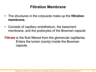
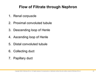
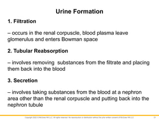
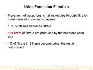
Because learning changes everything.®
Chapter 18
Urinary System and Fluid
Balance
Lecture Outline
Seeley’s ESSENTIALS OF
ANATOMY & PHYSIOLOGY
Eleventh Edition
Cinnamon VanPutte
Jennifer Regan
Andrew Russo
Copyright 2022 © McGraw Hill LLC. All rights reserved. No reproduction or distribution without the prior written consent of McGraw Hill LLC.
Urinary System
• The urinary system is the major excretory system of the
body.
• Some organs in other systems also eliminate wastes, but
they are not able to compensate in the case of kidney
failure.
2
Copyright 2022 © McGraw Hill LLC. All rights reserved. No reproduction or distribution without the prior written consent of McGraw Hill LLC.
Urinary System Functions
1. Excretion
2. Regulation of blood volume and pressure
3. Regulation of blood solute concentration
4. Regulation of extracellular fluid pH
5. Regulation of red blood cell synthesis
6. Regulation of Vitamin D synthesis
3
Copyright 2022 © McGraw Hill LLC. All rights reserved. No reproduction or distribution without the prior written consent of McGraw Hill LLC.
Kidney Structures
Renal capsule:
• connective tissue around each kidney
• protects and acts as a barrier
Hilum:
• indentation
• contains renal artery, veins, nerves, ureter
4
Copyright 2022 © McGraw Hill LLC. All rights reserved. No reproduction or distribution without the prior written consent of McGraw Hill LLC.
Kidney Structures
Renal sinus:
• contains renal pelvis, blood vessels, fat
Renal cortex:
• outer portion
Renal medulla:
• inner portion
5
Copyright 2022 © McGraw Hill LLC. All rights reserved. No reproduction or distribution without the prior written consent of McGraw Hill LLC.
Kidney Structures
Renal pyramid:
• Cone shaped structures in the medulla whose bases
project into the cortex
Renal papillae:
• tip of pyramids which drain into calyces
Renal pelvis:
• where calyces join together
• narrows to form ureter
6
Copyright 2022 © McGraw Hill LLC. All rights reserved. No reproduction or distribution without the prior written consent of McGraw Hill LLC.
Ureters and Urinary Bladder
Ureters:
• small tubes that carry urine from renal pelvis of kidney to
bladder
Urinary bladder:
• in pelvic cavity
• stores urine
• can hold a few ml to a maximum of 1000 milliliters
7
Copyright 2022 © McGraw Hill LLC. All rights reserved. No reproduction or distribution without the prior written consent of McGraw Hill LLC.
Urethra
Urethra:
• tube that exits bladder
• carries urine from urinary bladder to outside of body
Internal urethral sphincter
• smooth muscle surrounds urethra at the junction of the
urinary bladder and prevents urine from leaving the
bladder
8
Copyright 2022 © McGraw Hill LLC. All rights reserved. No reproduction or distribution without the prior written consent of McGraw Hill LLC.
Urethra
External urethral sphincter:
• formed of skeletal muscle surrounding the urethra near the
pelvic floor.
• allows a person to voluntarily start or stop the flow of urine
out of the urethra
9
Copyright 2022 © McGraw Hill LLC. All rights reserved. No reproduction or distribution without the prior written consent of McGraw Hill LLC.
Nephron
• The nephron is the functional unit of the kidney.
• Each kidney has over one million nephrons.
• The nephron includes the renal corpuscle, proximal
convoluted tubule, loop of Henle, distal convoluted tubule
and collecting duct
10
Copyright 2022 © McGraw Hill LLC. All rights reserved. No reproduction or distribution without the prior written consent of McGraw Hill LLC.
Renal Corpuscle
The filtration portion of the nephron
Glomerulus:
• A network of capillaries twisted around each other like a
ball of yarn
Bowman’s capsule:
• enlarged end of nephron surrounds glomerulus
• opens into proximal convoluted tubule
• contains podocytes (specialized cells around glomerular
capillaries)
11
Copyright 2022 © McGraw Hill LLC. All rights reserved. No reproduction or distribution without the prior written consent of McGraw Hill LLC.
Renal Corpuscle
• Bowman capsule consists of two layers:
• Outer layer - simple squamous epithelial cells that
become cube-shaped at the beginning of the proximal
convoluted tubule
• Inner layer - cells called podocytes, which wrap around
the glomerular capillaries
12
Copyright 2022 © McGraw Hill LLC. All rights reserved. No reproduction or distribution without the prior written consent of McGraw Hill LLC.
Proximal convoluted tubule:
• where filtrate passes first
• drains filtrate from Bowman capsule
Loop of Henle:
• contains descending and ascending loops
• water and solutes pass through thin walls by diffusion
13
Nephron Components
Copyright 2022 © McGraw Hill LLC. All rights reserved. No reproduction or distribution without the prior written consent of McGraw Hill LLC.
Nephron Components
Distal convoluted tubule:
• structure between Loop of Henle and collecting duct
Collecting duct:
• empties into calyces
• carry fluid from cortex through medulla
14
Copyright 2022 © McGraw Hill LLC. All rights reserved. No reproduction or distribution without the prior written consent of McGraw Hill LLC.
Types of Nephrons
1. Juxtamedullary nephrons
• renal corpuscles are deep in the cortex near the medulla
• long loops of Henle extend deep into the medulla
• Well adapted for water conservation
• About 15% of nephrons
15
Copyright 2022 © McGraw Hill LLC. All rights reserved. No reproduction or distribution without the prior written consent of McGraw Hill LLC.
Types of Nephrons
2. Cortical nephrons
• Renal corpuscles distributed throughout the cortex (outer
portion)
• Loops of Henle are shorter and closer to the outer edge of
the cortex than juxtamedullary nephrons
16
Copyright 2022 © McGraw Hill LLC. All rights reserved. No reproduction or distribution without the prior written consent of McGraw Hill LLC.
Characteristics of Renal Corpuscle
1. Porous capillaries
- highly permeable due to the presence of pores. Neither
large proteins nor blood cells can fit through them.
2. Porous inner layer of Bowman capsule
- A basement membrane lies between the endothelial cells
of the glomerular capillaries and the podocytes of the
Bowman capsule.
17
Copyright 2022 © McGraw Hill LLC. All rights reserved. No reproduction or distribution without the prior written consent of McGraw Hill LLC.
Characteristics of Renal Corpuscle
3. High pressure
An afferent arteriole supplies blood to the glomerulus for
filtration.
An efferent arteriole transports the filtered blood away from
the glomerulus.
Efferent arteriole has smaller diameter than afferent arteriole
creating a high pressure in the capillaries.
18
Copyright 2022 © McGraw Hill LLC. All rights reserved. No reproduction or distribution without the prior written consent of McGraw Hill LLC.
Filtration Membrane
• The structures in the corpuscle make up the filtration
membrane.
• Consists of capillary endothelium, the basement
membrane, and the podocytes of the Bowman capsule
Filtrate is the fluid filtered from the glomerular capillaries.
Enters the lumen (cavity) inside the Bowman
capsule.
19
Copyright 2022 © McGraw Hill LLC. All rights reserved. No reproduction or distribution without the prior written consent of McGraw Hill LLC.
Flow of Filtrate through Nephron
1. Renal corpuscle
2. Proximal convoluted tubule
3. Descending loop of Henle
4. Ascending loop of Henle
5. Distal convoluted tubule
6. Collecting duct
7. Papillary duct
20
Copyright 2022 © McGraw Hill LLC. All rights reserved. No reproduction or distribution without the prior written consent of McGraw Hill LLC.
Urine Formation
1. Filtration
– occurs in the renal corpuscle, blood plasma leave
glomerulus and enters Bowman space
2. Tubular Reabsorption
– involves removing substances from the filtrate and placing
them back into the blood
3. Secretion
– involves taking substances from the blood at a nephron
area other than the renal corpuscle and putting back into the
nephron tubule
21
Copyright 2022 © McGraw Hill LLC. All rights reserved. No reproduction or distribution without the prior written consent of McGraw Hill LLC.
Urine Formation-Filtration
• Movement of water, ions, small molecules through filtration
membrane into Bowman’s capsule
• 19% of plasma becomes filtrate
• 180 liters of filtrate are produced by the nephrons each
day
• 1% of filtrate (1.8 liters) become urine, the rest is
reabsorbed
22
Copyright 2022 © McGraw Hill LLC. All rights reserved. No reproduction or distribution without the prior written consent of McGraw Hill LLC.
Urine Production-Reabsorption
• 99% of filtrate is reabsorbed and reenters circulation
• The proximal convoluted tubule is the primary site for
reabsorption of solutes and water
• The descending Loop of Henle concentrates filtrate
• Reabsorption of water and solutes from distal convoluted
tubule and collecting duct is controlled by hormones
23
Copyright 2022 © McGraw Hill LLC. All rights reserved. No reproduction or distribution without the prior written consent of McGraw Hill LLC.
Urine Production—Secretion
• Ammonia secretion is passive.
• Secretion of H+, K+, creatinine, histamine, and penicillin is
by active transport.
• These substances are actively transported into the
nephron.
• The secretion of H+ plays an important role in regulating
the body fluid pH.
24
Copyright 2022 © McGraw Hill LLC. All rights reserved. No reproduction or distribution without the prior written consent of McGraw Hill LLC.
Regulation of Urine Concentration and Volume
Renin-angiotensin-aldosterone
- regulates blood pressure; controls fluid & electrolyte
balance
Antidiuretic hormone (ADH)
- helps blood vessels constrict; controls amount of water &
salts in the body
Atrial natriuretic hormone
- acts on kidney to decrease sodium reabsorption
25
Copyright 2022 © McGraw Hill LLC. All rights reserved. No reproduction or distribution without the prior written consent of McGraw Hill LLC.
Urine Movement
Micturition reflex:
• activated by stretch of urinary bladder wall
• action potentials are conducted from bladder to spinal cord
through pelvic nerves
• parasympathetic action potentials cause bladder to
contract
• stretching of bladder stimulates sensory neurons to inform
brain person needs to urinate
26

Anatomy and Physiology: Chapter 18- Urinary-System.pptx

  • 1.
    Because learning changeseverything.® Chapter 18 Urinary System and Fluid Balance Lecture Outline Seeley’s ESSENTIALS OF ANATOMY & PHYSIOLOGY Eleventh Edition Cinnamon VanPutte Jennifer Regan Andrew Russo Copyright 2022 © McGraw Hill LLC. All rights reserved. No reproduction or distribution without the prior written consent of McGraw Hill LLC.
  • 2.
    Copyright 2022 ©McGraw Hill LLC. All rights reserved. No reproduction or distribution without the prior written consent of McGraw Hill LLC. Urinary System • The urinary system is the major excretory system of the body. • Some organs in other systems also eliminate wastes, but they are not able to compensate in the case of kidney failure. 2
  • 3.
    Copyright 2022 ©McGraw Hill LLC. All rights reserved. No reproduction or distribution without the prior written consent of McGraw Hill LLC. Urinary System Functions 1. Excretion 2. Regulation of blood volume and pressure 3. Regulation of blood solute concentration 4. Regulation of extracellular fluid pH 5. Regulation of red blood cell synthesis 6. Regulation of Vitamin D synthesis 3
  • 4.
    Copyright 2022 ©McGraw Hill LLC. All rights reserved. No reproduction or distribution without the prior written consent of McGraw Hill LLC. Kidney Structures Renal capsule: • connective tissue around each kidney • protects and acts as a barrier Hilum: • indentation • contains renal artery, veins, nerves, ureter 4
  • 5.
    Copyright 2022 ©McGraw Hill LLC. All rights reserved. No reproduction or distribution without the prior written consent of McGraw Hill LLC. Kidney Structures Renal sinus: • contains renal pelvis, blood vessels, fat Renal cortex: • outer portion Renal medulla: • inner portion 5
  • 6.
    Copyright 2022 ©McGraw Hill LLC. All rights reserved. No reproduction or distribution without the prior written consent of McGraw Hill LLC. Kidney Structures Renal pyramid: • Cone shaped structures in the medulla whose bases project into the cortex Renal papillae: • tip of pyramids which drain into calyces Renal pelvis: • where calyces join together • narrows to form ureter 6
  • 7.
    Copyright 2022 ©McGraw Hill LLC. All rights reserved. No reproduction or distribution without the prior written consent of McGraw Hill LLC. Ureters and Urinary Bladder Ureters: • small tubes that carry urine from renal pelvis of kidney to bladder Urinary bladder: • in pelvic cavity • stores urine • can hold a few ml to a maximum of 1000 milliliters 7
  • 8.
    Copyright 2022 ©McGraw Hill LLC. All rights reserved. No reproduction or distribution without the prior written consent of McGraw Hill LLC. Urethra Urethra: • tube that exits bladder • carries urine from urinary bladder to outside of body Internal urethral sphincter • smooth muscle surrounds urethra at the junction of the urinary bladder and prevents urine from leaving the bladder 8
  • 9.
    Copyright 2022 ©McGraw Hill LLC. All rights reserved. No reproduction or distribution without the prior written consent of McGraw Hill LLC. Urethra External urethral sphincter: • formed of skeletal muscle surrounding the urethra near the pelvic floor. • allows a person to voluntarily start or stop the flow of urine out of the urethra 9
  • 10.
    Copyright 2022 ©McGraw Hill LLC. All rights reserved. No reproduction or distribution without the prior written consent of McGraw Hill LLC. Nephron • The nephron is the functional unit of the kidney. • Each kidney has over one million nephrons. • The nephron includes the renal corpuscle, proximal convoluted tubule, loop of Henle, distal convoluted tubule and collecting duct 10
  • 11.
    Copyright 2022 ©McGraw Hill LLC. All rights reserved. No reproduction or distribution without the prior written consent of McGraw Hill LLC. Renal Corpuscle The filtration portion of the nephron Glomerulus: • A network of capillaries twisted around each other like a ball of yarn Bowman’s capsule: • enlarged end of nephron surrounds glomerulus • opens into proximal convoluted tubule • contains podocytes (specialized cells around glomerular capillaries) 11
  • 12.
    Copyright 2022 ©McGraw Hill LLC. All rights reserved. No reproduction or distribution without the prior written consent of McGraw Hill LLC. Renal Corpuscle • Bowman capsule consists of two layers: • Outer layer - simple squamous epithelial cells that become cube-shaped at the beginning of the proximal convoluted tubule • Inner layer - cells called podocytes, which wrap around the glomerular capillaries 12
  • 13.
    Copyright 2022 ©McGraw Hill LLC. All rights reserved. No reproduction or distribution without the prior written consent of McGraw Hill LLC. Proximal convoluted tubule: • where filtrate passes first • drains filtrate from Bowman capsule Loop of Henle: • contains descending and ascending loops • water and solutes pass through thin walls by diffusion 13 Nephron Components
  • 14.
    Copyright 2022 ©McGraw Hill LLC. All rights reserved. No reproduction or distribution without the prior written consent of McGraw Hill LLC. Nephron Components Distal convoluted tubule: • structure between Loop of Henle and collecting duct Collecting duct: • empties into calyces • carry fluid from cortex through medulla 14
  • 15.
    Copyright 2022 ©McGraw Hill LLC. All rights reserved. No reproduction or distribution without the prior written consent of McGraw Hill LLC. Types of Nephrons 1. Juxtamedullary nephrons • renal corpuscles are deep in the cortex near the medulla • long loops of Henle extend deep into the medulla • Well adapted for water conservation • About 15% of nephrons 15
  • 16.
    Copyright 2022 ©McGraw Hill LLC. All rights reserved. No reproduction or distribution without the prior written consent of McGraw Hill LLC. Types of Nephrons 2. Cortical nephrons • Renal corpuscles distributed throughout the cortex (outer portion) • Loops of Henle are shorter and closer to the outer edge of the cortex than juxtamedullary nephrons 16
  • 17.
    Copyright 2022 ©McGraw Hill LLC. All rights reserved. No reproduction or distribution without the prior written consent of McGraw Hill LLC. Characteristics of Renal Corpuscle 1. Porous capillaries - highly permeable due to the presence of pores. Neither large proteins nor blood cells can fit through them. 2. Porous inner layer of Bowman capsule - A basement membrane lies between the endothelial cells of the glomerular capillaries and the podocytes of the Bowman capsule. 17
  • 18.
    Copyright 2022 ©McGraw Hill LLC. All rights reserved. No reproduction or distribution without the prior written consent of McGraw Hill LLC. Characteristics of Renal Corpuscle 3. High pressure An afferent arteriole supplies blood to the glomerulus for filtration. An efferent arteriole transports the filtered blood away from the glomerulus. Efferent arteriole has smaller diameter than afferent arteriole creating a high pressure in the capillaries. 18
  • 19.
    Copyright 2022 ©McGraw Hill LLC. All rights reserved. No reproduction or distribution without the prior written consent of McGraw Hill LLC. Filtration Membrane • The structures in the corpuscle make up the filtration membrane. • Consists of capillary endothelium, the basement membrane, and the podocytes of the Bowman capsule Filtrate is the fluid filtered from the glomerular capillaries. Enters the lumen (cavity) inside the Bowman capsule. 19
  • 20.
    Copyright 2022 ©McGraw Hill LLC. All rights reserved. No reproduction or distribution without the prior written consent of McGraw Hill LLC. Flow of Filtrate through Nephron 1. Renal corpuscle 2. Proximal convoluted tubule 3. Descending loop of Henle 4. Ascending loop of Henle 5. Distal convoluted tubule 6. Collecting duct 7. Papillary duct 20
  • 21.
    Copyright 2022 ©McGraw Hill LLC. All rights reserved. No reproduction or distribution without the prior written consent of McGraw Hill LLC. Urine Formation 1. Filtration – occurs in the renal corpuscle, blood plasma leave glomerulus and enters Bowman space 2. Tubular Reabsorption – involves removing substances from the filtrate and placing them back into the blood 3. Secretion – involves taking substances from the blood at a nephron area other than the renal corpuscle and putting back into the nephron tubule 21
  • 22.
    Copyright 2022 ©McGraw Hill LLC. All rights reserved. No reproduction or distribution without the prior written consent of McGraw Hill LLC. Urine Formation-Filtration • Movement of water, ions, small molecules through filtration membrane into Bowman’s capsule • 19% of plasma becomes filtrate • 180 liters of filtrate are produced by the nephrons each day • 1% of filtrate (1.8 liters) become urine, the rest is reabsorbed 22
  • 23.
    Copyright 2022 ©McGraw Hill LLC. All rights reserved. No reproduction or distribution without the prior written consent of McGraw Hill LLC. Urine Production-Reabsorption • 99% of filtrate is reabsorbed and reenters circulation • The proximal convoluted tubule is the primary site for reabsorption of solutes and water • The descending Loop of Henle concentrates filtrate • Reabsorption of water and solutes from distal convoluted tubule and collecting duct is controlled by hormones 23
  • 24.
    Copyright 2022 ©McGraw Hill LLC. All rights reserved. No reproduction or distribution without the prior written consent of McGraw Hill LLC. Urine Production—Secretion • Ammonia secretion is passive. • Secretion of H+, K+, creatinine, histamine, and penicillin is by active transport. • These substances are actively transported into the nephron. • The secretion of H+ plays an important role in regulating the body fluid pH. 24
  • 25.
    Copyright 2022 ©McGraw Hill LLC. All rights reserved. No reproduction or distribution without the prior written consent of McGraw Hill LLC. Regulation of Urine Concentration and Volume Renin-angiotensin-aldosterone - regulates blood pressure; controls fluid & electrolyte balance Antidiuretic hormone (ADH) - helps blood vessels constrict; controls amount of water & salts in the body Atrial natriuretic hormone - acts on kidney to decrease sodium reabsorption 25
  • 26.
    Copyright 2022 ©McGraw Hill LLC. All rights reserved. No reproduction or distribution without the prior written consent of McGraw Hill LLC. Urine Movement Micturition reflex: • activated by stretch of urinary bladder wall • action potentials are conducted from bladder to spinal cord through pelvic nerves • parasympathetic action potentials cause bladder to contract • stretching of bladder stimulates sensory neurons to inform brain person needs to urinate 26