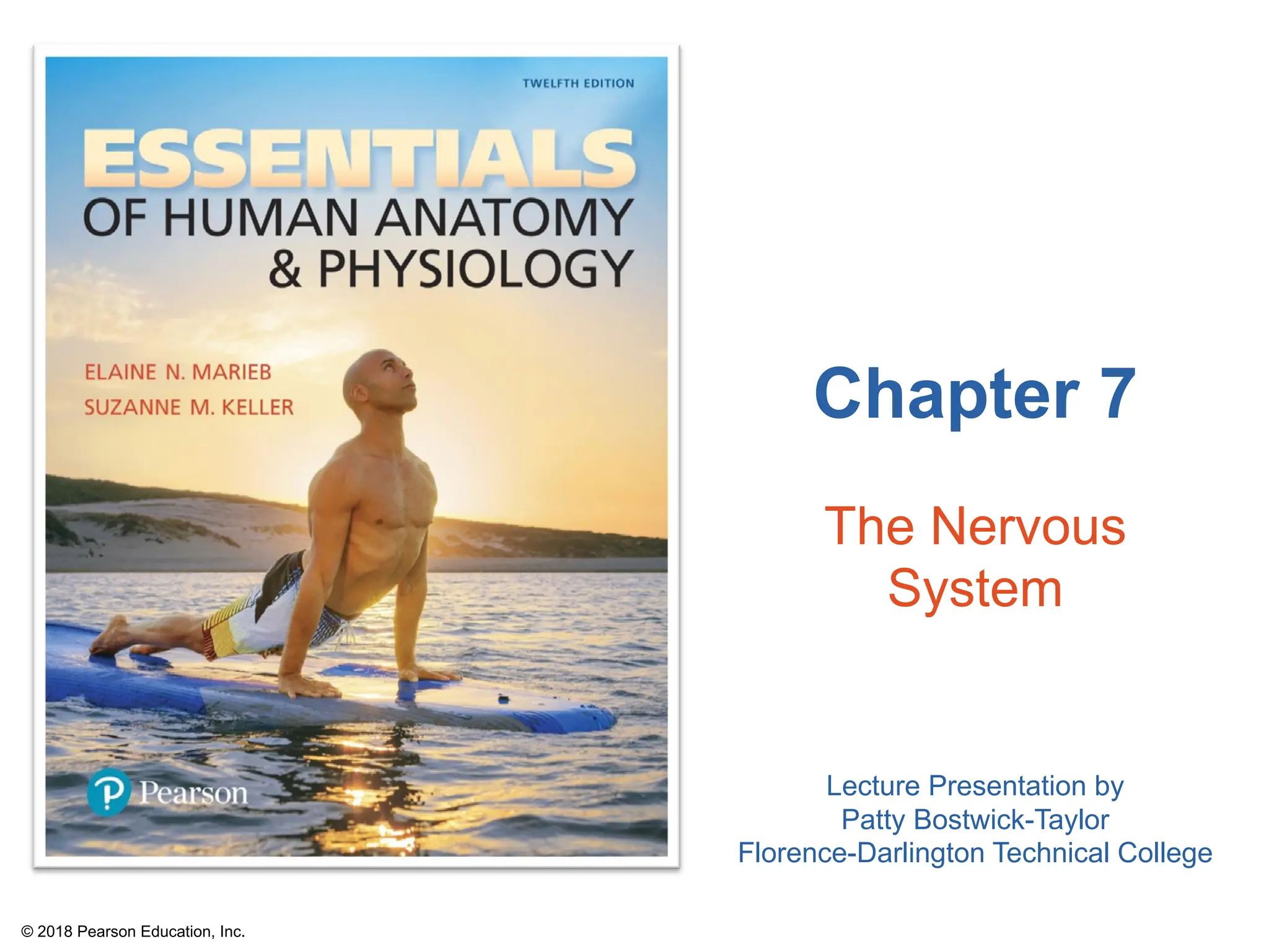
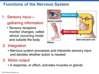
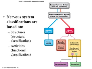
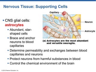
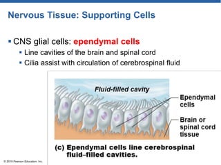
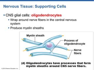
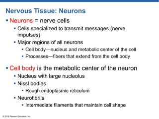
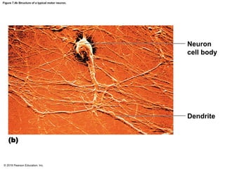
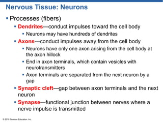
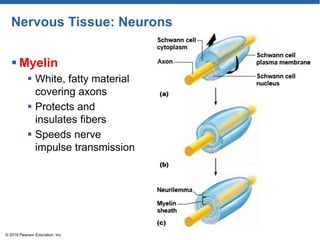
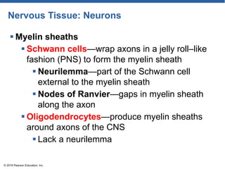
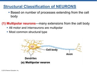
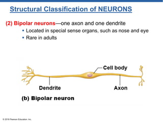
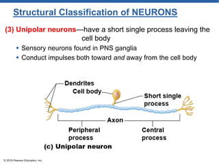
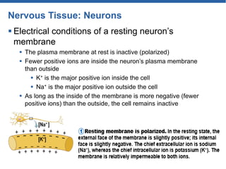
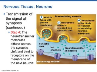
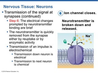
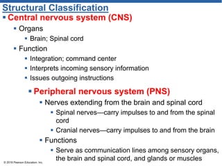
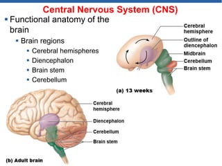
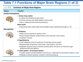
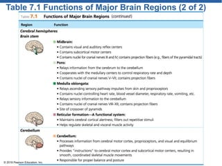
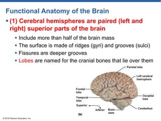
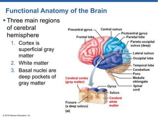
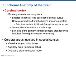
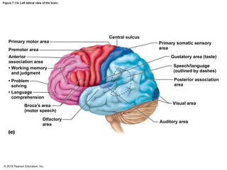
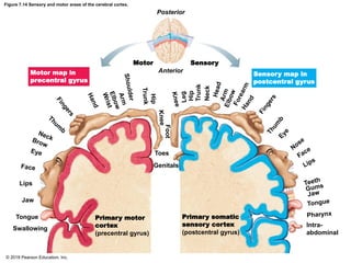
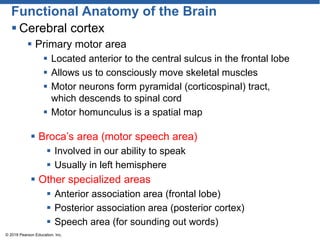
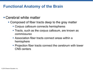
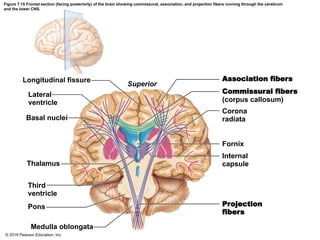
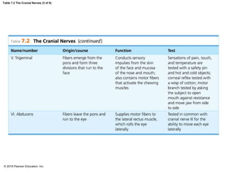
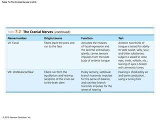
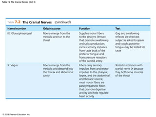
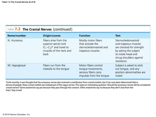
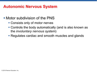
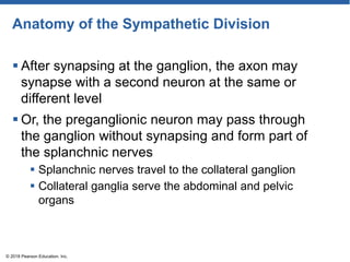
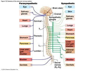
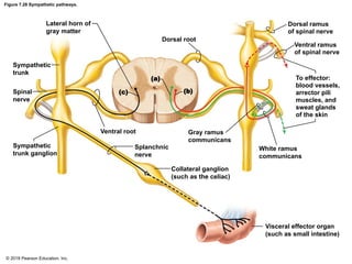
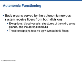
Chapter 7
The Nervous
System
Lecture Presentation by
Patty Bostwick-Taylor
Florence-Darlington Technical College
© 2018 Pearson Education, Inc.
Functions of the Nervous System
1. Sensory input—
gathering information
 Sensory receptors
monitor changes, called
stimuli, occurring inside
and outside the body
© 2018 Pearson Education, Inc.
2. Integration
 Nervous system processes and interprets sensory input
and decides whether action is needed
3. Motor output
 A response, or effect, activates muscles or glands
Figure 7.2 Organization of the nervous system.
Central Nervous System
(brain and spinal cord)
Peripheral Nervous System
(cranial and spinal nerves)
Sensory
(afferent)
Motor
(efferent)
Sense
organs
Somatic
(voluntary)
Skeletal
muscles
Autonomic
(involuntary)
Cardiac and
smooth muscle,
glands
Parasympathetic Sympathetic
© 2018 Pearson Education, Inc.
• Nervous system
classifications are
based on:
– Structures
(structural
classification)
– Activities
(functional
classification)
Nervous Tissue: Support Cells
 Support cells in the CNS are grouped together as
neuroglia
 General functions
 Support
 Insulate
 Protect neurons
© 2018 Pearson Education, Inc.
Nervous Tissue: Structure and Function
 Nervous tissue is made up of two principal cell
types
 Supporting cells (called neuroglia, or glial cells, or glia)
 Resemble neurons
 Unable to conduct nerve impulses
 Never lose the ability to divide
 Neurons
© 2018 Pearson Education, Inc.
Nervous Tissue: Supporting Cells
 CNS glial cells:
astrocytes
 Abundant, star-
shaped cells
 Brace and anchor
neurons to blood
capillaries
© 2018 Pearson Education, Inc.
 Determine permeability and exchanges between blood
capillaries and neurons
 Protect neurons from harmful substances in blood
 Control the chemical environment of the brain
Nervous Tissue: Supporting Cells
 CNS glial cells: microglia
 Spiderlike phagocytes
 Monitor health of nearby neurons
 Dispose of debris
© 2018 Pearson Education, Inc.
Nervous Tissue: Supporting Cells
 CNS glial cells: ependymal cells
 Line cavities of the brain and spinal cord
 Cilia assist with circulation of cerebrospinal fluid
© 2018 Pearson Education, Inc.
Nervous Tissue: Supporting Cells
 CNS glial cells: oligodendrocytes
 Wrap around nerve fibers in the central nervous
system
 Produce myelin sheaths
© 2018 Pearson Education, Inc.
Nervous Tissue: Supporting Cells
 PNS glial cells
 Schwann cells
 Form myelin sheath around nerve fibers in the PNS
 Satellite cells
 Protect and cushion neuron cell bodies
© 2018 Pearson Education, Inc.
Nervous
Tissue:
Neurons
© 2018 Pearson Education, Inc.
Nervous Tissue: Neurons
 Neurons = nerve cells
 Cells specialized to transmit messages (nerve
impulses)
 Major regions of all neurons
 Cell body—nucleus and metabolic center of the cell
 Processes—fibers that extend from the cell body
© 2018 Pearson Education, Inc.
 Cell body is the metabolic center of the neuron
 Nucleus with large nucleolus
 Nissl bodies
 Rough endoplasmic reticulum
 Neurofibrils
 Intermediate filaments that maintain cell shape
Figure 7.4b Structure of a typical motor neuron.
Neuron
cell body
Dendrite
(b)
© 2018 Pearson Education, Inc.
Nervous Tissue: Neurons
 Processes (fibers)
 Dendrites—conduct impulses toward the cell body
 Neurons may have hundreds of dendrites
 Axons—conduct impulses away from the cell body
 Neurons have only one axon arising from the cell body at
the axon hillock
 End in axon terminals, which contain vesicles with
neurotransmitters
 Axon terminals are separated from the next neuron by a
gap
 Synaptic cleft—gap between axon terminals and the next
neuron
 Synapse—functional junction between nerves where a
nerve impulse is transmitted
© 2018 Pearson Education, Inc.
Nervous Tissue: Neurons
 Myelin
 White, fatty material
covering axons
 Protects and
insulates fibers
 Speeds nerve
impulse transmission
© 2018 Pearson Education, Inc.
Nervous Tissue: Neurons
 Myelin sheaths
 Schwann cells—wrap axons in a jelly roll–like
fashion (PNS) to form the myelin sheath
 Neurilemma—part of the Schwann cell
external to the myelin sheath
 Nodes of Ranvier—gaps in myelin sheath
along the axon
 Oligodendrocytes—produce myelin sheaths
around axons of the CNS
 Lack a neurilemma
© 2018 Pearson Education, Inc.
Nervous Tissue: Neurons
 Terminology
 Nuclei—clusters of cell bodies in the CNS
 Ganglia—collections of cell bodies outside the
CNS in the PNS
 Tracts—bundles of nerve fibers in the CNS
 Nerves—bundles of nerve fibers in the PNS
 White matter—collections of myelinated fibers
(tracts)
 Gray matter—mostly unmyelinated fibers and
cell bodies
© 2018 Pearson Education, Inc.
Figure 7.6 Neurons classified by function.
Cell
body
Ganglion
Dendrites Peripheral
process (axon)
Afferent
transmission
Receptors Peripheral
nervous
system
Central process (axon)
Sensory
neuron Spinal cord
(central nervous system)
Interneuron
(association
neuron)
Efferent transmission
Motor neuron
To effectors
(muscles and glands)
© 2018 Pearson Education, Inc.
Functional Classification of NEURONS
 (1) Sensory (afferent) neuron
 Carry impulses from the sensory receptors to the
CNS
 Receptors include:
 Cutaneous sense organs in skin
 Proprioceptors in muscles and tendons
© 2018 Pearson Education, Inc.
 (2) Motor (efferent) neuron
 Carry impulses from the central nervous system
to viscera and/or muscles and glands
 (3) Interneurons (association neurons)
 Cell bodies located in the CNS
 Connect sensory and motor neurons
Figure 7.7a Types of sensory receptors.
(a) Free nerve endings (pain
and temperature receptors)
© 2018 Pearson Education, Inc.
Figure 7.7b Types of sensory receptors.
(b) Meissner’s corpuscle
(touch receptor)
© 2018 Pearson Education, Inc.
Figure 7.7c Types of sensory receptors.
(c) Lamellar corpuscle (deep
pressure receptor)
© 2018 Pearson Education, Inc.
Figure 7.7d Types of sensory receptors.
(d) Golgi tendon organ (proprioceptor)
© 2018 Pearson Education, Inc.
Figure 7.7e Types of sensory receptors.
(e) Muscle spindle (proprioceptor)
© 2018 Pearson Education, Inc.
Structural Classification of NEURONS
(1) Multipolar neurons—many extensions from the cell body
 All motor and interneurons are multipolar
 Most common structural type
© 2018 Pearson Education, Inc.
 Based on number of processes extending from the cell
body
(2) Bipolar neurons—one axon and one dendrite
 Located in special sense organs, such as nose and eye
 Rare in adults
© 2018 Pearson Education, Inc.
Structural Classification of NEURONS
(3) Unipolar neurons—have a short single process leaving the
cell body
 Sensory neurons found in PNS ganglia
 Conduct impulses both toward and away from the cell body
© 2018 Pearson Education, Inc.
Structural Classification of NEURONS
Functional properties of neurons
 Irritability
 Ability to respond to a stimulus and convert it to a nerve
impulse
 Conductivity
 Ability to transmit the impulse to other neurons,
muscles, or glands
© 2018 Pearson Education, Inc.
Nervous Tissue: Neurons
 Electrical conditions of a resting neuron’s
membrane
 The plasma membrane at rest is inactive (polarized)
 Fewer positive ions are inside the neuron’s plasma membrane
than outside
 K+ is the major positive ion inside the cell
 Na+ is the major positive ion outside the cell
 As long as the inside of the membrane is more negative (fewer
positive ions) than the outside, the cell remains inactive
Nervous Tissue: Neurons
 Action potential initiation and generation
 A stimulus changes the permeability of the neuron’s
membrane to sodium ions
 Sodium channels now open, and sodium (Na+) diffuses
into the neuron
 The inward rush of sodium ions changes the polarity at
that site and is called depolarization
© 2018 Pearson Education, Inc.
Nervous Tissue: Neurons
 Action potential initiation and generation
(continued)
 A graded potential (localized depolarization) exists
where the inside of the membrane is more positive and
the outside is less positive
 If the stimulus is strong enough and sodium influx
great enough, local depolarization activates the neuron
to conduct an action potential (nerve impulse)
© 2018 Pearson Education, Inc.
Nervous Tissue: Neurons
 Propagation of the action potential
 If enough sodium enters the cell, the action potential
(nerve impulse) starts and is propagated over the
entire axon
 All-or-none response means the nerve impulse either
is propagated or is not
 Fibers with myelin sheaths conduct nerve impulses
more quickly
© 2018 Pearson Education, Inc.
Nervous Tissue: Neurons
 Repolarization
 Membrane permeability changes again—becoming
impermeable to sodium ions and permeable to
potassium ions
 Potassium ions rapidly diffuse out of the neuron,
repolarizing the membrane
 Repolarization involves restoring the inside of the
membrane to a negative charge and the outer surface
to a positive charge
© 2018 Pearson Education, Inc.
Nervous Tissue: Neurons
 Repolarization (continued)
 Initial conditions of sodium and potassium ions are
restored using the sodium-potassium pump
 This pump, using ATP, restores the original
configuration
 Three sodium ions are ejected from the cell while two
potassium ions are returned to the cell
 Until repolarization is complete, a neuron cannot
conduct another nerve impulse
© 2018 Pearson Education, Inc.
Nervous Tissue: Neurons
 Transmission of the
signal at synapses
 Step 1: When the
action potential
reaches the axon
terminal, the
electrical charge
opens calcium
channels
© 2018 Pearson Education, Inc.
Nervous Tissue: Neurons
 Transmission of
the signal at
synapses
(continued)
 Step 2: Calcium,
in turn, causes
the tiny vesicles
containing the
neurotransmitter
chemical to fuse
with the axonal
membrane
© 2018 Pearson Education, Inc.
Nervous Tissue: Neurons
 Transmission of
the signal at
synapses
(continued)
 Step 3: The entry
of calcium into the
axon terminal
causes porelike
openings to form,
releasing the
neurotransmitter
into the synaptic
cleft
© 2018 Pearson Education, Inc.
Nervous Tissue: Neurons
 Transmission of
the signal at
synapses
(continued)
 Step 4: The
neurotransmitter
molecules
diffuse across
the synaptic
cleft and bind to
receptors on the
membrane of
the next neuron
© 2018 Pearson Education, Inc.
Nervous Tissue: Neurons
 Transmission of the signal
at synapses (continued)
 Step 5: If enough
neurotransmitter is
released, a graded potential
will be generated
 Eventually an action
potential (nerve impulse)
will occur in the neuron
beyond the synapse
© 2018 Pearson Education, Inc.
Nervous Tissue: Neurons
 Transmission of the signal at
synapses (continued)
 Step 6: The electrical changes
prompted by neurotransmitter
binding are brief
 The neurotransmitter is quickly
removed from the synapse
either by reuptake or by
enzymatic activity
 Transmission of an impulse is
electrochemical
 Transmission down neuron is
electrical
 Transmission to next neuron
is chemical
© 2018 Pearson Education, Inc.
BioFlix: How Synapses Work
© 2018 Pearson Education, Inc.
REFLEXES
 Reflexes are rapid, predictable, and involuntary
responses to stimuli
 Reflexes occur over neural pathways called reflex
arcs
 Two types of reflexes
 Somatic reflexes
 Autonomic reflexes
© 2018 Pearson Education, Inc.
2 TYPES of REFLEXES
 Somatic reflexes
 Reflexes that stimulate the skeletal muscles
 Involuntary, although skeletal muscle is normally under
voluntary control
 Example: pulling your hand away from a hot object
 Autonomic reflexes
 Regulate the activity of smooth muscles, the heart,
and glands
 Example: regulation of smooth muscles, heart and
blood pressure, glands, digestive system
© 2018 Pearson Education, Inc.
Five elements of a reflex arc
1. Sensory receptor—reacts to a stimulus
2. Sensory neuron—carries message to the integration center
3. Integration center (CNS)—processes information and
directs motor output
4. Motor neuron—carries message to an effector
5. Effector organ—is the muscle or gland to be stimulated
© 2018 Pearson Education, Inc.
TYPES of REFLEXES ARCS
 Two-neuron reflex arcs
 Simplest type
 Example: patellar (knee-jerk) reflex
TYPES of REFLEXES ARCS
 Three-neuron reflex arcs
 Consists of five elements: receptor, sensory neuron,
interneuron, motor neuron, and effector
 Example: flexor (withdrawal) reflex
Structural Classification
 Central nervous system (CNS)
 Organs
 Brain; Spinal cord
 Function
 Integration; command center
 Interprets incoming sensory information
 Issues outgoing instructions
© 2018 Pearson Education, Inc.
 Peripheral nervous system (PNS)
 Nerves extending from the brain and spinal cord
 Spinal nerves—carry impulses to and from the spinal
cord
 Cranial nerves—carry impulses to and from the brain
 Functions
 Serve as communication lines among sensory organs,
the brain and spinal cord, and glands or muscles
Central Nervous System (CNS)
 Functional anatomy of the
brain
 Brain regions
 Cerebral hemispheres
 Diencephalon
 Brain stem
 Cerebellum
© 2018 Pearson Education, Inc.
Table 7.1 Functions of Major Brain Regions (1 of 2)
© 2018 Pearson Education, Inc.
Table 7.1 Functions of Major Brain Regions (2 of 2)
© 2018 Pearson Education, Inc.
Functional Anatomy of the Brain
 (1) Cerebral hemispheres are paired (left and
right) superior parts of the brain
© 2018 Pearson Education, Inc.
 Include more than half of the brain mass
 The surface is made of ridges (gyri) and grooves (sulci)
 Fissures are deeper grooves
 Lobes are named for the cranial bones that lie over them
Functional Anatomy of the Brain
 Three main regions
of cerebral
hemisphere
1. Cortex is
superficial gray
matter
2. White matter
3. Basal nuclei are
deep pockets of
gray matter
© 2018 Pearson Education, Inc.
Functional Anatomy of the Brain
 Cerebral cortex
 Primary somatic sensory area
 Located in parietal lobe posterior to central sulcus
 Receives impulses from the body’s sensory receptors
 Pain, temperature, light touch (except for special senses)
 Sensory homunculus is a spatial map
 Left side of the primary somatic sensory area receives
impulses from right side (and vice versa)
© 2018 Pearson Education, Inc.
 Cerebral areas involved in special senses
 Visual area (occipital lobe)
 Auditory area (temporal lobe)
 Olfactory area (temporal lobe)
Figure 7.13c Left lateral view of the brain.
Primary motor area
Premotor area
Anterior
association area
• Working memory
and judgment
• Problem
solving
• Language
comprehension
Broca’s area
(motor speech)
Olfactory
area
(c)
Central sulcus
Primary somatic sensory
area
Gustatory area (taste)
Speech/language
(outlined by dashes)
Posterior association
area
Visual area
Auditory area
© 2018 Pearson Education, Inc.
Figure 7.14 Sensory and motor areas of the cerebral cortex.
Posterior
Motor
Anterior
Sensory
Toes
Genitals
Lips
Jaw
Tongue
Swallowing
Primary motor
cortex
(precentral gyrus)
Primary somatic
sensory cortex
(postcentral gyrus)
Intra-
abdominal
Motor map in
precentral gyrus
Sensory map in
postcentral gyrus
Foot
Hip
Trunk
Neck
© 2018 Pearson Education, Inc.
Functional Anatomy of the Brain
 Cerebral cortex
 Primary motor area
 Located anterior to the central sulcus in the frontal lobe
 Allows us to consciously move skeletal muscles
 Motor neurons form pyramidal (corticospinal) tract,
which descends to spinal cord
 Motor homunculus is a spatial map
© 2018 Pearson Education, Inc.
 Broca’s area (motor speech area)
 Involved in our ability to speak
 Usually in left hemisphere
 Other specialized areas
 Anterior association area (frontal lobe)
 Posterior association area (posterior cortex)
 Speech area (for sounding out words)
Functional Anatomy of the Brain
 Cerebral white matter
 Composed of fiber tracts deep to the gray matter
 Corpus callosum connects hemispheres
 Tracts, such as the corpus callosum, are known as
commissures
 Association fiber tracts connect areas within a
hemisphere
 Projection fiber tracts connect the cerebrum with lower
CNS centers
© 2018 Pearson Education, Inc.
Figure 7.15 Frontal section (facing posteriorly) of the brain showing commissural, association, and projection fibers running through the cerebrum
and the lower CNS.
Longitudinal fissure
Lateral
ventricle
Basal nuclei
Superior
Association fibers
Commissural fibers
(corpus callosum)
Corona
radiata
Fornix
Thalamus
Third
ventricle
Pons
Medulla oblongata
Internal
capsule
Projection
fibers
© 2018 Pearson Education, Inc.
Functional Anatomy of the Brain
 Basal nuclei
 ―Islands‖ of gray matter buried deep within the white
matter of the cerebrum
 Regulate voluntary motor activities by modifying
instructions sent to skeletal muscles by the primary
motor cortex
© 2018 Pearson Education, Inc.
Functional Anatomy of the Brain
 (2) Diencephalon
 Sits on top of the brain stem
 Enclosed by the cerebral hemispheres
 Made of three structures
1. Thalamus
2. Hypothalamus
3. Epithalamus
© 2018 Pearson Education, Inc.
Figure 7.16a Diencephalon and brain stem structures.
Cerebral hemisphere
Third ventricle
Corpus callosum
Choroid plexus of third
ventricle
Occipital lobe of
cerebral hemisphere
Anterior
commissure
Hypothalamus
Optic chiasma
Pituitary gland
Mammillary body
Pons
Medulla oblongata
(a)
Thalamus
(encloses third ventricle)
Pineal gland
(part of epithalamus)
Corpora
quadrigemina
Cerebral
aqueduct
Cerebral
peduncle
Fourth ventricle
Choroid plexus
(part of epithalamus)
Cerebellum
Midbrain
Spinal cord
© 2018 Pearson Education, Inc.
Functional Anatomy of the Brain
 Diencephalon: thalamus
 Encloses the third ventricle
 Relay station for sensory impulses passing upward to
the cerebral cortex
 Transfers impulses to the correct part of the cortex for
localization and interpretation
© 2018 Pearson Education, Inc.
Functional Anatomy of the Brain
 Diencephalon: hypothalamus
 Makes up the floor of the diencephalon
 Important autonomic nervous system center
 Regulates body temperature
 Regulates water balance
 Regulates metabolism
 Houses the limbic center for emotions
 Regulates the nearby pituitary gland
 Houses mammillary bodies for olfaction (smell)
© 2018 Pearson Education, Inc.
Functional Anatomy of the Brain
 Diencephalon: epithalamus
 Forms the roof of the third ventricle
 Houses the pineal body (an endocrine gland)
 Includes the choroid plexus—forms cerebrospinal fluid
© 2018 Pearson Education, Inc.
Functional Anatomy of the Brain
 (3) Brain stem
 Attaches to the spinal cord
 Parts of the brain stem
© 2018 Pearson Education, Inc.
1. Midbrain
2. Pons
3. Medulla
oblongata
Functional Anatomy of the Brain
 Brain stem: midbrain
 Extends from the mammillary bodies to the pons
inferiorly
 Cerebral aqueduct (tiny canal) connects the third and
fourth ventricles
 Two bulging fiber tracts, cerebral peduncles, convey
ascending and descending impulses
 Four rounded protrusions, corpora quadrigemina, are
visual and auditory reflex centers
© 2018 Pearson Education, Inc.
Functional Anatomy of the Brain
 Brain stem: pons
 The rounded structure protruding just below the
midbrain
 Mostly composed of fiber tracts
 Includes nuclei involved in the control of breathing
© 2018 Pearson Education, Inc.
Functional Anatomy of the Brain
 Brain stem: medulla oblongata
 The most inferior part of the brain stem that merges
into the spinal cord
 Includes important fiber tracts
 Contains important centers that control:
 Heart rate
 Blood pressure
 Breathing
 Swallowing
 Vomiting
 Fourth ventricle lies posterior to pons and medulla
© 2018 Pearson Education, Inc.
Functional Anatomy of the Brain
 Brain stem: reticular formation
 Diffuse mass of gray matter along the brain stem
 Involved in motor control of visceral organs
 Reticular activating system (RAS)
 Plays a role in
awake/sleep
cycles and
consciousness
 Filter for incoming
sensory information
© 2018 Pearson Education, Inc.
Functional Anatomy of the Brain
 (4) Cerebrum
 Two hemispheres with convoluted surfaces
 Outer cortex of gray matter and inner region of white
matter
 Controls balance
 Provides precise
timing for skeletal
muscle activity and
coordination of body
movements
© 2018 Pearson Education, Inc.
Protection of the Central Nervous System
 Meninges
 Cerebrospinal fluid (CSF)
 Blood-brain barrier
Figure 7.17b Meninges of the brain.
Skull
Scalp
Superior
sagittal sinus
Dura mater
Transverse
sinus
Temporal
bone
Occipital lobe
Tentorium
cerebelli
Cerebellum
Arachnoid mater
over medulla oblongata
(b)
© 2018 Pearson Education, Inc.
Protection of the Central Nervous System
 Meninges
 Dura mater
 Outermost leathery layer
 Double-layered external covering
 Periosteum—attached to inner surface of the skull
 Meningeal layer—outer covering of the brain
 Folds inward in several areas
 Falx cerebri
 Tentorium cerebelli
 Arachnoid layer
 Middle layer
 Subarachnoid space is filled with cerebrospinal fluid
 Arachnoid granulations protrude through the dura mater and
absorb cerebrospinal fluid into venous blood
 Pia mater
 Internal layer
 Clings to the surface of the brain and spinal cord
Protection of the Central Nervous System
 Cerebrospinal fluid
 Similar to blood plasma in composition
 Formed continually by the choroid plexuses
 Choroid plexuses—capillaries in the ventricles of the
brain
 CSF forms a watery cushion to protect the brain and
spinal cord
 Circulated in the arachnoid space, ventricles, and
central canal of the spinal cord
© 2018 Pearson Education, Inc.
Figure 7.18c Ventricles and location of the cerebrospinal fluid.
Superior
sagittal sinus
Choroid plexuses
of lateral and
third ventricles
Corpus callosum
Interventricular
foramen
Third ventricle
Cerebral aqueduct
Lateral aperture
Fourth ventricle
Median aperture
Choroid plexus
of fourth ventricle
(c) CSF circulation
Arachnoid granulation
Subarachnoid space
Arachnoid mater
Meningeal dura mater
Periosteal dura mater
Right lateral ventricle
(deep to cut)
Central canal
of spinal cord
CSF is produced by the
choroid plexus of each
ventricle.
CSF flows through the ventricles
and into the subarachnoid space via
the median and lateral apertures.
Some CSF flows through the central
canal of the spinal cord.
CSF flows through the
subarachnoid space.
CSF is absorbed into the dural
venous sinuses via the arachnoid
granulations.
2
1
3
4
2
1
3
4
© 2018 Pearson Education, Inc.
Protection of the Central Nervous System
 Blood-brain barrier
 Includes the least permeable capillaries of the body
 Allows water, glucose, and amino acids to pass
through the capillary walls
 Excludes many potentially harmful substances from
entering the brain, such as wastes
 Useless as a barrier against some substances
© 2018 Pearson Education, Inc.
Brain Dysfunctions
 Traumatic brain injuries
 Concussion
 Slight brain injury
 Typically little permanent brain damage occurs
 Contusion
 Marked nervous tissue destruction occurs
 Coma may occur
 Death may occur after head blows due to:
 Intracranial hemorrhage
 Cerebral edema
© 2018 Pearson Education, Inc.
Brain Dysfunctions
 Cerebrovascular accident (CVA), or stroke
 Results when blood circulation to a brain area is
blocked and brain tissue dies
 Loss of some functions or death may result
 Hemiplegia—one-sided paralysis
 Aphasia—damage to speech center in left hemisphere
 Transient ischemic attack (TIA)
 Temporary brain ischemia (restriction of blood flow)
 Numbness, temporary paralysis, impaired speech
© 2018 Pearson Education, Inc.
Spinal Cord
 Extends from the foramen magnum of the skull to
the first or second lumbar vertebra
 Cauda equina is a collection of spinal nerves at
the inferior end
 Provides a two-way conduction pathway to and
from the brain
 31 pairs of spinal nerves arise from the spinal
cord
© 2018 Pearson Education, Inc.
Figure 7.19 Anatomy of the spinal cord, posterior view.
Cervical
enlargement
Cervical
spinal nerves
C8
Dura and
arachnoid
mater
Lumbar
enlargement
Thoracic
spinal nerves
T12
End of spinal cord
Cauda
equina
End of
meningeal
coverings
Lumbar
spinal nerves
L5
S1 Sacral
spinal nerves
S5
© 2018 Pearson Education, Inc.
Figure 7.20 Spinal cord with meninges (three-dimensional, anterior view).
Dorsal root
ganglion
Central canal
White matter Dorsal (posterior)
horn of gray matter
Lateral horn of
gray matter
Spinal nerve
Dorsal root of
spinal nerve
Ventral root
of spinal nerve
Ventral (anterior)
horn of gray matter
Pia mater
Arachnoid mater
Dura mater
© 2018 Pearson Education, Inc.
Figure 7.21 Schematic of ascending (sensory) and descending (motor) pathways between the brain and the spinal cord.
Interneuron carrying sensory
information to cerebral cortex
Integration (processing and
interpretation of sensory input)
occurs
Interneuron carrying
response to
motor neurons
Cerebrum
Cerebral cortex
(gray matter)
White matter
Thalamus
Interneuron
carrying response
to motor neuron
Cell body of sensory
neuron in sensory
ganglion
Nerve
Skin
Sensory
receptors
Muscle
Motor output
Motor neuron
cell body
Brain stem
Interneuron carrying
sensory information to
cerebral cortex
Cervical spinal cord
White matter
Gray matter
Interneuron
© 2018 Pearson Education, Inc.
Peripheral Nervous System (PNS)
 PNS consists of nerves and ganglia outside the
CNS
© 2018 Pearson Education, Inc.
Structure of a Nerve
 Nerves are bundles of neurons found outside the
CNS
 Endoneurium is a connective tissue sheath that
surrounds each fiber
 Perineurium wraps groups of fibers bound into a
fascicle
 Epineurium binds groups of fascicles
Figure 7.22 Structure of a nerve.
Axon
Myelin sheath
Endoneurium
Perineurium
Epineurium
Fascicle
Blood
vessels
© 2018 Pearson Education, Inc.
Structure of a Nerve
 Mixed nerves
 Contain both sensory and motor fibers
 Sensory (afferent) nerves
 Carry impulses toward the CNS
 Motor (efferent) nerves
 Carry impulses away from the CNS
© 2018 Pearson Education, Inc.
Cranial Nerves
 12 pairs of nerves serve mostly the head and
neck
 Only the pair of vagus nerves extends to thoracic
and abdominal cavities
 Most are mixed nerves, but three are sensory
only
1. Optic
2. Olfactory
3. Vestibulocochlear
© 2018 Pearson Education, Inc.
Cranial Nerves Mnemonic Device
 Oh – Olfactory
 Oh – Optic
 Oh – Oculomotor
 To – Trochlear
 Touch – Trigeminal
 And – Abducens
 Feel – Facial
 Very – Vestibulocochlear
 Green – Glossopharyngeal
 Vegetables – Vagus
 A – Accessory
 H – Hypoglossal
© 2018 Pearson Education, Inc.
Figure 7.23 Distribution of cranial nerves.
III Oculomotor
IV Trochlear
VI Abducens
I Olfactory II Optic
V Trigeminal V Trigeminal
VII Facial
Vestibular
branch
Cochlear
branch
VIII Vestibulocochlear
X Vagus
IX Glossopharyngeal
XII Hypoglossal XI Accessory
© 2018 Pearson Education, Inc.
Table 7.2 The Cranial Nerves (1 of 6)
© 2018 Pearson Education, Inc.
Table 7.2 The Cranial Nerves (2 of 6)
© 2018 Pearson Education, Inc.
Table 7.2 The Cranial Nerves (3 of 6)
© 2018 Pearson Education, Inc.
Table 7.2 The Cranial Nerves (4 of 6)
© 2018 Pearson Education, Inc.
Table 7.2 The Cranial Nerves (5 of 6)
© 2018 Pearson Education, Inc.
Table 7.2 The Cranial Nerves (6 of 6)
© 2018 Pearson Education, Inc.
Spinal Nerves
 Spinal nerves
 31 pairs
 Formed by the combination of the ventral and dorsal
roots of the spinal cord
 Named for the region of the spinal cord from which
they arise
© 2018 Pearson Education, Inc.
Figure 7.24a Spinal nerves.
C1
2
3
4
5
6
7
8*
T1
2
3
4
5
6
7
8
9
10
11
12
L1
2
3
4
5
S1
2
3
4
Cervical
nerves
Thoracic
nerves
Lumbar
nerves
Sacral
nerves
Ventral rami form
cervical plexus
(C1 – C5)
Ventral rami form
brachial plexus
(C5 – C8; T1)
No plexus
formed
(intercostal
nerves)
(T2 – T12)
Ventral rami form
lumbar plexus
(L1 – L4)
Ventral rami form
sacral plexus
(L4 – L5; S1 – S4)
(a)
*Note that the cervical nerve C8 emerges inferior to the C7 vertebra, while the other seven cervical nerves
emerge superior to the vertebrae for which they are named.
© 2018 Pearson Education, Inc.
Spinal Nerves
 Spinal nerves divide soon after leaving the spinal
cord into a dorsal ramus and a ventral ramus
 Ramus—branch of a spinal nerve; contains both motor
and sensory fibers
 Dorsal rami—serve the skin and muscles of the
posterior trunk
 Ventral rami (T1–T12) —form the intercostal nerves that
supply muscles and skin of the ribs and trunk
 Ventral rami (except T1–T12)—form a complex of
networks (plexus) for the anterior
© 2018 Pearson Education, Inc.
Figure 7.24b Spinal nerves.
Dorsal root
Dorsal root
ganglion
Spinal
cord
Ventral
root
Spinal nerve
Dorsal
ramus
Ventral
ramus
(b)
© 2018 Pearson Education, Inc.
Spinal Nerves
 Plexus—networks of nerves serving motor and
sensory needs of the limbs
 Form from ventral rami of spinal nerves in the
cervical, lumbar, and sacral regions
 Four plexuses
1. Cervical
2. Brachial
3. Lumbar
4. Sacral
© 2018 Pearson Education, Inc.
Table 7.3 Spinal Nerve Plexuses (1 of 3)
© 2018 Pearson Education, Inc.
Figure 7.25a Distribution of the major peripheral nerves of the upper and lower limbs.
Axillary nerve
Humerus
Radial
nerve
Musculo-
cutaneous
nerve
Ulna
Radius
Ulnar nerve
Median
nerve
(a) Brachial plexus,
anterior view
© 2018 Pearson Education, Inc.
Table 7.3 Spinal Nerve Plexuses (2 of 3)
© 2018 Pearson Education, Inc.
Figure 7.25b Distribution of the major peripheral nerves of the upper and lower limbs.
Femoral nerve
Lateral femoral
cutaneous nerve
Obturator nerve
Femur
Anterior femoral
cutaneous nerve
Saphenous nerve
(b) Lumbar plexus,
anterior view
© 2018 Pearson Education, Inc.
Table 7.3 Spinal Nerve Plexuses (3 of 3)
© 2018 Pearson Education, Inc.
Figure 7.25c Distribution of the major peripheral nerves of the upper and lower limbs.
Superior gluteal
nerve
Inferior gluteal
nerve
Sciatic nerve
Posterior femoral
cutaneous nerve
Common fibular
nerve
Tibial nerve
Sural (cut) nerve
Deep fibular
nerve
Superficial fibular
nerve
Plantar branches
(c) Sacral plexus, posterior view
© 2018 Pearson Education, Inc.
Autonomic Nervous System
 Motor subdivision of the PNS
 Consists only of motor nerves
 Controls the body automatically (and is also known as
the involuntary nervous system)
 Regulates cardiac and smooth muscles and glands
© 2018 Pearson Education, Inc.
Somatic and Autonomic Nervous Systems
Compared
 Somatic nervous system
 Motor neuron cell bodies originate inside the CNS
 Axons extends to skeletal muscles that are served
 Autonomic nervous system
 Chain of two motor neurons
 Preganglionic neuron is in the brain or spinal cord
 Postganglionic neuron extends to the organ
 Has two arms
 Sympathetic division
 Parasympathetic division
© 2018 Pearson Education, Inc.
Figure 7.26 Comparison of the somatic and autonomic nervous systems.
Central
nervous system Peripheral nervous system
Acetylcholine
Effector organs
Somatic nervous system Skeletal muscle
Acetylcholine Norepinephrine Smooth muscle
(e.g., in stomach)
Sympathetic
division
Autonomic
nervous
system
Ganglion
Acetylcholine Epinephrine and
norepinephrine
Blood
vessel
Glands
Adrenal medulla
Acetylcholine
Parasympathetic
division
Cardiac
muscle
Ganglion
KEY:
Preganglionic
axons
(sympathetic)
Postganglionic
axons
(sympathetic)
Myelination Preganglionic
axons
(parasympathetic)
Postganglionic
axons
(parasympathetic)
© 2018 Pearson Education, Inc.
Anatomy of the Parasympathetic Division
 Parasympathetic division is also known as the
craniosacral division
 Preganglionic neurons originate in:
 Cranial nerves III, VII, IX, and X
 S2 through S4 regions of the spinal cord
 Preganglionic neurons synapse with terminal
ganglia; from there, postganglionic axons extend
to organs that are served
© 2018 Pearson Education, Inc.
Anatomy of the Sympathetic Division
 Sympathetic division is also known as the
thoracolumbar division
 Preganglionic neurons originate from T1 through
L2
 Axons pass through a ramus communicans to enter a
sympathetic trunk ganglion
 Sympathetic trunk, or chain, lies near the spinal cord
© 2018 Pearson Education, Inc.
Anatomy of the Sympathetic Division
 After synapsing at the ganglion, the axon may
synapse with a second neuron at the same or
different level
 Or, the preganglionic neuron may pass through
the ganglion without synapsing and form part of
the splanchnic nerves
 Splanchnic nerves travel to the collateral ganglion
 Collateral ganglia serve the abdominal and pelvic
organs
© 2018 Pearson Education, Inc.
Figure 7.27 Anatomy of the autonomic nervous system.
Parasympathetic
Eye
Salivary
glands
Heart
Lungs
T1
Thoracic
Sympathetic
Brain stem
Cranial
Eye
Skin
Sympathetic
ganglia
Salivary
glands
Lungs
Heart
Stomach
Pancreas
Liver
and gall-
bladder
Adrenal
gland
Cervical
Stomach
Pancreas
Liver and
gall-
bladder
Bladder
Genitals
L1
Lumbar
Bladder
Sacral
nerves
(S2–S4)
Genitals
© 2018 Pearson Education, Inc.
Figure 7.28 Sympathetic pathways.
Lateral horn of
gray matter
Dorsal root
Dorsal ramus
of spinal nerve
Ventral ramus
of spinal nerve
Sympathetic
trunk
Spinal
nerve
(c)
(a)
(b)
To effector:
blood vessels,
arrector pili
muscles, and
sweat glands
of the skin
Ventral root
Sympathetic
trunk ganglion
Splanchnic
nerve
Gray ramus
communicans
White ramus
communicans
Collateral ganglion
(such as the celiac)
Visceral effector organ
(such as small intestine)
© 2018 Pearson Education, Inc.
Autonomic Functioning
 Body organs served by the autonomic nervous
system receive fibers from both divisions
 Exceptions: blood vessels, structures of the skin, some
glands, and the adrenal medulla
 These exceptions receive only sympathetic fibers
© 2018 Pearson Education, Inc.
Autonomic Functioning
 When body divisions serve the same organ, they
cause antagonistic effects due to different
neurotransmitters
 Parasympathetic (cholinergic) fibers release
acetylcholine
 Sympathetic postganglionic (adrenergic) fibers release
norepinephrine
 Preganglionic axons of both divisions release
acetycholine
© 2018 Pearson Education, Inc.
Autonomic Functioning
 Sympathetic—―fight or flight‖ division
 Response to unusual stimulus when emotionally or
physically stressed or threatened
 Takes over to increase activities
 Remember as the ―E‖ division
 Exercise
 Excitement
 Emergency
 Embarrassment
© 2018 Pearson Education, Inc.
Autonomic Functioning
 Parasympathetic—―housekeeping‖ activites
 ―Rest-and-digest‖ system
 Conserves energy
 Maintains daily necessary body functions
 Remember as the ―D‖ division
 Digestion
 Defecation
 Diuresis
© 2018 Pearson Education, Inc.
Table 7.4 Effects of the Sympathetic and Parasympathetic Divisions of the Autonomic Nervous System (1 of 2)
© 2018 Pearson Education, Inc.
Table 7.4 Effects of the Sympathetic and Parasympathetic Divisions of the Autonomic Nervous System (2 of 2)
© 2018 Pearson Education, Inc.
Developmental Aspects of the Nervous
System
 The nervous system is formed during the first
month of embryonic development
 Any maternal infection can have extremely
harmful effects
 Oxygen deprivation destroys brain cells
 The hypothalamus is one of the last areas of the
brain to develop
© 2018 Pearson Education, Inc.
Developmental Aspects of the Nervous
System
 Severe congenital brain diseases include:
 Cerebral palsy
 Anencephaly
 Hydrocephalus
 Spina bifida
© 2018 Pearson Education, Inc.
Developmental Aspects of the Nervous
System
 Premature babies have trouble regulating body
temperature because the hypothalamus is one of
the last brain areas to mature prenatally
 Development of motor control indicates the
progressive myelination and maturation of a
child’s nervous system
© 2018 Pearson Education, Inc.
Developmental Aspects of the Nervous
System
 Brain growth ends in young adulthood. Neurons die
throughout life and are not replaced; thus, brain
mass declines with age
 Orthostatic hypotension is low blood pressure due
to changes in body position
 Healthy aged people maintain nearly optimal
intellectual function
 Disease—particularly cardiovascular disease—is
the major cause of declining mental function with
age
 Arteriosclerosis is decreased elasticity of blood vessels
© 2018 Pearson Education, Inc.

CHAPTER 7 Nervous System anaphysiology.pdf

  • 1.
    Chapter 7 The Nervous System LecturePresentation by Patty Bostwick-Taylor Florence-Darlington Technical College © 2018 Pearson Education, Inc.
  • 2.
    Functions of theNervous System 1. Sensory input— gathering information  Sensory receptors monitor changes, called stimuli, occurring inside and outside the body © 2018 Pearson Education, Inc. 2. Integration  Nervous system processes and interprets sensory input and decides whether action is needed 3. Motor output  A response, or effect, activates muscles or glands
  • 3.
    Figure 7.2 Organizationof the nervous system. Central Nervous System (brain and spinal cord) Peripheral Nervous System (cranial and spinal nerves) Sensory (afferent) Motor (efferent) Sense organs Somatic (voluntary) Skeletal muscles Autonomic (involuntary) Cardiac and smooth muscle, glands Parasympathetic Sympathetic © 2018 Pearson Education, Inc. • Nervous system classifications are based on: – Structures (structural classification) – Activities (functional classification)
  • 4.
    Nervous Tissue: SupportCells  Support cells in the CNS are grouped together as neuroglia  General functions  Support  Insulate  Protect neurons © 2018 Pearson Education, Inc.
  • 5.
    Nervous Tissue: Structureand Function  Nervous tissue is made up of two principal cell types  Supporting cells (called neuroglia, or glial cells, or glia)  Resemble neurons  Unable to conduct nerve impulses  Never lose the ability to divide  Neurons © 2018 Pearson Education, Inc.
  • 6.
    Nervous Tissue: SupportingCells  CNS glial cells: astrocytes  Abundant, star- shaped cells  Brace and anchor neurons to blood capillaries © 2018 Pearson Education, Inc.  Determine permeability and exchanges between blood capillaries and neurons  Protect neurons from harmful substances in blood  Control the chemical environment of the brain
  • 7.
    Nervous Tissue: SupportingCells  CNS glial cells: microglia  Spiderlike phagocytes  Monitor health of nearby neurons  Dispose of debris © 2018 Pearson Education, Inc.
  • 8.
    Nervous Tissue: SupportingCells  CNS glial cells: ependymal cells  Line cavities of the brain and spinal cord  Cilia assist with circulation of cerebrospinal fluid © 2018 Pearson Education, Inc.
  • 9.
    Nervous Tissue: SupportingCells  CNS glial cells: oligodendrocytes  Wrap around nerve fibers in the central nervous system  Produce myelin sheaths © 2018 Pearson Education, Inc.
  • 10.
    Nervous Tissue: SupportingCells  PNS glial cells  Schwann cells  Form myelin sheath around nerve fibers in the PNS  Satellite cells  Protect and cushion neuron cell bodies © 2018 Pearson Education, Inc.
  • 11.
  • 12.
    Nervous Tissue: Neurons Neurons = nerve cells  Cells specialized to transmit messages (nerve impulses)  Major regions of all neurons  Cell body—nucleus and metabolic center of the cell  Processes—fibers that extend from the cell body © 2018 Pearson Education, Inc.  Cell body is the metabolic center of the neuron  Nucleus with large nucleolus  Nissl bodies  Rough endoplasmic reticulum  Neurofibrils  Intermediate filaments that maintain cell shape
  • 13.
    Figure 7.4b Structureof a typical motor neuron. Neuron cell body Dendrite (b) © 2018 Pearson Education, Inc.
  • 14.
    Nervous Tissue: Neurons Processes (fibers)  Dendrites—conduct impulses toward the cell body  Neurons may have hundreds of dendrites  Axons—conduct impulses away from the cell body  Neurons have only one axon arising from the cell body at the axon hillock  End in axon terminals, which contain vesicles with neurotransmitters  Axon terminals are separated from the next neuron by a gap  Synaptic cleft—gap between axon terminals and the next neuron  Synapse—functional junction between nerves where a nerve impulse is transmitted © 2018 Pearson Education, Inc.
  • 15.
    Nervous Tissue: Neurons Myelin  White, fatty material covering axons  Protects and insulates fibers  Speeds nerve impulse transmission © 2018 Pearson Education, Inc.
  • 16.
    Nervous Tissue: Neurons Myelin sheaths  Schwann cells—wrap axons in a jelly roll–like fashion (PNS) to form the myelin sheath  Neurilemma—part of the Schwann cell external to the myelin sheath  Nodes of Ranvier—gaps in myelin sheath along the axon  Oligodendrocytes—produce myelin sheaths around axons of the CNS  Lack a neurilemma © 2018 Pearson Education, Inc.
  • 17.
    Nervous Tissue: Neurons Terminology  Nuclei—clusters of cell bodies in the CNS  Ganglia—collections of cell bodies outside the CNS in the PNS  Tracts—bundles of nerve fibers in the CNS  Nerves—bundles of nerve fibers in the PNS  White matter—collections of myelinated fibers (tracts)  Gray matter—mostly unmyelinated fibers and cell bodies © 2018 Pearson Education, Inc.
  • 18.
    Figure 7.6 Neuronsclassified by function. Cell body Ganglion Dendrites Peripheral process (axon) Afferent transmission Receptors Peripheral nervous system Central process (axon) Sensory neuron Spinal cord (central nervous system) Interneuron (association neuron) Efferent transmission Motor neuron To effectors (muscles and glands) © 2018 Pearson Education, Inc.
  • 19.
    Functional Classification ofNEURONS  (1) Sensory (afferent) neuron  Carry impulses from the sensory receptors to the CNS  Receptors include:  Cutaneous sense organs in skin  Proprioceptors in muscles and tendons © 2018 Pearson Education, Inc.  (2) Motor (efferent) neuron  Carry impulses from the central nervous system to viscera and/or muscles and glands  (3) Interneurons (association neurons)  Cell bodies located in the CNS  Connect sensory and motor neurons
  • 20.
    Figure 7.7a Typesof sensory receptors. (a) Free nerve endings (pain and temperature receptors) © 2018 Pearson Education, Inc.
  • 21.
    Figure 7.7b Typesof sensory receptors. (b) Meissner’s corpuscle (touch receptor) © 2018 Pearson Education, Inc.
  • 22.
    Figure 7.7c Typesof sensory receptors. (c) Lamellar corpuscle (deep pressure receptor) © 2018 Pearson Education, Inc.
  • 23.
    Figure 7.7d Typesof sensory receptors. (d) Golgi tendon organ (proprioceptor) © 2018 Pearson Education, Inc.
  • 24.
    Figure 7.7e Typesof sensory receptors. (e) Muscle spindle (proprioceptor) © 2018 Pearson Education, Inc.
  • 25.
    Structural Classification ofNEURONS (1) Multipolar neurons—many extensions from the cell body  All motor and interneurons are multipolar  Most common structural type © 2018 Pearson Education, Inc.  Based on number of processes extending from the cell body
  • 26.
    (2) Bipolar neurons—oneaxon and one dendrite  Located in special sense organs, such as nose and eye  Rare in adults © 2018 Pearson Education, Inc. Structural Classification of NEURONS
  • 27.
    (3) Unipolar neurons—havea short single process leaving the cell body  Sensory neurons found in PNS ganglia  Conduct impulses both toward and away from the cell body © 2018 Pearson Education, Inc. Structural Classification of NEURONS
  • 28.
    Functional properties ofneurons  Irritability  Ability to respond to a stimulus and convert it to a nerve impulse  Conductivity  Ability to transmit the impulse to other neurons, muscles, or glands © 2018 Pearson Education, Inc.
  • 29.
    Nervous Tissue: Neurons Electrical conditions of a resting neuron’s membrane  The plasma membrane at rest is inactive (polarized)  Fewer positive ions are inside the neuron’s plasma membrane than outside  K+ is the major positive ion inside the cell  Na+ is the major positive ion outside the cell  As long as the inside of the membrane is more negative (fewer positive ions) than the outside, the cell remains inactive
  • 30.
    Nervous Tissue: Neurons Action potential initiation and generation  A stimulus changes the permeability of the neuron’s membrane to sodium ions  Sodium channels now open, and sodium (Na+) diffuses into the neuron  The inward rush of sodium ions changes the polarity at that site and is called depolarization © 2018 Pearson Education, Inc.
  • 31.
    Nervous Tissue: Neurons Action potential initiation and generation (continued)  A graded potential (localized depolarization) exists where the inside of the membrane is more positive and the outside is less positive  If the stimulus is strong enough and sodium influx great enough, local depolarization activates the neuron to conduct an action potential (nerve impulse) © 2018 Pearson Education, Inc.
  • 32.
    Nervous Tissue: Neurons Propagation of the action potential  If enough sodium enters the cell, the action potential (nerve impulse) starts and is propagated over the entire axon  All-or-none response means the nerve impulse either is propagated or is not  Fibers with myelin sheaths conduct nerve impulses more quickly © 2018 Pearson Education, Inc.
  • 33.
    Nervous Tissue: Neurons Repolarization  Membrane permeability changes again—becoming impermeable to sodium ions and permeable to potassium ions  Potassium ions rapidly diffuse out of the neuron, repolarizing the membrane  Repolarization involves restoring the inside of the membrane to a negative charge and the outer surface to a positive charge © 2018 Pearson Education, Inc.
  • 34.
    Nervous Tissue: Neurons Repolarization (continued)  Initial conditions of sodium and potassium ions are restored using the sodium-potassium pump  This pump, using ATP, restores the original configuration  Three sodium ions are ejected from the cell while two potassium ions are returned to the cell  Until repolarization is complete, a neuron cannot conduct another nerve impulse © 2018 Pearson Education, Inc.
  • 35.
    Nervous Tissue: Neurons Transmission of the signal at synapses  Step 1: When the action potential reaches the axon terminal, the electrical charge opens calcium channels © 2018 Pearson Education, Inc.
  • 36.
    Nervous Tissue: Neurons Transmission of the signal at synapses (continued)  Step 2: Calcium, in turn, causes the tiny vesicles containing the neurotransmitter chemical to fuse with the axonal membrane © 2018 Pearson Education, Inc.
  • 37.
    Nervous Tissue: Neurons Transmission of the signal at synapses (continued)  Step 3: The entry of calcium into the axon terminal causes porelike openings to form, releasing the neurotransmitter into the synaptic cleft © 2018 Pearson Education, Inc.
  • 38.
    Nervous Tissue: Neurons Transmission of the signal at synapses (continued)  Step 4: The neurotransmitter molecules diffuse across the synaptic cleft and bind to receptors on the membrane of the next neuron © 2018 Pearson Education, Inc.
  • 39.
    Nervous Tissue: Neurons Transmission of the signal at synapses (continued)  Step 5: If enough neurotransmitter is released, a graded potential will be generated  Eventually an action potential (nerve impulse) will occur in the neuron beyond the synapse © 2018 Pearson Education, Inc.
  • 40.
    Nervous Tissue: Neurons Transmission of the signal at synapses (continued)  Step 6: The electrical changes prompted by neurotransmitter binding are brief  The neurotransmitter is quickly removed from the synapse either by reuptake or by enzymatic activity  Transmission of an impulse is electrochemical  Transmission down neuron is electrical  Transmission to next neuron is chemical © 2018 Pearson Education, Inc.
  • 41.
    BioFlix: How SynapsesWork © 2018 Pearson Education, Inc.
  • 42.
    REFLEXES  Reflexes arerapid, predictable, and involuntary responses to stimuli  Reflexes occur over neural pathways called reflex arcs  Two types of reflexes  Somatic reflexes  Autonomic reflexes © 2018 Pearson Education, Inc.
  • 43.
    2 TYPES ofREFLEXES  Somatic reflexes  Reflexes that stimulate the skeletal muscles  Involuntary, although skeletal muscle is normally under voluntary control  Example: pulling your hand away from a hot object  Autonomic reflexes  Regulate the activity of smooth muscles, the heart, and glands  Example: regulation of smooth muscles, heart and blood pressure, glands, digestive system © 2018 Pearson Education, Inc.
  • 44.
    Five elements ofa reflex arc 1. Sensory receptor—reacts to a stimulus 2. Sensory neuron—carries message to the integration center 3. Integration center (CNS)—processes information and directs motor output 4. Motor neuron—carries message to an effector 5. Effector organ—is the muscle or gland to be stimulated © 2018 Pearson Education, Inc.
  • 45.
    TYPES of REFLEXESARCS  Two-neuron reflex arcs  Simplest type  Example: patellar (knee-jerk) reflex
  • 46.
    TYPES of REFLEXESARCS  Three-neuron reflex arcs  Consists of five elements: receptor, sensory neuron, interneuron, motor neuron, and effector  Example: flexor (withdrawal) reflex
  • 47.
    Structural Classification  Centralnervous system (CNS)  Organs  Brain; Spinal cord  Function  Integration; command center  Interprets incoming sensory information  Issues outgoing instructions © 2018 Pearson Education, Inc.  Peripheral nervous system (PNS)  Nerves extending from the brain and spinal cord  Spinal nerves—carry impulses to and from the spinal cord  Cranial nerves—carry impulses to and from the brain  Functions  Serve as communication lines among sensory organs, the brain and spinal cord, and glands or muscles
  • 48.
    Central Nervous System(CNS)  Functional anatomy of the brain  Brain regions  Cerebral hemispheres  Diencephalon  Brain stem  Cerebellum © 2018 Pearson Education, Inc.
  • 49.
    Table 7.1 Functionsof Major Brain Regions (1 of 2) © 2018 Pearson Education, Inc.
  • 50.
    Table 7.1 Functionsof Major Brain Regions (2 of 2) © 2018 Pearson Education, Inc.
  • 51.
    Functional Anatomy ofthe Brain  (1) Cerebral hemispheres are paired (left and right) superior parts of the brain © 2018 Pearson Education, Inc.  Include more than half of the brain mass  The surface is made of ridges (gyri) and grooves (sulci)  Fissures are deeper grooves  Lobes are named for the cranial bones that lie over them
  • 52.
    Functional Anatomy ofthe Brain  Three main regions of cerebral hemisphere 1. Cortex is superficial gray matter 2. White matter 3. Basal nuclei are deep pockets of gray matter © 2018 Pearson Education, Inc.
  • 53.
    Functional Anatomy ofthe Brain  Cerebral cortex  Primary somatic sensory area  Located in parietal lobe posterior to central sulcus  Receives impulses from the body’s sensory receptors  Pain, temperature, light touch (except for special senses)  Sensory homunculus is a spatial map  Left side of the primary somatic sensory area receives impulses from right side (and vice versa) © 2018 Pearson Education, Inc.  Cerebral areas involved in special senses  Visual area (occipital lobe)  Auditory area (temporal lobe)  Olfactory area (temporal lobe)
  • 54.
    Figure 7.13c Leftlateral view of the brain. Primary motor area Premotor area Anterior association area • Working memory and judgment • Problem solving • Language comprehension Broca’s area (motor speech) Olfactory area (c) Central sulcus Primary somatic sensory area Gustatory area (taste) Speech/language (outlined by dashes) Posterior association area Visual area Auditory area © 2018 Pearson Education, Inc.
  • 55.
    Figure 7.14 Sensoryand motor areas of the cerebral cortex. Posterior Motor Anterior Sensory Toes Genitals Lips Jaw Tongue Swallowing Primary motor cortex (precentral gyrus) Primary somatic sensory cortex (postcentral gyrus) Intra- abdominal Motor map in precentral gyrus Sensory map in postcentral gyrus Foot Hip Trunk Neck © 2018 Pearson Education, Inc.
  • 56.
    Functional Anatomy ofthe Brain  Cerebral cortex  Primary motor area  Located anterior to the central sulcus in the frontal lobe  Allows us to consciously move skeletal muscles  Motor neurons form pyramidal (corticospinal) tract, which descends to spinal cord  Motor homunculus is a spatial map © 2018 Pearson Education, Inc.  Broca’s area (motor speech area)  Involved in our ability to speak  Usually in left hemisphere  Other specialized areas  Anterior association area (frontal lobe)  Posterior association area (posterior cortex)  Speech area (for sounding out words)
  • 57.
    Functional Anatomy ofthe Brain  Cerebral white matter  Composed of fiber tracts deep to the gray matter  Corpus callosum connects hemispheres  Tracts, such as the corpus callosum, are known as commissures  Association fiber tracts connect areas within a hemisphere  Projection fiber tracts connect the cerebrum with lower CNS centers © 2018 Pearson Education, Inc.
  • 58.
    Figure 7.15 Frontalsection (facing posteriorly) of the brain showing commissural, association, and projection fibers running through the cerebrum and the lower CNS. Longitudinal fissure Lateral ventricle Basal nuclei Superior Association fibers Commissural fibers (corpus callosum) Corona radiata Fornix Thalamus Third ventricle Pons Medulla oblongata Internal capsule Projection fibers © 2018 Pearson Education, Inc.
  • 59.
    Functional Anatomy ofthe Brain  Basal nuclei  ―Islands‖ of gray matter buried deep within the white matter of the cerebrum  Regulate voluntary motor activities by modifying instructions sent to skeletal muscles by the primary motor cortex © 2018 Pearson Education, Inc.
  • 60.
    Functional Anatomy ofthe Brain  (2) Diencephalon  Sits on top of the brain stem  Enclosed by the cerebral hemispheres  Made of three structures 1. Thalamus 2. Hypothalamus 3. Epithalamus © 2018 Pearson Education, Inc.
  • 61.
    Figure 7.16a Diencephalonand brain stem structures. Cerebral hemisphere Third ventricle Corpus callosum Choroid plexus of third ventricle Occipital lobe of cerebral hemisphere Anterior commissure Hypothalamus Optic chiasma Pituitary gland Mammillary body Pons Medulla oblongata (a) Thalamus (encloses third ventricle) Pineal gland (part of epithalamus) Corpora quadrigemina Cerebral aqueduct Cerebral peduncle Fourth ventricle Choroid plexus (part of epithalamus) Cerebellum Midbrain Spinal cord © 2018 Pearson Education, Inc.
  • 62.
    Functional Anatomy ofthe Brain  Diencephalon: thalamus  Encloses the third ventricle  Relay station for sensory impulses passing upward to the cerebral cortex  Transfers impulses to the correct part of the cortex for localization and interpretation © 2018 Pearson Education, Inc.
  • 63.
    Functional Anatomy ofthe Brain  Diencephalon: hypothalamus  Makes up the floor of the diencephalon  Important autonomic nervous system center  Regulates body temperature  Regulates water balance  Regulates metabolism  Houses the limbic center for emotions  Regulates the nearby pituitary gland  Houses mammillary bodies for olfaction (smell) © 2018 Pearson Education, Inc.
  • 64.
    Functional Anatomy ofthe Brain  Diencephalon: epithalamus  Forms the roof of the third ventricle  Houses the pineal body (an endocrine gland)  Includes the choroid plexus—forms cerebrospinal fluid © 2018 Pearson Education, Inc.
  • 65.
    Functional Anatomy ofthe Brain  (3) Brain stem  Attaches to the spinal cord  Parts of the brain stem © 2018 Pearson Education, Inc. 1. Midbrain 2. Pons 3. Medulla oblongata
  • 66.
    Functional Anatomy ofthe Brain  Brain stem: midbrain  Extends from the mammillary bodies to the pons inferiorly  Cerebral aqueduct (tiny canal) connects the third and fourth ventricles  Two bulging fiber tracts, cerebral peduncles, convey ascending and descending impulses  Four rounded protrusions, corpora quadrigemina, are visual and auditory reflex centers © 2018 Pearson Education, Inc.
  • 67.
    Functional Anatomy ofthe Brain  Brain stem: pons  The rounded structure protruding just below the midbrain  Mostly composed of fiber tracts  Includes nuclei involved in the control of breathing © 2018 Pearson Education, Inc.
  • 68.
    Functional Anatomy ofthe Brain  Brain stem: medulla oblongata  The most inferior part of the brain stem that merges into the spinal cord  Includes important fiber tracts  Contains important centers that control:  Heart rate  Blood pressure  Breathing  Swallowing  Vomiting  Fourth ventricle lies posterior to pons and medulla © 2018 Pearson Education, Inc.
  • 69.
    Functional Anatomy ofthe Brain  Brain stem: reticular formation  Diffuse mass of gray matter along the brain stem  Involved in motor control of visceral organs  Reticular activating system (RAS)  Plays a role in awake/sleep cycles and consciousness  Filter for incoming sensory information © 2018 Pearson Education, Inc.
  • 70.
    Functional Anatomy ofthe Brain  (4) Cerebrum  Two hemispheres with convoluted surfaces  Outer cortex of gray matter and inner region of white matter  Controls balance  Provides precise timing for skeletal muscle activity and coordination of body movements © 2018 Pearson Education, Inc.
  • 71.
    Protection of theCentral Nervous System  Meninges  Cerebrospinal fluid (CSF)  Blood-brain barrier
  • 72.
    Figure 7.17b Meningesof the brain. Skull Scalp Superior sagittal sinus Dura mater Transverse sinus Temporal bone Occipital lobe Tentorium cerebelli Cerebellum Arachnoid mater over medulla oblongata (b) © 2018 Pearson Education, Inc.
  • 73.
    Protection of theCentral Nervous System  Meninges  Dura mater  Outermost leathery layer  Double-layered external covering  Periosteum—attached to inner surface of the skull  Meningeal layer—outer covering of the brain  Folds inward in several areas  Falx cerebri  Tentorium cerebelli  Arachnoid layer  Middle layer  Subarachnoid space is filled with cerebrospinal fluid  Arachnoid granulations protrude through the dura mater and absorb cerebrospinal fluid into venous blood  Pia mater  Internal layer  Clings to the surface of the brain and spinal cord
  • 74.
    Protection of theCentral Nervous System  Cerebrospinal fluid  Similar to blood plasma in composition  Formed continually by the choroid plexuses  Choroid plexuses—capillaries in the ventricles of the brain  CSF forms a watery cushion to protect the brain and spinal cord  Circulated in the arachnoid space, ventricles, and central canal of the spinal cord © 2018 Pearson Education, Inc.
  • 75.
    Figure 7.18c Ventriclesand location of the cerebrospinal fluid. Superior sagittal sinus Choroid plexuses of lateral and third ventricles Corpus callosum Interventricular foramen Third ventricle Cerebral aqueduct Lateral aperture Fourth ventricle Median aperture Choroid plexus of fourth ventricle (c) CSF circulation Arachnoid granulation Subarachnoid space Arachnoid mater Meningeal dura mater Periosteal dura mater Right lateral ventricle (deep to cut) Central canal of spinal cord CSF is produced by the choroid plexus of each ventricle. CSF flows through the ventricles and into the subarachnoid space via the median and lateral apertures. Some CSF flows through the central canal of the spinal cord. CSF flows through the subarachnoid space. CSF is absorbed into the dural venous sinuses via the arachnoid granulations. 2 1 3 4 2 1 3 4 © 2018 Pearson Education, Inc.
  • 76.
    Protection of theCentral Nervous System  Blood-brain barrier  Includes the least permeable capillaries of the body  Allows water, glucose, and amino acids to pass through the capillary walls  Excludes many potentially harmful substances from entering the brain, such as wastes  Useless as a barrier against some substances © 2018 Pearson Education, Inc.
  • 77.
    Brain Dysfunctions  Traumaticbrain injuries  Concussion  Slight brain injury  Typically little permanent brain damage occurs  Contusion  Marked nervous tissue destruction occurs  Coma may occur  Death may occur after head blows due to:  Intracranial hemorrhage  Cerebral edema © 2018 Pearson Education, Inc.
  • 78.
    Brain Dysfunctions  Cerebrovascularaccident (CVA), or stroke  Results when blood circulation to a brain area is blocked and brain tissue dies  Loss of some functions or death may result  Hemiplegia—one-sided paralysis  Aphasia—damage to speech center in left hemisphere  Transient ischemic attack (TIA)  Temporary brain ischemia (restriction of blood flow)  Numbness, temporary paralysis, impaired speech © 2018 Pearson Education, Inc.
  • 79.
    Spinal Cord  Extendsfrom the foramen magnum of the skull to the first or second lumbar vertebra  Cauda equina is a collection of spinal nerves at the inferior end  Provides a two-way conduction pathway to and from the brain  31 pairs of spinal nerves arise from the spinal cord © 2018 Pearson Education, Inc.
  • 80.
    Figure 7.19 Anatomyof the spinal cord, posterior view. Cervical enlargement Cervical spinal nerves C8 Dura and arachnoid mater Lumbar enlargement Thoracic spinal nerves T12 End of spinal cord Cauda equina End of meningeal coverings Lumbar spinal nerves L5 S1 Sacral spinal nerves S5 © 2018 Pearson Education, Inc.
  • 81.
    Figure 7.20 Spinalcord with meninges (three-dimensional, anterior view). Dorsal root ganglion Central canal White matter Dorsal (posterior) horn of gray matter Lateral horn of gray matter Spinal nerve Dorsal root of spinal nerve Ventral root of spinal nerve Ventral (anterior) horn of gray matter Pia mater Arachnoid mater Dura mater © 2018 Pearson Education, Inc.
  • 82.
    Figure 7.21 Schematicof ascending (sensory) and descending (motor) pathways between the brain and the spinal cord. Interneuron carrying sensory information to cerebral cortex Integration (processing and interpretation of sensory input) occurs Interneuron carrying response to motor neurons Cerebrum Cerebral cortex (gray matter) White matter Thalamus Interneuron carrying response to motor neuron Cell body of sensory neuron in sensory ganglion Nerve Skin Sensory receptors Muscle Motor output Motor neuron cell body Brain stem Interneuron carrying sensory information to cerebral cortex Cervical spinal cord White matter Gray matter Interneuron © 2018 Pearson Education, Inc.
  • 83.
    Peripheral Nervous System(PNS)  PNS consists of nerves and ganglia outside the CNS © 2018 Pearson Education, Inc. Structure of a Nerve  Nerves are bundles of neurons found outside the CNS  Endoneurium is a connective tissue sheath that surrounds each fiber  Perineurium wraps groups of fibers bound into a fascicle  Epineurium binds groups of fascicles
  • 84.
    Figure 7.22 Structureof a nerve. Axon Myelin sheath Endoneurium Perineurium Epineurium Fascicle Blood vessels © 2018 Pearson Education, Inc.
  • 85.
    Structure of aNerve  Mixed nerves  Contain both sensory and motor fibers  Sensory (afferent) nerves  Carry impulses toward the CNS  Motor (efferent) nerves  Carry impulses away from the CNS © 2018 Pearson Education, Inc.
  • 86.
    Cranial Nerves  12pairs of nerves serve mostly the head and neck  Only the pair of vagus nerves extends to thoracic and abdominal cavities  Most are mixed nerves, but three are sensory only 1. Optic 2. Olfactory 3. Vestibulocochlear © 2018 Pearson Education, Inc.
  • 87.
    Cranial Nerves MnemonicDevice  Oh – Olfactory  Oh – Optic  Oh – Oculomotor  To – Trochlear  Touch – Trigeminal  And – Abducens  Feel – Facial  Very – Vestibulocochlear  Green – Glossopharyngeal  Vegetables – Vagus  A – Accessory  H – Hypoglossal © 2018 Pearson Education, Inc.
  • 88.
    Figure 7.23 Distributionof cranial nerves. III Oculomotor IV Trochlear VI Abducens I Olfactory II Optic V Trigeminal V Trigeminal VII Facial Vestibular branch Cochlear branch VIII Vestibulocochlear X Vagus IX Glossopharyngeal XII Hypoglossal XI Accessory © 2018 Pearson Education, Inc.
  • 89.
    Table 7.2 TheCranial Nerves (1 of 6) © 2018 Pearson Education, Inc.
  • 90.
    Table 7.2 TheCranial Nerves (2 of 6) © 2018 Pearson Education, Inc.
  • 91.
    Table 7.2 TheCranial Nerves (3 of 6) © 2018 Pearson Education, Inc.
  • 92.
    Table 7.2 TheCranial Nerves (4 of 6) © 2018 Pearson Education, Inc.
  • 93.
    Table 7.2 TheCranial Nerves (5 of 6) © 2018 Pearson Education, Inc.
  • 94.
    Table 7.2 TheCranial Nerves (6 of 6) © 2018 Pearson Education, Inc.
  • 95.
    Spinal Nerves  Spinalnerves  31 pairs  Formed by the combination of the ventral and dorsal roots of the spinal cord  Named for the region of the spinal cord from which they arise © 2018 Pearson Education, Inc.
  • 96.
    Figure 7.24a Spinalnerves. C1 2 3 4 5 6 7 8* T1 2 3 4 5 6 7 8 9 10 11 12 L1 2 3 4 5 S1 2 3 4 Cervical nerves Thoracic nerves Lumbar nerves Sacral nerves Ventral rami form cervical plexus (C1 – C5) Ventral rami form brachial plexus (C5 – C8; T1) No plexus formed (intercostal nerves) (T2 – T12) Ventral rami form lumbar plexus (L1 – L4) Ventral rami form sacral plexus (L4 – L5; S1 – S4) (a) *Note that the cervical nerve C8 emerges inferior to the C7 vertebra, while the other seven cervical nerves emerge superior to the vertebrae for which they are named. © 2018 Pearson Education, Inc.
  • 97.
    Spinal Nerves  Spinalnerves divide soon after leaving the spinal cord into a dorsal ramus and a ventral ramus  Ramus—branch of a spinal nerve; contains both motor and sensory fibers  Dorsal rami—serve the skin and muscles of the posterior trunk  Ventral rami (T1–T12) —form the intercostal nerves that supply muscles and skin of the ribs and trunk  Ventral rami (except T1–T12)—form a complex of networks (plexus) for the anterior © 2018 Pearson Education, Inc.
  • 98.
    Figure 7.24b Spinalnerves. Dorsal root Dorsal root ganglion Spinal cord Ventral root Spinal nerve Dorsal ramus Ventral ramus (b) © 2018 Pearson Education, Inc.
  • 99.
    Spinal Nerves  Plexus—networksof nerves serving motor and sensory needs of the limbs  Form from ventral rami of spinal nerves in the cervical, lumbar, and sacral regions  Four plexuses 1. Cervical 2. Brachial 3. Lumbar 4. Sacral © 2018 Pearson Education, Inc.
  • 100.
    Table 7.3 SpinalNerve Plexuses (1 of 3) © 2018 Pearson Education, Inc.
  • 101.
    Figure 7.25a Distributionof the major peripheral nerves of the upper and lower limbs. Axillary nerve Humerus Radial nerve Musculo- cutaneous nerve Ulna Radius Ulnar nerve Median nerve (a) Brachial plexus, anterior view © 2018 Pearson Education, Inc.
  • 102.
    Table 7.3 SpinalNerve Plexuses (2 of 3) © 2018 Pearson Education, Inc.
  • 103.
    Figure 7.25b Distributionof the major peripheral nerves of the upper and lower limbs. Femoral nerve Lateral femoral cutaneous nerve Obturator nerve Femur Anterior femoral cutaneous nerve Saphenous nerve (b) Lumbar plexus, anterior view © 2018 Pearson Education, Inc.
  • 104.
    Table 7.3 SpinalNerve Plexuses (3 of 3) © 2018 Pearson Education, Inc.
  • 105.
    Figure 7.25c Distributionof the major peripheral nerves of the upper and lower limbs. Superior gluteal nerve Inferior gluteal nerve Sciatic nerve Posterior femoral cutaneous nerve Common fibular nerve Tibial nerve Sural (cut) nerve Deep fibular nerve Superficial fibular nerve Plantar branches (c) Sacral plexus, posterior view © 2018 Pearson Education, Inc.
  • 106.
    Autonomic Nervous System Motor subdivision of the PNS  Consists only of motor nerves  Controls the body automatically (and is also known as the involuntary nervous system)  Regulates cardiac and smooth muscles and glands © 2018 Pearson Education, Inc.
  • 107.
    Somatic and AutonomicNervous Systems Compared  Somatic nervous system  Motor neuron cell bodies originate inside the CNS  Axons extends to skeletal muscles that are served  Autonomic nervous system  Chain of two motor neurons  Preganglionic neuron is in the brain or spinal cord  Postganglionic neuron extends to the organ  Has two arms  Sympathetic division  Parasympathetic division © 2018 Pearson Education, Inc.
  • 108.
    Figure 7.26 Comparisonof the somatic and autonomic nervous systems. Central nervous system Peripheral nervous system Acetylcholine Effector organs Somatic nervous system Skeletal muscle Acetylcholine Norepinephrine Smooth muscle (e.g., in stomach) Sympathetic division Autonomic nervous system Ganglion Acetylcholine Epinephrine and norepinephrine Blood vessel Glands Adrenal medulla Acetylcholine Parasympathetic division Cardiac muscle Ganglion KEY: Preganglionic axons (sympathetic) Postganglionic axons (sympathetic) Myelination Preganglionic axons (parasympathetic) Postganglionic axons (parasympathetic) © 2018 Pearson Education, Inc.
  • 109.
    Anatomy of theParasympathetic Division  Parasympathetic division is also known as the craniosacral division  Preganglionic neurons originate in:  Cranial nerves III, VII, IX, and X  S2 through S4 regions of the spinal cord  Preganglionic neurons synapse with terminal ganglia; from there, postganglionic axons extend to organs that are served © 2018 Pearson Education, Inc.
  • 110.
    Anatomy of theSympathetic Division  Sympathetic division is also known as the thoracolumbar division  Preganglionic neurons originate from T1 through L2  Axons pass through a ramus communicans to enter a sympathetic trunk ganglion  Sympathetic trunk, or chain, lies near the spinal cord © 2018 Pearson Education, Inc.
  • 111.
    Anatomy of theSympathetic Division  After synapsing at the ganglion, the axon may synapse with a second neuron at the same or different level  Or, the preganglionic neuron may pass through the ganglion without synapsing and form part of the splanchnic nerves  Splanchnic nerves travel to the collateral ganglion  Collateral ganglia serve the abdominal and pelvic organs © 2018 Pearson Education, Inc.
  • 112.
    Figure 7.27 Anatomyof the autonomic nervous system. Parasympathetic Eye Salivary glands Heart Lungs T1 Thoracic Sympathetic Brain stem Cranial Eye Skin Sympathetic ganglia Salivary glands Lungs Heart Stomach Pancreas Liver and gall- bladder Adrenal gland Cervical Stomach Pancreas Liver and gall- bladder Bladder Genitals L1 Lumbar Bladder Sacral nerves (S2–S4) Genitals © 2018 Pearson Education, Inc.
  • 113.
    Figure 7.28 Sympatheticpathways. Lateral horn of gray matter Dorsal root Dorsal ramus of spinal nerve Ventral ramus of spinal nerve Sympathetic trunk Spinal nerve (c) (a) (b) To effector: blood vessels, arrector pili muscles, and sweat glands of the skin Ventral root Sympathetic trunk ganglion Splanchnic nerve Gray ramus communicans White ramus communicans Collateral ganglion (such as the celiac) Visceral effector organ (such as small intestine) © 2018 Pearson Education, Inc.
  • 114.
    Autonomic Functioning  Bodyorgans served by the autonomic nervous system receive fibers from both divisions  Exceptions: blood vessels, structures of the skin, some glands, and the adrenal medulla  These exceptions receive only sympathetic fibers © 2018 Pearson Education, Inc.
  • 115.
    Autonomic Functioning  Whenbody divisions serve the same organ, they cause antagonistic effects due to different neurotransmitters  Parasympathetic (cholinergic) fibers release acetylcholine  Sympathetic postganglionic (adrenergic) fibers release norepinephrine  Preganglionic axons of both divisions release acetycholine © 2018 Pearson Education, Inc.
  • 116.
    Autonomic Functioning  Sympathetic—―fightor flight‖ division  Response to unusual stimulus when emotionally or physically stressed or threatened  Takes over to increase activities  Remember as the ―E‖ division  Exercise  Excitement  Emergency  Embarrassment © 2018 Pearson Education, Inc.
  • 117.
    Autonomic Functioning  Parasympathetic—―housekeeping‖activites  ―Rest-and-digest‖ system  Conserves energy  Maintains daily necessary body functions  Remember as the ―D‖ division  Digestion  Defecation  Diuresis © 2018 Pearson Education, Inc.
  • 118.
    Table 7.4 Effectsof the Sympathetic and Parasympathetic Divisions of the Autonomic Nervous System (1 of 2) © 2018 Pearson Education, Inc.
  • 119.
    Table 7.4 Effectsof the Sympathetic and Parasympathetic Divisions of the Autonomic Nervous System (2 of 2) © 2018 Pearson Education, Inc.
  • 120.
    Developmental Aspects ofthe Nervous System  The nervous system is formed during the first month of embryonic development  Any maternal infection can have extremely harmful effects  Oxygen deprivation destroys brain cells  The hypothalamus is one of the last areas of the brain to develop © 2018 Pearson Education, Inc.
  • 121.
    Developmental Aspects ofthe Nervous System  Severe congenital brain diseases include:  Cerebral palsy  Anencephaly  Hydrocephalus  Spina bifida © 2018 Pearson Education, Inc.
  • 122.
    Developmental Aspects ofthe Nervous System  Premature babies have trouble regulating body temperature because the hypothalamus is one of the last brain areas to mature prenatally  Development of motor control indicates the progressive myelination and maturation of a child’s nervous system © 2018 Pearson Education, Inc.
  • 123.
    Developmental Aspects ofthe Nervous System  Brain growth ends in young adulthood. Neurons die throughout life and are not replaced; thus, brain mass declines with age  Orthostatic hypotension is low blood pressure due to changes in body position  Healthy aged people maintain nearly optimal intellectual function  Disease—particularly cardiovascular disease—is the major cause of declining mental function with age  Arteriosclerosis is decreased elasticity of blood vessels © 2018 Pearson Education, Inc.