Recommended
PPT
PPTX
Nervous System Day 1.pptx
PPTX
DOC-20230617-WA0003..pptx
PPTX
PPT
PPT
PPT
PPT
PDF
ELAINE_N_MARIEB_The_Nervous_System Anaphy
PPT
nervous system.ppt777777777777777777777777
PPT
PPT
Ch7appt nervous function and cells
PDF
CHAPTER 7 Nervous System anaphysiology.pdf
PPT
PDF
Anatomy-Physiol-Lecture08-Nervous-System
PPTX
UNIT VII. THE NERVOUS SYSTEM ANATOMY.pptx
PPT
Nervous System BSN 2nd semester KMU ....
PPT
Introduction to nervous system, structure and functions of neurons
PDF
system organ The human body is made up of
PDF
PPT
02 central-nervous-systemppt692 (4)
PPT
02 central-nervous-systemppt692
PDF
nervous_system_ppt.pdf anatomy and physiology
PPTX
Anatomy-Nervous-System Anatomy and Physiology updated.pptx
PPT
163 ch 08_lecture_presentation
PPT
chapter-9-powerpoint-le.ppt
PPTX
PPT
PPT
anaesthesia and postoperativeSFXDGCFHGVJKLKL.ppt
PPT
3.DUTIES OF THE SURGICAL TEAM NSC 318.ppt
More Related Content
PPT
PPTX
Nervous System Day 1.pptx
PPTX
DOC-20230617-WA0003..pptx
PPTX
PPT
PPT
PPT
PPT
Similar to Esuyjfnhdtbgsfvxdfvbghjsential Marieb (1).ppt
PDF
ELAINE_N_MARIEB_The_Nervous_System Anaphy
PPT
nervous system.ppt777777777777777777777777
PPT
PPT
Ch7appt nervous function and cells
PDF
CHAPTER 7 Nervous System anaphysiology.pdf
PPT
PDF
Anatomy-Physiol-Lecture08-Nervous-System
PPTX
UNIT VII. THE NERVOUS SYSTEM ANATOMY.pptx
PPT
Nervous System BSN 2nd semester KMU ....
PPT
Introduction to nervous system, structure and functions of neurons
PDF
system organ The human body is made up of
PDF
PPT
02 central-nervous-systemppt692 (4)
PPT
02 central-nervous-systemppt692
PDF
nervous_system_ppt.pdf anatomy and physiology
PPTX
Anatomy-Nervous-System Anatomy and Physiology updated.pptx
PPT
163 ch 08_lecture_presentation
PPT
chapter-9-powerpoint-le.ppt
PPTX
PPT
More from FREDRICK CIIRA
PPT
anaesthesia and postoperativeSFXDGCFHGVJKLKL.ppt
PPT
3.DUTIES OF THE SURGICAL TEAM NSC 318.ppt
PPTX
4.ANAEFXDGCFHGVJBHKJNL;MK,L;VSTHESIA.pptx
PPTX
micsrdtfyguhijklrdtcfyvgubhjnkml;ro b.pptx
PPTX
microbiolorxdtcfygvuihjnomk,'l;.gy(1)-1-1.pptx
PPTX
Phlebotvcbcfhgvjbhjnkl;jhlg,tomy notes.pptx
PPTX
STI- SEXUALLxdfgcY TRANSMITTED INFECTIONS & HIV AIDS.pptx
PPTX
theatre techzsdfxdgcfhgvjbhkjnlkm;nical services.pptx
PPT
DZSXFCGJHKL;';LKJHGFDGJKACUTE DIARRHOEA.ppt
PPT
GJHL;K'LP;OHILKJCGACUTE GLOMERULONEPHRITIS (AGN).ppt
PPTX
ADFGHGJIL;KOP'LDVANCED NURSING PROCEDURES.pptx
PPTX
TRAUMA AZSCFHP'LJHKDFGUHJIND EMERGENCY-1.pptx
PPTX
OCTOBER BREAST CANCER AWARENESS MONTH SELF BREAST EXAMINATION ( SBE).pptx
PPTX
SAFETY AND PRECAUTIONSHUKGYJFTDRGXFHGJHKJL;KOJL WEEK7.pptx
PPTX
4aesrdtfygujiopkojifydtr control of blood glucose.pptx
PPTX
zsrxdcfgvubhjnkmlzsxrdcfgbjhnkxdfSATA.pptx
PPT
Chapter_5_Integumentary_System_Power_Point.ppt
PPTX
HSM- LEADBAVFjhvf hawmb vkamw e ERSHIP & MANAGEMENT.pptx
PPTX
STIs HIV & wctq4gqcg4qc34gg4vgcw5eAIDS.pptx
PPTX
BOne TUMORS Group EFCEfxaefwfaexf1 (1).pptx
Recently uploaded
PPTX
FOLEY urinarybladder CATHETERISATION.pptx
PDF
Rohit_Suri_Resume_0126.pdf Rohit Suri Resume 2026
PDF
180. Reviewer Certificate in Archives of Current Research International
PDF
Muzammil Khan presentation .pdf etc sab kuch
PDF
asofphadfahsfhap hdsfoah fapohf fhsdapfhdaspohf psdfh hafpodhofhf
PPTX
Madura14e_Ch03_Final.pptx international financial market
PPTX
PPT-FOR-ACC-2.p all revised code ...excellence
PPT
Methods and Technique for Goal setting_Ok (2).PPT
PPTX
Careers & Education Pathways in Aviation Engineering (1).pptx
PPTX
Presentation2.pptxppppppppgggggqqqqqqqqp
PPTX
wheatstone_bridge_basic.pptxejjejejejejjejej
PDF
Top 10 Best Tutoring Platforms for Indian Tutors to connect with NRI Students...
PPTX
Namo-E-Waste-H2-FY25-Investor-Presentation.pptx
PPTX
ECON-365 farming systems law of variable proportion.pptx
PPTX
Presentation3.pptxpppppppxxxwwgppppppppph
PPTX
CODE BLUE SLIDE PRESENTATION BY ALEX TUMUHAISE
PDF
ecofriendly agriculture practices.pdf 11
PPTX
Coustomer preference on UPI transaction in Sivakasi
PDF
normal hematopoiesis,blood cell morphology
PPT
Rohit ppt for presentation for promotion
Esuyjfnhdtbgsfvxdfvbghjsential Marieb (1).ppt 1. Essentials of Human Anatomy & Physiology
Copyright © 2003 Pearson Education, Inc. publishing as Benjamin Cummings
Seventh Edition
Elaine N. Marieb
Chapter 7
The Nervous System
2. Functions of the Nervous System
Functions of the Nervous System
Slide 7.1a
Copyright © 2003 Pearson Education, Inc. publishing as Benjamin Cummings
1. Sensory input – gathering information
To monitor changes occurring inside and
outside the body (changes = stimuli)
2. Integration –
to process and interpret sensory input
and decide if action is needed.
3. Motor output
A response to integrated stimuli
The response activates muscles or glands
3. Structural Classification of the
Structural Classification of the
Nervous System
Nervous System
Slide 7.2
Copyright © 2003 Pearson Education, Inc. publishing as Benjamin Cummings
Central nervous system (CNS)
Brain
Spinal cord
Peripheral nervous system (PNS)
Nerve outside the brain and spinal cord
4. Functional Classification of the
Functional Classification of the
Peripheral Nervous System
Peripheral Nervous System
Slide 7.3a
Copyright © 2003 Pearson Education, Inc. publishing as Benjamin Cummings
Sensory (afferent) division
Nerve fibers that carry information to the
central nervous system
Figure 7.1
5. Functional Classification of the
Functional Classification of the
Peripheral Nervous System
Peripheral Nervous System
Slide 7.3b
Copyright © 2003 Pearson Education, Inc. publishing as Benjamin Cummings
Motor (efferent) division
Nerve fibers that carry impulses away from
the central nervous system
Figure 7.1
6. Functional Classification of the
Functional Classification of the
Peripheral Nervous System
Peripheral Nervous System
Slide 7.3c
Copyright © 2003 Pearson Education, Inc. publishing as Benjamin Cummings
Motor (efferent) division
Two subdivisions
Somatic nervous system = voluntary
Autonomic nervous system = involuntary
Figure 7.1
7. Organization of the Nervous
Organization of the Nervous
System
System
Slide 7.4
Copyright © 2003 Pearson Education, Inc. publishing as Benjamin Cummings
Figure 7.2
8. Nervous Tissue: Support Cells
Nervous Tissue: Support Cells
(Neuroglia or Glia)
(Neuroglia or Glia)
Slide 7.5
Copyright © 2003 Pearson Education, Inc. publishing as Benjamin Cummings
Astrocytes
Abundant, star-shaped cells
Brace neurons
Form barrier
between capillaries
and neurons
Control the chemical
environment of
the brain (CNS)
Figure 7.3a
9. Nervous Tissue: Support Cells
Nervous Tissue: Support Cells
Slide 7.6
Copyright © 2003 Pearson Education, Inc. publishing as Benjamin Cummings
Microglia (CNS)
Spider-like phagocytes
Dispose of debris
Ependymal cells
(CNS)
Line cavities of the
brain and spinal cord
Circulate
cerebrospinal
fluid
Figure 7.3b, c
10. Nervous Tissue: Support Cells
Nervous Tissue: Support Cells
Slide 7.7a
Copyright © 2003 Pearson Education, Inc. publishing as Benjamin Cummings
Oligodendrocytes
(CNS)
Produce myelin
sheath around
nerve fibers in the
central nervous
system Figure 7.3d
11. Neuroglia vs. Neurons
• Neuroglia divide.
• Neurons do not.
• Most brain tumors are “gliomas.”
• Most brain tumors involve the neuroglia
cells, not the neurons.
• Consider the role of cell division in cancer!
12. Support Cells of the PNS
Support Cells of the PNS
Slide 7.7b
Copyright © 2003 Pearson Education, Inc. publishing as Benjamin Cummings
Satellite cells
Protect neuron cell bodies
Schwann cells
Form myelin sheath in the peripheral
nervous system
Figure 7.3e
13. Nervous Tissue: Neurons
Nervous Tissue: Neurons
Slide 7.8
Copyright © 2003 Pearson Education, Inc. publishing as Benjamin Cummings
Neurons = nerve cells
Cells specialized to transmit messages
Major regions of neurons
Cell body – nucleus and metabolic center
of the cell
Processes – fibers that extend from the
cell body (dendrites and axons)
14. 15. Neuron Anatomy
Neuron Anatomy
Slide 7.10
Copyright © 2003 Pearson Education, Inc. publishing as Benjamin Cummings
Extensions
outside the cell
body
Dendrites –
conduct
impulses toward
the cell body
Axons – conduct
impulses away
from the cell
body (only 1!)
Figure 7.4a
16. Axons and Nerve Impulses
Axons and Nerve Impulses
Slide 7.11
Copyright © 2003 Pearson Education, Inc. publishing as Benjamin Cummings
Axons end in axonal terminals
Axonal terminals contain vesicles with
neurotransmitters
Axonal terminals are separated from the
next neuron by a gap
Synaptic cleft – gap between adjacent
neurons
Synapse – junction between nerves
18. Nerve Fiber Coverings
Nerve Fiber Coverings
Slide 7.12
Copyright © 2003 Pearson Education, Inc. publishing as Benjamin Cummings
Schwann cells –
produce myelin
sheaths in jelly-roll
like fashion
Nodes of Ranvier –
gaps in myelin
sheath along the
axon
Figure 7.5
19. Application
• In Multiple Scleroses the myelin sheath is
destroyed.
• The myelin sheath hardens to a tissue called
the scleroses.
• This is considered an autoimmune disease.
• Why does MS appear to affect the muscles?
20. Neuron Cell Body Location
Neuron Cell Body Location
Slide 7.13
Copyright © 2003 Pearson Education, Inc. publishing as Benjamin Cummings
Most are found in the central nervous
system
Gray matter – cell bodies and unmylenated
fibers
Nuclei – clusters of cell bodies within the
white matter of the central nervous system
Ganglia – collections of cell bodies
outside the central nervous system
21. Functional Classification of
Functional Classification of
Neurons
Neurons
Slide
Copyright © 2003 Pearson Education, Inc. publishing as Benjamin Cummings
Sensory (afferent) neurons
Carry impulses from the sensory receptors
Cutaneous sense organs
Proprioceptors – detect stretch or tension
Motor (efferent) neurons
Carry impulses from the central nervous
system
22. Functional Classification of
Functional Classification of
Neurons
Neurons
Slide
Copyright © 2003 Pearson Education, Inc. publishing as Benjamin Cummings
Interneurons (association neurons)
Found in neural pathways in the central
nervous system
Connect sensory and motor neurons
23. 24. Structural Classification of Neurons
Structural Classification of Neurons
Slide
Copyright © 2003 Pearson Education, Inc. publishing as Benjamin Cummings
Multipolar neurons – many extensions
from the cell body
Figure 7.8a
25. Structural Classification of Neurons
Structural Classification of Neurons
Slide
Copyright © 2003 Pearson Education, Inc. publishing as Benjamin Cummings
Bipolar neurons – one axon and one
dendrite
Figure 7.8b
26. Structural Classification of Neurons
Structural Classification of Neurons
Slide
Copyright © 2003 Pearson Education, Inc. publishing as Benjamin Cummings
Unipolar neurons – have a short single
process leaving the cell body
Figure 7.8c
27. How Neurons Function
How Neurons Function
(Physiology)
(Physiology)
Slide 7.17
Copyright © 2003 Pearson Education, Inc. publishing as Benjamin Cummings
Irritability – ability to respond to stimuli
Conductivity – ability to transmit an
impulse
The plasma membrane at rest is
polarized
Fewer positive ions are inside the cell than
outside the cell
28. Starting a Nerve Impulse
Starting a Nerve Impulse
Slide 7.18
Copyright © 2003 Pearson Education, Inc. publishing as Benjamin Cummings
Depolarization – a
stimulus depolarizes the
neuron’s membrane
A deploarized
membrane allows
sodium (Na+
) to flow
inside the membrane
The exchange of ions
initiates an action
potential in the neuron
Figure 7.9a–c
29. The Action Potential
The Action Potential
Slide 7.19
Copyright © 2003 Pearson Education, Inc. publishing as Benjamin Cummings
If the action potential (nerve impulse)
starts, it is propagated over the entire
axon
Potassium ions rush out of the neuron
after sodium ions rush in, which
repolarizes the membrane
The sodium-potassium pump restores
the original configuration
This action requires ATP
30. Nerve Impulse Propagation
Nerve Impulse Propagation
Slide 7.20
Copyright © 2003 Pearson Education, Inc. publishing as Benjamin Cummings
The impulse
continues to move
toward the cell body
Impulses travel
faster when fibers
have a myelin
sheath
Figure 7.9c–e
31. Continuation of the Nerve Impulse
Continuation of the Nerve Impulse
between Neurons
between Neurons
Slide 7.21
Copyright © 2003 Pearson Education, Inc. publishing as Benjamin Cummings
Impulses are able to cross the synapse
to another nerve
Neurotransmitter is released from a nerve’s
axon terminal
The dendrite of the next neuron has
receptors that are stimulated by the
neurotransmitter
An action potential is started in the dendrite
32. How Neurons Communicate at
How Neurons Communicate at
Synapses
Synapses
Slide 7.22
Copyright © 2003 Pearson Education, Inc. publishing as Benjamin Cummings
Figure 7.10
33. The Reflex Arc
The Reflex Arc
Slide 7.23
Copyright © 2003 Pearson Education, Inc. publishing as Benjamin Cummings
Reflex – rapid, predictable, and
involuntary responses to stimuli
Reflex arc – direct route from a sensory
neuron, to an interneuron, to an effector
Figure 7.11a
34. Simple Reflex Arc
Simple Reflex Arc
Slide 7.24
Copyright © 2003 Pearson Education, Inc. publishing as Benjamin Cummings
Figure 7.11b, c
35. Types of Reflexes and Regulation
Types of Reflexes and Regulation
Slide 7.25
Copyright © 2003 Pearson Education, Inc. publishing as Benjamin Cummings
Autonomic reflexes
Smooth muscle regulation
Heart and blood pressure regulation
Regulation of glands
Digestive system regulation
Somatic reflexes
Activation of skeletal muscles
36. Central Nervous System (CNS)
Central Nervous System (CNS)
Slide 7.26
Copyright © 2003 Pearson Education, Inc. publishing as Benjamin Cummings
CNS develops from the embryonic
neural tube
The neural tube becomes the brain and
spinal cord
The opening of the neural tube becomes
the ventricles
Four chambers within the brain
Filled with cerebrospinal fluid
37. Regions of the Brain
Regions of the Brain
Slide 7.27
Copyright © 2003 Pearson Education, Inc. publishing as Benjamin Cummings
Cerebral
hemispheres
Diencephalon
Brain stem
Cerebellum Figure 7.12
38. Cerebral Hemispheres (Cerebrum)
Cerebral Hemispheres (Cerebrum)
Slide
Copyright © 2003 Pearson Education, Inc. publishing as Benjamin Cummings
Paired (left
and right)
superior parts
of the brain
Include more
than half of
the brain
mass
Figure 7.13a
39. Cerebral Hemispheres (Cerebrum)
Cerebral Hemispheres (Cerebrum)
Slide
Copyright © 2003 Pearson Education, Inc. publishing as Benjamin Cummings
The surface
is made of
ridges (gyri)
and grooves
(sulci)
Figure 7.13a
40. Lobes of the Cerebrum
Lobes of the Cerebrum
Slide
Copyright © 2003 Pearson Education, Inc. publishing as Benjamin Cummings
Fissures (deep grooves) divide the
cerebrum into lobes
Surface lobes of the cerebrum
Frontal lobe
Parietal lobe
Occipital lobe
Temporal lobe
41. Lobes of the Cerebrum
Lobes of the Cerebrum
Slide
Copyright © 2003 Pearson Education, Inc. publishing as Benjamin Cummings
Figure 7.15a
42. Specialized Areas of the Cerebrum
Specialized Areas of the Cerebrum
Slide 7.30
Copyright © 2003 Pearson Education, Inc. publishing as Benjamin Cummings
Somatic sensory area – receives
impulses from the body’s sensory
receptors
Primary motor area – sends impulses to
skeletal muscles
Broca’s area – involved in our ability to
speak
44. Sensory and Motor Areas of the
Sensory and Motor Areas of the
Cerebral Cortex
Cerebral Cortex
Slide 7.31
Copyright © 2003 Pearson Education, Inc. publishing as Benjamin Cummings
Figure 7.14
45. Specialized Area of the Cerebrum
Specialized Area of the Cerebrum
Slide
Copyright © 2003 Pearson Education, Inc. publishing as Benjamin Cummings
Cerebral areas involved in special
senses
Gustatory area (taste)
Visual area
Auditory area
Olfactory area
46. Specialized Area of the Cerebrum
Specialized Area of the Cerebrum
Slide
Copyright © 2003 Pearson Education, Inc. publishing as Benjamin Cummings
Interpretation areas of the cerebrum
Speech/language region
Language comprehension region
General interpretation area
47. Specialized Area of the Cerebrum
Specialized Area of the Cerebrum
Slide
Copyright © 2003 Pearson Education, Inc. publishing as Benjamin Cummings
Figure 7.13c
48. Layers of the Cerebrum
Layers of the Cerebrum
Slide
Copyright © 2003 Pearson Education, Inc. publishing as Benjamin Cummings
Gray matter
Outer layer
Composed
mostly of neuron
cell bodies
Figure 7.13a
49. Layers of the Cerebrum
Layers of the Cerebrum
Slide
Copyright © 2003 Pearson Education, Inc. publishing as Benjamin Cummings
White matter
Fiber tracts
inside the gray
matter
Example:
corpus callosum
connects
hemispheres
Figure 7.13a
50. Layers of the Cerebrum
Layers of the Cerebrum
Slide
Copyright © 2003 Pearson Education, Inc. publishing as Benjamin Cummings
Basal nuclei – internal
islands of gray matter
Regulates voluntary
motor activities by
modifying info sent to
the motor cortex
Problems = ie unable to
control muscles,
spastic, jerky
Involved in
Huntington’s and
Parkinson’s Disease
Figure 7.13a
51. 52. 53. Thalamus
Thalamus
Slide 7.35
Copyright © 2003 Pearson Education, Inc. publishing as Benjamin Cummings
Surrounds the third ventricle
The relay station for sensory impulses
Transfers impulses to the correct part of
the cortex for localization and
interpretation
54. Hypothalamus
Hypothalamus
Slide
Copyright © 2003 Pearson Education, Inc. publishing as Benjamin Cummings
Under the thalamus
Important autonomic nervous system
center
Helps regulate body temperature
Controls water balance
Regulates metabolism
55. 56. Epithalamus
Epithalamus
Slide 7.37
Copyright © 2003 Pearson Education, Inc. publishing as Benjamin Cummings
Forms the roof of the third ventricle
Houses the pineal body (an endocrine
gland)
Includes the choroid plexus – forms
cerebrospinal fluid
57. Brain Stem
Brain Stem
Slide
Copyright © 2003 Pearson Education, Inc. publishing as Benjamin Cummings
Attaches to the spinal cord
Parts of the brain stem
Midbrain
Pons
Medulla oblongata
58. 59. Midbrain
Midbrain
Slide 7.39
Copyright © 2003 Pearson Education, Inc. publishing as Benjamin Cummings
Mostly composed of tracts of nerve
fibers
Reflex centers for vision and hearing
Cerebral aquaduct – 3rd
-4th
ventricles
60. Pons
Pons
Slide 7.40
Copyright © 2003 Pearson Education, Inc. publishing as Benjamin Cummings
The bulging center part of the brain
stem
Mostly composed of fiber tracts
Includes nuclei involved in the control of
breathing
61. Medulla Oblongata
Medulla Oblongata
Slide 7.41
Copyright © 2003 Pearson Education, Inc. publishing as Benjamin Cummings
The lowest part of the brain stem
Merges into the spinal cord
Includes important fiber tracts
Contains important control centers
Heart rate control
Blood pressure regulation
Breathing
Swallowing
Vomiting
62. 63. 64. Protection of the Central Nervous
Protection of the Central Nervous
System
System
Slide
Copyright © 2003 Pearson Education, Inc. publishing as Benjamin Cummings
Scalp and skin
Skull and vertebral column
Meninges
Figure 7.16a
65. Protection of the Central Nervous
Protection of the Central Nervous
System
System
Slide
Copyright © 2003 Pearson Education, Inc. publishing as Benjamin Cummings
Cerebrospinal fluid
Blood brain barrier
Figure 7.16a
66. Meninges
Meninges
Slide
Copyright © 2003 Pearson Education, Inc. publishing as Benjamin Cummings
Dura mater
Double-layered external covering
Periosteum – attached to surface of the
skull
Meningeal layer – outer covering of the
brain
Folds inward in several areas
67. Meninges
Meninges
Slide
Copyright © 2003 Pearson Education, Inc. publishing as Benjamin Cummings
Arachnoid layer
Middle layer
Web-like
Pia mater
Internal layer
Clings to the surface of the brain
68. Cerebrospinal Fluid
Cerebrospinal Fluid
Slide 7.46
Copyright © 2003 Pearson Education, Inc. publishing as Benjamin Cummings
Similar to blood plasma composition
Formed by the choroid plexus
Forms a watery cushion to protect the
brain
Circulated in arachnoid space,
ventricles, and central canal of the
spinal cord
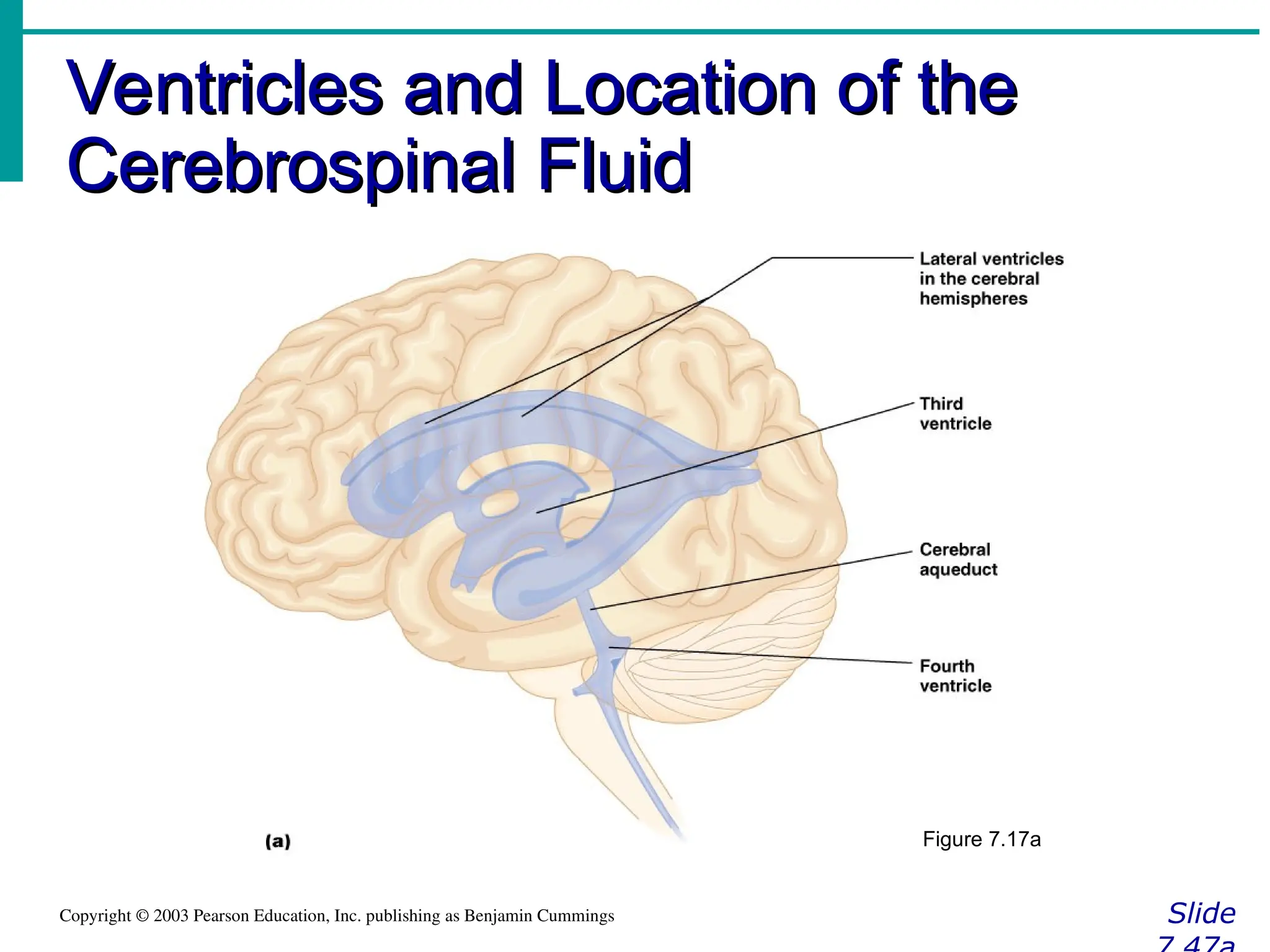
69. Ventricles and Location of the
Ventricles and Location of the
Cerebrospinal Fluid
Cerebrospinal Fluid
Slide
Copyright © 2003 Pearson Education, Inc. publishing as Benjamin Cummings
Figure 7.17a
70. Ventricles and Location of the
Ventricles and Location of the
Cerebrospinal Fluid
Cerebrospinal Fluid
Slide
Copyright © 2003 Pearson Education, Inc. publishing as Benjamin Cummings
Figure 7.17b
71. Blood Brain Barrier
Blood Brain Barrier
Slide 7.48
Copyright © 2003 Pearson Education, Inc. publishing as Benjamin Cummings
Includes the least permeable capillaries
of the body
Excludes many potentially harmful
substances
Useless against some substances
Fats and fat soluble molecules
Respiratory gases
Alcohol
Nicotine
Anesthesia
72. Traumatic Brain Injuries
Traumatic Brain Injuries
Slide 7.49
Copyright © 2003 Pearson Education, Inc. publishing as Benjamin Cummings
Concussion
Slight brain injury
No permanent brain damage
Contusion
Nervous tissue destruction occurs
Nervous tissue does not regenerate
Cerebral edema
Swelling from the inflammatory response
May compress and kill brain tissue
73. Cerebrovascular Accident (CVA)
Cerebrovascular Accident (CVA)
Slide 7.50
Copyright © 2003 Pearson Education, Inc. publishing as Benjamin Cummings
Commonly called a stroke
The result of a ruptured blood vessel
supplying a region of the brain
Brain tissue supplied with oxygen from
that blood source dies
Loss of some functions or death may
result
74. Spinal Cord
Spinal Cord
Slide 7.52
Copyright © 2003 Pearson Education, Inc. publishing as Benjamin Cummings
Extends from the
medulla oblongata to
the region of T12
Below T12 is the cauda
equina (a collection of
spinal nerves)
Enlargements occur in
the cervical and lumbar
regions
Figure 7.18
75. Alzheimer’s Disease
Alzheimer’s Disease
Slide 7.51
Copyright © 2003 Pearson Education, Inc. publishing as Benjamin Cummings
Progressive degenerative brain disease
Mostly seen in the elderly, but may
begin in middle age
Structural changes in the brain include
abnormal protein deposits and twisted
fibers within neurons
Victims experience memory loss,
irritability, confusion and ultimately,
hallucinations and death
76. Spinal Cord Anatomy
Spinal Cord Anatomy
Slide
Copyright © 2003 Pearson Education, Inc. publishing as Benjamin Cummings
Exterior white mater – conduction tracts
Figure 7.19
77. Spinal Cord Anatomy
Spinal Cord Anatomy
Slide
Copyright © 2003 Pearson Education, Inc. publishing as Benjamin Cummings
Internal gray matter - mostly cell bodies
Dorsal (posterior) horns
Anterior (ventral) horns
Figure 7.19
78. Spinal Cord Anatomy
Spinal Cord Anatomy
Slide
Copyright © 2003 Pearson Education, Inc. publishing as Benjamin Cummings
Central canal filled with cerebrospinal
fluid
Figure 7.19
79. Spinal Cord Anatomy
Spinal Cord Anatomy
Slide 7.54
Copyright © 2003 Pearson Education, Inc. publishing as Benjamin Cummings
Meninges cover the spinal cord
Nerves leave at the level of each
vertebrae
Dorsal root
Associated with the dorsal root ganglia –
collections of cell bodies outside the central
nervous system
Ventral root
80. Peripheral Nervous System
Peripheral Nervous System
Slide 7.55
Copyright © 2003 Pearson Education, Inc. publishing as Benjamin Cummings
Nerves and ganglia outside the central
nervous system
Nerve = bundle of neuron fibers
Neuron fibers are bundled by
connective tissue
81. Structure of a Nerve
Structure of a Nerve
Slide 7.56
Copyright © 2003 Pearson Education, Inc. publishing as Benjamin Cummings
Endoneurium
surrounds each fiber
Groups of fibers are
bound into fascicles
by perineurium
Fascicles are bound
together by
epineurium
Figure 7.20
82. Classification of Nerves
Classification of Nerves
Slide 7.57
Copyright © 2003 Pearson Education, Inc. publishing as Benjamin Cummings
Mixed nerves – both sensory and motor
fibers
Afferent (sensory) nerves – carry
impulses toward the CNS
Efferent (motor) nerves – carry impulses
away from the CNS
83. Spinal Nerves
Spinal Nerves
Slide 7.63
Copyright © 2003 Pearson Education, Inc. publishing as Benjamin Cummings
There is a pair of spinal nerves at the
level of each vertebrae for a total of 31
pairs
84. 85. Autonomic Nervous System
Autonomic Nervous System
Slide 7.67
Copyright © 2003 Pearson Education, Inc. publishing as Benjamin Cummings
The involuntary branch of the nervous
system
Consists of only motor nerves
Divided into two divisions
Sympathetic division
Parasympathetic division
86. Comparison of Somatic and
Comparison of Somatic and
Autonomic Nervous Systems
Autonomic Nervous Systems
Slide 7.69
Copyright © 2003 Pearson Education, Inc. publishing as Benjamin Cummings Figure 7.24
87. Anatomy of the Autonomic Nervous
Anatomy of the Autonomic Nervous
System
System
Slide 7.73
Copyright © 2003 Pearson Education, Inc. publishing as Benjamin Cummings
Figure 7.25
88. Autonomic Functioning
Autonomic Functioning
Slide
Copyright © 2003 Pearson Education, Inc. publishing as Benjamin Cummings
Sympathetic – “fight-or-flight”
Response to unusual stimulus
Takes over to increase activities
Remember as the “E” division = exercise,
excitement, emergency, and
embarrassment
89. Autonomic Functioning
Autonomic Functioning
Slide
Copyright © 2003 Pearson Education, Inc. publishing as Benjamin Cummings
Parasympathetic – housekeeping
activites
Conserves energy
Maintains daily necessary body functions
Remember as the “D” division - digestion,
defecation, and diuresis
90. Development Aspects of the
Development Aspects of the
Nervous System
Nervous System
Slide
Copyright © 2003 Pearson Education, Inc. publishing as Benjamin Cummings
The nervous system is formed during
the first month of embryonic
development
Any maternal infection can have
extremely harmful effects
The hypothalamus is one of the last
areas of the brain to develop
91. Development Aspects of the
Development Aspects of the
Nervous System
Nervous System
Slide
Copyright © 2003 Pearson Education, Inc. publishing as Benjamin Cummings
No more neurons are formed after birth,
but growth and maturation continues for
several years (new evidence!)
The brain reaches maximum weight as
a young adult
However, we can always grow
dendrites!