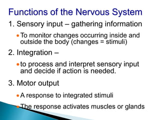
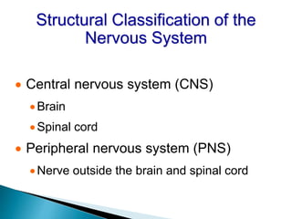
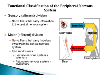
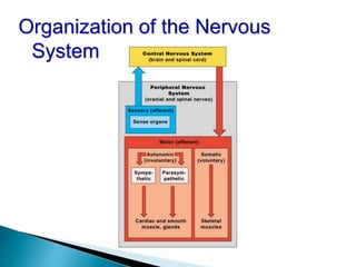
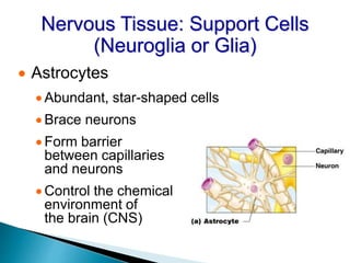
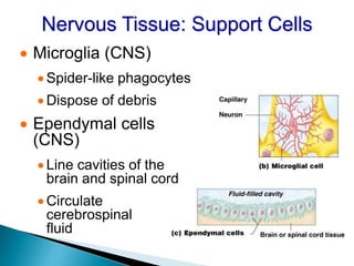
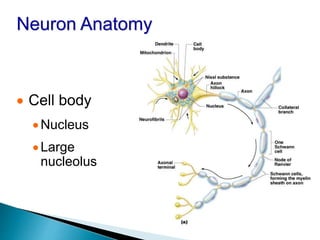
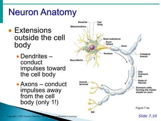
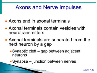
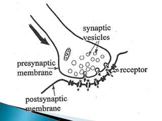
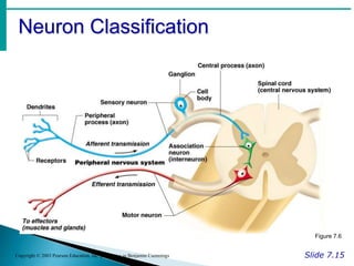
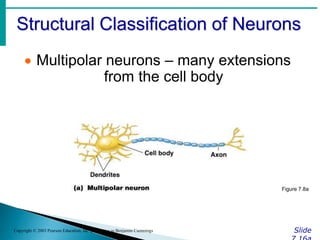
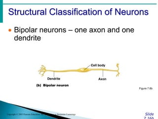
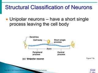
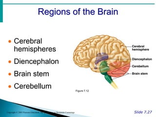
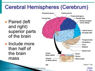
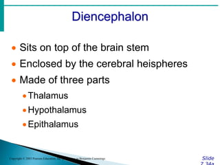
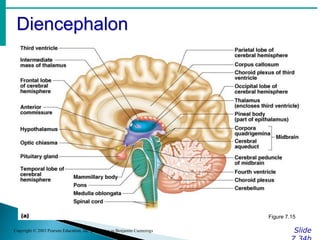
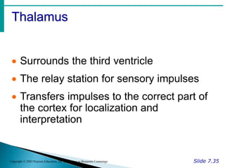
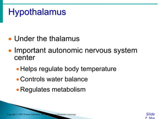
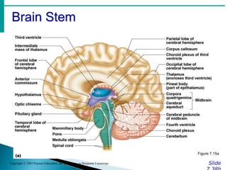
INTRODUCTION TO NERVIOUS SYSTEM
By Unis
Functions of the Nervous System
1. Sensory input – gathering information
To monitor changes occurring inside and
outside the body (changes = stimuli)
2. Integration –
to process and interpret sensory input
and decide if action is needed.
3. Motor output
A response to integrated stimuli
The response activates muscles or glands
Structural Classification of the
Nervous System
 Central nervous system (CNS)
Brain
Spinal cord
 Peripheral nervous system (PNS)
Nerve outside the brain and spinal cord
Functional Classification of the Peripheral Nervous
System
 Sensory (afferent) division
 Nerve fibers that carry information
to the central nervous system
 Motor (efferent) division
 Nerve fibers that carry impulses
away from the central nervous
system
 Two subdivisions
 Somatic nervous system =
voluntary
 Autonomic nervous system =
involuntary
Organization of the Nervous
System
Nervous Tissue: Support Cells
(Neuroglia or Glia)
 Astrocytes
 Abundant, star-shaped cells
 Brace neurons
 Form barrier
between capillaries
and neurons
 Control the chemical
environment of
the brain (CNS)
Nervous Tissue: Support Cells
 Microglia (CNS)
 Spider-like phagocytes
 Dispose of debris
 Ependymal cells
(CNS)
 Line cavities of the
brain and spinal cord
 Circulate
cerebrospinal
fluid
Nervous Tissue: Support Cells
 Oligodendrocytes
(CNS)
Produce myelin
sheath around
nerve fibers in the
central nervous
system Figure 7.3d
 Neuroglia divide.
 Neurons do not.
 Most brain tumors are “gliomas.”
 Most brain tumors involve the neuroglia cells,
not the neurons.
 Consider the role of cell division in cancer!
Support Cells of the PNS
 Satellite cells
Protect neuron cell bodies
 Schwann cells
Form myelin sheath in the peripheral
nervous system
Figure 7.3e
Nervous Tissue: Neurons
 Neurons = nerve cells
Cells specialized to transmit messages
Major regions of neurons
Cell body – nucleus and metabolic center
of the cell
Processes – fibers that extend from the
cell body (dendrites and axons)
Neuron Anatomy
 Cell body
Nucleus
Large
nucleolus
Neuron Anatomy
Slide 7.10
Copyright © 2003 Pearson Education, Inc. publishing as Benjamin Cummings
 Extensions
outside the cell
body
 Dendrites –
conduct
impulses toward
the cell body
 Axons – conduct
impulses away
from the cell
body (only 1!)
Figure 7.4a
Axons and Nerve Impulses
Slide 7.11
Copyright © 2003 Pearson Education, Inc. publishing as Benjamin Cummings
 Axons end in axonal terminals
 Axonal terminals contain vesicles with
neurotransmitters
 Axonal terminals are separated from the
next neuron by a gap
Synaptic cleft – gap between adjacent
neurons
Synapse – junction between nerves
Nerve Fiber Coverings
Slide 7.12
Copyright © 2003 Pearson Education, Inc. publishing as Benjamin Cummings
 Schwann cells –
produce myelin
sheaths in jelly-roll
like fashion
 Nodes of Ranvier –
gaps in myelin
sheath along the
axon
Figure 7.5
Neuron Cell Body Location
Slide 7.13
Copyright © 2003 Pearson Education, Inc. publishing as Benjamin Cummings
 Most are found in the central nervous
system
Gray matter – cell bodies and unmylenated
fibers
Nuclei – clusters of cell bodies within the
white matter of the central nervous system
 Ganglia – collections of cell bodies
outside the central nervous system
Functional Classification of
Neurons
Slide
Copyright © 2003 Pearson Education, Inc. publishing as Benjamin Cummings
 Sensory (afferent) neurons
Carry impulses from the sensory receptors
Cutaneous sense organs
Proprioceptors – detect stretch or tension
 Motor (efferent) neurons
Carry impulses from the central nervous
system
Functional Classification of
Neurons
Slide
Copyright © 2003 Pearson Education, Inc. publishing as Benjamin Cummings
 Interneurons (association neurons)
Found in neural pathways in the central
nervous system
Connect sensory and motor neurons
Neuron Classification
Slide 7.15
Copyright © 2003 Pearson Education, Inc. publishing as Benjamin Cummings
Figure 7.6
Structural Classification of Neurons
Slide
Copyright © 2003 Pearson Education, Inc. publishing as Benjamin Cummings
 Multipolar neurons – many extensions
from the cell body
Figure 7.8a
Structural Classification of Neurons
Slide
Copyright © 2003 Pearson Education, Inc. publishing as Benjamin Cummings
 Bipolar neurons – one axon and one
dendrite
Figure 7.8b
Structural Classification of Neurons
Slide
Copyright © 2003 Pearson Education, Inc. publishing as Benjamin Cummings
 Unipolar neurons – have a short single
process leaving the cell body
Figure 7.8c
How Neurons Function
(Physiology)
Slide 7.17
Copyright © 2003 Pearson Education, Inc. publishing as Benjamin Cummings
 Irritability – ability to respond to stimuli
 Conductivity – ability to transmit an
impulse
 The plasma membrane at rest is
polarized
Fewer positive ions are inside the cell than
outside the cell
Starting a Nerve Impulse
Slide 7.18
Copyright © 2003 Pearson Education, Inc. publishing as Benjamin Cummings
 Depolarization – a
stimulus depolarizes the
neuron’s membrane
 A deploarized
membrane allows
sodium (Na+) to flow
inside the membrane
 The exchange of ions
initiates an action
potential in the neuron
Figure 7.9a–c
The Action Potential
Slide 7.19
Copyright © 2003 Pearson Education, Inc. publishing as Benjamin Cummings
 If the action potential (nerve impulse)
starts, it is propagated over the entire
axon
 Potassium ions rush out of the neuron
after sodium ions rush in, which
repolarizes the membrane
 The sodium-potassium pump restores
the original configuration
This action requires ATP
Nerve Impulse Propagation
Slide 7.20
Copyright © 2003 Pearson Education, Inc. publishing as Benjamin Cummings
 The impulse
continues to move
toward the cell body
 Impulses travel
faster when fibers
have a myelin
sheath
Figure 7.9c–e
Continuation of the Nerve Impulse
between Neurons
Slide 7.21
Copyright © 2003 Pearson Education, Inc. publishing as Benjamin Cummings
 Impulses are able to cross the synapse
to another nerve
Neurotransmitter is released from a nerve’s
axon terminal
The dendrite of the next neuron has
receptors that are stimulated by the
neurotransmitter
An action potential is started in the dendrite
How Neurons Communicate at
Synapses
Slide 7.22
Copyright © 2003 Pearson Education, Inc. publishing as Benjamin Cummings
Figure 7.10
The Reflex Arc
Slide 7.23
Copyright © 2003 Pearson Education, Inc. publishing as Benjamin Cummings
 Reflex – rapid, predictable, and
involuntary responses to stimuli
 Reflex arc – direct route from a sensory
neuron, to an interneuron, to an effector
Figure 7.11a
Simple Reflex Arc
Slide 7.24
Copyright © 2003 Pearson Education, Inc. publishing as Benjamin Cummings
Figure 7.11b, c
Types of Reflexes and Regulation
Slide 7.25
Copyright © 2003 Pearson Education, Inc. publishing as Benjamin Cummings
 Autonomic reflexes
Smooth muscle regulation
Heart and blood pressure regulation
Regulation of glands
Digestive system regulation
 Somatic reflexes
Activation of skeletal muscles
Central Nervous System (CNS)
Slide 7.26
Copyright © 2003 Pearson Education, Inc. publishing as Benjamin Cummings
 CNS develops from the embryonic
neural tube
The neural tube becomes the brain and
spinal cord
The opening of the neural tube becomes
the ventricles
Four chambers within the brain
Filled with cerebrospinal fluid
Regions of the Brain
Slide 7.27
Copyright © 2003 Pearson Education, Inc. publishing as Benjamin Cummings
 Cerebral
hemispheres
 Diencephalon
 Brain stem
 Cerebellum Figure 7.12
Cerebral Hemispheres (Cerebrum)
Slide
Copyright © 2003 Pearson Education, Inc. publishing as Benjamin Cummings
 Paired (left
and right)
superior parts
of the brain
 Include more
than half of
the brain
mass
Figure 7.13a
Cerebral Hemispheres (Cerebrum)
Slide
Copyright © 2003 Pearson Education, Inc. publishing as Benjamin Cummings
 The surface
is made of
ridges (gyri)
and grooves
(sulci)
Figure 7.13a
Lobes of the Cerebrum
Slide
Copyright © 2003 Pearson Education, Inc. publishing as Benjamin Cummings
 Fissures (deep grooves) divide the
cerebrum into lobes
 Surface lobes of the cerebrum
Frontal lobe
Parietal lobe
Occipital lobe
Temporal lobe
Lobes of the Cerebrum
Slide
Copyright © 2003 Pearson Education, Inc. publishing as Benjamin Cummings
Figure 7.15a
Specialized Areas of the Cerebrum
Slide 7.30
Copyright © 2003 Pearson Education, Inc. publishing as Benjamin Cummings
 Somatic sensory area – receives
impulses from the body’s sensory
receptors
 Primary motor area – sends impulses to
skeletal muscles
 Broca’s area – involved in our ability to
speak
Sensory and Motor Areas of the
Cerebral Cortex
Slide 7.31
Copyright © 2003 Pearson Education, Inc. publishing as Benjamin Cummings
Figure 7.14
Specialized Area of the Cerebrum
Slide
Copyright © 2003 Pearson Education, Inc. publishing as Benjamin Cummings
 Cerebral areas involved in special
senses
Gustatory area (taste)
Visual area
Auditory area
Olfactory area
Specialized Area of the Cerebrum
Slide
Copyright © 2003 Pearson Education, Inc. publishing as Benjamin Cummings
 Interpretation areas of the cerebrum
Speech/language region
Language comprehension region
General interpretation area
Specialized Area of the Cerebrum
Slide
Copyright © 2003 Pearson Education, Inc. publishing as Benjamin Cummings
Figure 7.13c
Layers of the Cerebrum
Slide
Copyright © 2003 Pearson Education, Inc. publishing as Benjamin Cummings
 Gray matter
Outer layer
Composed
mostly of neuron
cell bodies
Figure 7.13a
Layers of the Cerebrum
Slide
Copyright © 2003 Pearson Education, Inc. publishing as Benjamin Cummings
 White matter
Fiber tracts
inside the gray
matter
Example:
corpus callosum
connects
hemispheres
Figure 7.13a
Layers of the Cerebrum
Slide
Copyright © 2003 Pearson Education, Inc. publishing as Benjamin Cummings
 Basal nuclei – internal
islands of gray matter
 Regulates voluntary
motor activities by
modifying info sent to
the motor cortex
 Problems = ie unable
to control muscles,
spastic, jerky
 Involved in
Huntington’s and
Parkinson’s Disease
Figure 7.13a
Diencephalon
Slide
Copyright © 2003 Pearson Education, Inc. publishing as Benjamin Cummings
 Sits on top of the brain stem
 Enclosed by the cerebral heispheres
 Made of three parts
Thalamus
Hypothalamus
Epithalamus
Diencephalon
Slide
Copyright © 2003 Pearson Education, Inc. publishing as Benjamin Cummings
Figure 7.15
Thalamus
Slide 7.35
Copyright © 2003 Pearson Education, Inc. publishing as Benjamin Cummings
 Surrounds the third ventricle
 The relay station for sensory impulses
 Transfers impulses to the correct part of
the cortex for localization and
interpretation
Hypothalamus
Slide
Copyright © 2003 Pearson Education, Inc. publishing as Benjamin Cummings
 Under the thalamus
 Important autonomic nervous system
center
Helps regulate body temperature
Controls water balance
Regulates metabolism
Hypothalamus
Slide
Copyright © 2003 Pearson Education, Inc. publishing as Benjamin Cummings
 An important part of the limbic system
(emotions)
 The pituitary gland is attached to the
hypothalamus
Epithalamus
Slide 7.37
Copyright © 2003 Pearson Education, Inc. publishing as Benjamin Cummings
 Forms the roof of the third ventricle
 Houses the pineal body (an endocrine
gland)
 Includes the choroid plexus – forms
cerebrospinal fluid
Brain Stem
Slide
Copyright © 2003 Pearson Education, Inc. publishing as Benjamin Cummings
 Attaches to the spinal cord
 Parts of the brain stem
Midbrain
Pons
Medulla oblongata
Brain Stem
Slide
Copyright © 2003 Pearson Education, Inc. publishing as Benjamin Cummings
Figure 7.15a
Midbrain
Slide 7.39
Copyright © 2003 Pearson Education, Inc. publishing as Benjamin Cummings
 Mostly composed of tracts of nerve
fibers
Reflex centers for vision and hearing
Cerebral aquaduct – 3rd-4th ventricles
Pons
Slide 7.40
Copyright © 2003 Pearson Education, Inc. publishing as Benjamin Cummings
 The bulging center part of the brain
stem
 Mostly composed of fiber tracts
 Includes nuclei involved in the control of
breathing
Medulla Oblongata
Slide 7.41
Copyright © 2003 Pearson Education, Inc. publishing as Benjamin Cummings
 The lowest part of the brain stem
 Merges into the spinal cord
 Includes important fiber tracts
 Contains important control centers
Heart rate control
Blood pressure regulation
Breathing
Swallowing
Vomiting
Cerebellum
Slide
Copyright © 2003 Pearson Education, Inc. publishing as Benjamin Cummings
 Two hemispheres with convoluted
surfaces
 Provides involuntary coordination of
body movements
Cerebellum
Slide
Copyright © 2003 Pearson Education, Inc. publishing as Benjamin Cummings
Figure 7.15a
Protection of the Central Nervous
System
Slide
Copyright © 2003 Pearson Education, Inc. publishing as Benjamin Cummings
 Scalp and skin
 Skull and vertebral column
 Meninges
Figure 7.16a
Protection of the Central Nervous
System
Slide
Copyright © 2003 Pearson Education, Inc. publishing as Benjamin Cummings
 Cerebrospinal fluid
 Blood brain barrier
Figure 7.16a
Meninges
Slide
Copyright © 2003 Pearson Education, Inc. publishing as Benjamin Cummings
 Dura mater
Double-layered external covering
Periosteum – attached to surface of the
skull
Meningeal layer – outer covering of the
brain
Folds inward in several areas
Meninges
Slide
Copyright © 2003 Pearson Education, Inc. publishing as Benjamin Cummings
 Arachnoid layer
Middle layer
Web-like
 Pia mater
Internal layer
Clings to the surface of the brain
Cerebrospinal Fluid
Slide 7.46
Copyright © 2003 Pearson Education, Inc. publishing as Benjamin Cummings
 Similar to blood plasma composition
 Formed by the choroid plexus
 Forms a watery cushion to protect the
brain
 Circulated in arachnoid space,
ventricles, and central canal of the
spinal cord
Ventricles and Location of the
Cerebrospinal Fluid
Slide
Copyright © 2003 Pearson Education, Inc. publishing as Benjamin Cummings
Figure 7.17a
Ventricles and Location of the
Cerebrospinal Fluid
Slide
Copyright © 2003 Pearson Education, Inc. publishing as Benjamin Cummings
Figure 7.17b
Blood Brain Barrier
Slide 7.48
Copyright © 2003 Pearson Education, Inc. publishing as Benjamin Cummings
 Includes the least permeable capillaries
of the body
 Excludes many potentially harmful
substances
 Useless against some substances
Fats and fat soluble molecules
Respiratory gases
Alcohol
Nicotine
Anesthesia
Traumatic Brain Injuries
Slide 7.49
Copyright © 2003 Pearson Education, Inc. publishing as Benjamin Cummings
 Concussion
Slight brain injury
No permanent brain damage
 Contusion
Nervous tissue destruction occurs
Nervous tissue does not regenerate
 Cerebral edema
Swelling from the inflammatory response
May compress and kill brain tissue
Cerebrovascular Accident (CVA)
Slide 7.50
Copyright © 2003 Pearson Education, Inc. publishing as Benjamin Cummings
 Commonly called a stroke
 The result of a ruptured blood vessel
supplying a region of the brain
 Brain tissue supplied with oxygen from
that blood source dies
 Loss of some functions or death may
result
Spinal Cord
Slide 7.52
Copyright © 2003 Pearson Education, Inc. publishing as Benjamin Cummings
 Extends from the
medulla oblongata to
the region of T12
 Below T12 is the cauda
equina (a collection of
spinal nerves)
 Enlargements occur in
the cervical and lumbar
regions
Figure 7.18
Alzheimer’s Disease
Slide 7.51
Copyright © 2003 Pearson Education, Inc. publishing as Benjamin Cummings
 Progressive degenerative brain disease
 Mostly seen in the elderly, but may
begin in middle age
 Structural changes in the brain include
abnormal protein deposits and twisted
fibers within neurons
 Victims experience memory loss,
irritability, confusion and ultimately,
hallucinations and death
Spinal Cord Anatomy
Slide
Copyright © 2003 Pearson Education, Inc. publishing as Benjamin Cummings
 Exterior white mater – conduction tracts
Figure 7.19
Spinal Cord Anatomy
Slide
Copyright © 2003 Pearson Education, Inc. publishing as Benjamin Cummings
 Internal gray matter - mostly cell bodies
Dorsal (posterior) horns
Anterior (ventral) horns
Figure 7.19
Spinal Cord Anatomy
Slide
Copyright © 2003 Pearson Education, Inc. publishing as Benjamin Cummings
 Central canal filled with cerebrospinal
fluid
Figure 7.19
Spinal Cord Anatomy
Slide 7.54
Copyright © 2003 Pearson Education, Inc. publishing as Benjamin Cummings
 Meninges cover the spinal cord
 Nerves leave at the level of each
vertebrae
Dorsal root
 Associated with the dorsal root ganglia –
collections of cell bodies outside the central
nervous system
Ventral root
Peripheral Nervous System
Slide 7.55
Copyright © 2003 Pearson Education, Inc. publishing as Benjamin Cummings
 Nerves and ganglia outside the central
nervous system
 Nerve = bundle of neuron fibers
 Neuron fibers are bundled by
connective tissue
Structure of a Nerve
Slide 7.56
Copyright © 2003 Pearson Education, Inc. publishing as Benjamin Cummings
 Endoneurium
surrounds each fiber
 Groups of fibers are
bound into fascicles
by perineurium
 Fascicles are bound
together by
epineurium
Figure 7.20
Classification of Nerves
Slide 7.57
Copyright © 2003 Pearson Education, Inc. publishing as Benjamin Cummings
 Mixed nerves – both sensory and motor
fibers
 Afferent (sensory) nerves – carry
impulses toward the CNS
 Efferent (motor) nerves – carry impulses
away from the CNS
Spinal Nerves
Slide 7.63
Copyright © 2003 Pearson Education, Inc. publishing as Benjamin Cummings
 There is a pair of spinal nerves at the
level of each vertebrae for a total of 31
pairs
Spinal Nerves
Slide 7.64
Copyright © 2003 Pearson Education, Inc. publishing as Benjamin Cummings
Figure 7.22a
Autonomic Nervous System
Slide 7.67
Copyright © 2003 Pearson Education, Inc. publishing as Benjamin Cummings
 The involuntary branch of the nervous
system
 Consists of only motor nerves
 Divided into two divisions
Sympathetic division
Parasympathetic division
Comparison of Somatic and
Autonomic Nervous Systems
Slide 7.69
Copyright © 2003 Pearson Education, Inc. publishing as Benjamin Cummings Figure 7.24
Anatomy of the Autonomic Nervous
System
Autonomic Functioning
 Sympathetic – “fight-or-flight”
Response to unusual stimulus
Takes over to increase activities
Remember as the “E” division = exercise,
excitement, emergency, and
embarrassment
Autonomic Functioning
 Parasympathetic – housekeeping
activites
Conserves energy
Maintains daily necessary body functions
Remember as the “D” division - digestion,
defecation, and diuresis
Development Aspects of the
Nervous System
 The nervous system is formed during
the first month of embryonic
development
 Any maternal infection can have
extremely harmful effects
 The hypothalamus is one of the last
areas of the brain to develop
Development Aspects of the
Nervous System
 No more neurons are formed after birth,
but growth and maturation continues for
several years (new evidence!)
 The brain reaches maximum weight as
a young adult
 However, we can always grow
dendrites!

nervous system.ppt777777777777777777777777

  • 1.
  • 2.
    Functions of theNervous System 1. Sensory input – gathering information To monitor changes occurring inside and outside the body (changes = stimuli) 2. Integration – to process and interpret sensory input and decide if action is needed. 3. Motor output A response to integrated stimuli The response activates muscles or glands
  • 3.
    Structural Classification ofthe Nervous System  Central nervous system (CNS) Brain Spinal cord  Peripheral nervous system (PNS) Nerve outside the brain and spinal cord
  • 4.
    Functional Classification ofthe Peripheral Nervous System  Sensory (afferent) division  Nerve fibers that carry information to the central nervous system  Motor (efferent) division  Nerve fibers that carry impulses away from the central nervous system  Two subdivisions  Somatic nervous system = voluntary  Autonomic nervous system = involuntary
  • 5.
    Organization of theNervous System
  • 6.
    Nervous Tissue: SupportCells (Neuroglia or Glia)  Astrocytes  Abundant, star-shaped cells  Brace neurons  Form barrier between capillaries and neurons  Control the chemical environment of the brain (CNS)
  • 7.
    Nervous Tissue: SupportCells  Microglia (CNS)  Spider-like phagocytes  Dispose of debris  Ependymal cells (CNS)  Line cavities of the brain and spinal cord  Circulate cerebrospinal fluid
  • 8.
    Nervous Tissue: SupportCells  Oligodendrocytes (CNS) Produce myelin sheath around nerve fibers in the central nervous system Figure 7.3d
  • 9.
     Neuroglia divide. Neurons do not.  Most brain tumors are “gliomas.”  Most brain tumors involve the neuroglia cells, not the neurons.  Consider the role of cell division in cancer!
  • 10.
    Support Cells ofthe PNS  Satellite cells Protect neuron cell bodies  Schwann cells Form myelin sheath in the peripheral nervous system Figure 7.3e
  • 11.
    Nervous Tissue: Neurons Neurons = nerve cells Cells specialized to transmit messages Major regions of neurons Cell body – nucleus and metabolic center of the cell Processes – fibers that extend from the cell body (dendrites and axons)
  • 12.
    Neuron Anatomy  Cellbody Nucleus Large nucleolus
  • 13.
    Neuron Anatomy Slide 7.10 Copyright© 2003 Pearson Education, Inc. publishing as Benjamin Cummings  Extensions outside the cell body  Dendrites – conduct impulses toward the cell body  Axons – conduct impulses away from the cell body (only 1!) Figure 7.4a
  • 14.
    Axons and NerveImpulses Slide 7.11 Copyright © 2003 Pearson Education, Inc. publishing as Benjamin Cummings  Axons end in axonal terminals  Axonal terminals contain vesicles with neurotransmitters  Axonal terminals are separated from the next neuron by a gap Synaptic cleft – gap between adjacent neurons Synapse – junction between nerves
  • 16.
    Nerve Fiber Coverings Slide7.12 Copyright © 2003 Pearson Education, Inc. publishing as Benjamin Cummings  Schwann cells – produce myelin sheaths in jelly-roll like fashion  Nodes of Ranvier – gaps in myelin sheath along the axon Figure 7.5
  • 17.
    Neuron Cell BodyLocation Slide 7.13 Copyright © 2003 Pearson Education, Inc. publishing as Benjamin Cummings  Most are found in the central nervous system Gray matter – cell bodies and unmylenated fibers Nuclei – clusters of cell bodies within the white matter of the central nervous system  Ganglia – collections of cell bodies outside the central nervous system
  • 18.
    Functional Classification of Neurons Slide Copyright© 2003 Pearson Education, Inc. publishing as Benjamin Cummings  Sensory (afferent) neurons Carry impulses from the sensory receptors Cutaneous sense organs Proprioceptors – detect stretch or tension  Motor (efferent) neurons Carry impulses from the central nervous system
  • 19.
    Functional Classification of Neurons Slide Copyright© 2003 Pearson Education, Inc. publishing as Benjamin Cummings  Interneurons (association neurons) Found in neural pathways in the central nervous system Connect sensory and motor neurons
  • 20.
    Neuron Classification Slide 7.15 Copyright© 2003 Pearson Education, Inc. publishing as Benjamin Cummings Figure 7.6
  • 21.
    Structural Classification ofNeurons Slide Copyright © 2003 Pearson Education, Inc. publishing as Benjamin Cummings  Multipolar neurons – many extensions from the cell body Figure 7.8a
  • 22.
    Structural Classification ofNeurons Slide Copyright © 2003 Pearson Education, Inc. publishing as Benjamin Cummings  Bipolar neurons – one axon and one dendrite Figure 7.8b
  • 23.
    Structural Classification ofNeurons Slide Copyright © 2003 Pearson Education, Inc. publishing as Benjamin Cummings  Unipolar neurons – have a short single process leaving the cell body Figure 7.8c
  • 24.
    How Neurons Function (Physiology) Slide7.17 Copyright © 2003 Pearson Education, Inc. publishing as Benjamin Cummings  Irritability – ability to respond to stimuli  Conductivity – ability to transmit an impulse  The plasma membrane at rest is polarized Fewer positive ions are inside the cell than outside the cell
  • 25.
    Starting a NerveImpulse Slide 7.18 Copyright © 2003 Pearson Education, Inc. publishing as Benjamin Cummings  Depolarization – a stimulus depolarizes the neuron’s membrane  A deploarized membrane allows sodium (Na+) to flow inside the membrane  The exchange of ions initiates an action potential in the neuron Figure 7.9a–c
  • 26.
    The Action Potential Slide7.19 Copyright © 2003 Pearson Education, Inc. publishing as Benjamin Cummings  If the action potential (nerve impulse) starts, it is propagated over the entire axon  Potassium ions rush out of the neuron after sodium ions rush in, which repolarizes the membrane  The sodium-potassium pump restores the original configuration This action requires ATP
  • 27.
    Nerve Impulse Propagation Slide7.20 Copyright © 2003 Pearson Education, Inc. publishing as Benjamin Cummings  The impulse continues to move toward the cell body  Impulses travel faster when fibers have a myelin sheath Figure 7.9c–e
  • 28.
    Continuation of theNerve Impulse between Neurons Slide 7.21 Copyright © 2003 Pearson Education, Inc. publishing as Benjamin Cummings  Impulses are able to cross the synapse to another nerve Neurotransmitter is released from a nerve’s axon terminal The dendrite of the next neuron has receptors that are stimulated by the neurotransmitter An action potential is started in the dendrite
  • 29.
    How Neurons Communicateat Synapses Slide 7.22 Copyright © 2003 Pearson Education, Inc. publishing as Benjamin Cummings Figure 7.10
  • 30.
    The Reflex Arc Slide7.23 Copyright © 2003 Pearson Education, Inc. publishing as Benjamin Cummings  Reflex – rapid, predictable, and involuntary responses to stimuli  Reflex arc – direct route from a sensory neuron, to an interneuron, to an effector Figure 7.11a
  • 31.
    Simple Reflex Arc Slide7.24 Copyright © 2003 Pearson Education, Inc. publishing as Benjamin Cummings Figure 7.11b, c
  • 32.
    Types of Reflexesand Regulation Slide 7.25 Copyright © 2003 Pearson Education, Inc. publishing as Benjamin Cummings  Autonomic reflexes Smooth muscle regulation Heart and blood pressure regulation Regulation of glands Digestive system regulation  Somatic reflexes Activation of skeletal muscles
  • 33.
    Central Nervous System(CNS) Slide 7.26 Copyright © 2003 Pearson Education, Inc. publishing as Benjamin Cummings  CNS develops from the embryonic neural tube The neural tube becomes the brain and spinal cord The opening of the neural tube becomes the ventricles Four chambers within the brain Filled with cerebrospinal fluid
  • 34.
    Regions of theBrain Slide 7.27 Copyright © 2003 Pearson Education, Inc. publishing as Benjamin Cummings  Cerebral hemispheres  Diencephalon  Brain stem  Cerebellum Figure 7.12
  • 35.
    Cerebral Hemispheres (Cerebrum) Slide Copyright© 2003 Pearson Education, Inc. publishing as Benjamin Cummings  Paired (left and right) superior parts of the brain  Include more than half of the brain mass Figure 7.13a
  • 36.
    Cerebral Hemispheres (Cerebrum) Slide Copyright© 2003 Pearson Education, Inc. publishing as Benjamin Cummings  The surface is made of ridges (gyri) and grooves (sulci) Figure 7.13a
  • 37.
    Lobes of theCerebrum Slide Copyright © 2003 Pearson Education, Inc. publishing as Benjamin Cummings  Fissures (deep grooves) divide the cerebrum into lobes  Surface lobes of the cerebrum Frontal lobe Parietal lobe Occipital lobe Temporal lobe
  • 38.
    Lobes of theCerebrum Slide Copyright © 2003 Pearson Education, Inc. publishing as Benjamin Cummings Figure 7.15a
  • 39.
    Specialized Areas ofthe Cerebrum Slide 7.30 Copyright © 2003 Pearson Education, Inc. publishing as Benjamin Cummings  Somatic sensory area – receives impulses from the body’s sensory receptors  Primary motor area – sends impulses to skeletal muscles  Broca’s area – involved in our ability to speak
  • 41.
    Sensory and MotorAreas of the Cerebral Cortex Slide 7.31 Copyright © 2003 Pearson Education, Inc. publishing as Benjamin Cummings Figure 7.14
  • 42.
    Specialized Area ofthe Cerebrum Slide Copyright © 2003 Pearson Education, Inc. publishing as Benjamin Cummings  Cerebral areas involved in special senses Gustatory area (taste) Visual area Auditory area Olfactory area
  • 43.
    Specialized Area ofthe Cerebrum Slide Copyright © 2003 Pearson Education, Inc. publishing as Benjamin Cummings  Interpretation areas of the cerebrum Speech/language region Language comprehension region General interpretation area
  • 44.
    Specialized Area ofthe Cerebrum Slide Copyright © 2003 Pearson Education, Inc. publishing as Benjamin Cummings Figure 7.13c
  • 45.
    Layers of theCerebrum Slide Copyright © 2003 Pearson Education, Inc. publishing as Benjamin Cummings  Gray matter Outer layer Composed mostly of neuron cell bodies Figure 7.13a
  • 46.
    Layers of theCerebrum Slide Copyright © 2003 Pearson Education, Inc. publishing as Benjamin Cummings  White matter Fiber tracts inside the gray matter Example: corpus callosum connects hemispheres Figure 7.13a
  • 47.
    Layers of theCerebrum Slide Copyright © 2003 Pearson Education, Inc. publishing as Benjamin Cummings  Basal nuclei – internal islands of gray matter  Regulates voluntary motor activities by modifying info sent to the motor cortex  Problems = ie unable to control muscles, spastic, jerky  Involved in Huntington’s and Parkinson’s Disease Figure 7.13a
  • 48.
    Diencephalon Slide Copyright © 2003Pearson Education, Inc. publishing as Benjamin Cummings  Sits on top of the brain stem  Enclosed by the cerebral heispheres  Made of three parts Thalamus Hypothalamus Epithalamus
  • 49.
    Diencephalon Slide Copyright © 2003Pearson Education, Inc. publishing as Benjamin Cummings Figure 7.15
  • 50.
    Thalamus Slide 7.35 Copyright ©2003 Pearson Education, Inc. publishing as Benjamin Cummings  Surrounds the third ventricle  The relay station for sensory impulses  Transfers impulses to the correct part of the cortex for localization and interpretation
  • 51.
    Hypothalamus Slide Copyright © 2003Pearson Education, Inc. publishing as Benjamin Cummings  Under the thalamus  Important autonomic nervous system center Helps regulate body temperature Controls water balance Regulates metabolism
  • 52.
    Hypothalamus Slide Copyright © 2003Pearson Education, Inc. publishing as Benjamin Cummings  An important part of the limbic system (emotions)  The pituitary gland is attached to the hypothalamus
  • 53.
    Epithalamus Slide 7.37 Copyright ©2003 Pearson Education, Inc. publishing as Benjamin Cummings  Forms the roof of the third ventricle  Houses the pineal body (an endocrine gland)  Includes the choroid plexus – forms cerebrospinal fluid
  • 54.
    Brain Stem Slide Copyright ©2003 Pearson Education, Inc. publishing as Benjamin Cummings  Attaches to the spinal cord  Parts of the brain stem Midbrain Pons Medulla oblongata
  • 55.
    Brain Stem Slide Copyright ©2003 Pearson Education, Inc. publishing as Benjamin Cummings Figure 7.15a
  • 56.
    Midbrain Slide 7.39 Copyright ©2003 Pearson Education, Inc. publishing as Benjamin Cummings  Mostly composed of tracts of nerve fibers Reflex centers for vision and hearing Cerebral aquaduct – 3rd-4th ventricles
  • 57.
    Pons Slide 7.40 Copyright ©2003 Pearson Education, Inc. publishing as Benjamin Cummings  The bulging center part of the brain stem  Mostly composed of fiber tracts  Includes nuclei involved in the control of breathing
  • 58.
    Medulla Oblongata Slide 7.41 Copyright© 2003 Pearson Education, Inc. publishing as Benjamin Cummings  The lowest part of the brain stem  Merges into the spinal cord  Includes important fiber tracts  Contains important control centers Heart rate control Blood pressure regulation Breathing Swallowing Vomiting
  • 59.
    Cerebellum Slide Copyright © 2003Pearson Education, Inc. publishing as Benjamin Cummings  Two hemispheres with convoluted surfaces  Provides involuntary coordination of body movements
  • 60.
    Cerebellum Slide Copyright © 2003Pearson Education, Inc. publishing as Benjamin Cummings Figure 7.15a
  • 61.
    Protection of theCentral Nervous System Slide Copyright © 2003 Pearson Education, Inc. publishing as Benjamin Cummings  Scalp and skin  Skull and vertebral column  Meninges Figure 7.16a
  • 62.
    Protection of theCentral Nervous System Slide Copyright © 2003 Pearson Education, Inc. publishing as Benjamin Cummings  Cerebrospinal fluid  Blood brain barrier Figure 7.16a
  • 63.
    Meninges Slide Copyright © 2003Pearson Education, Inc. publishing as Benjamin Cummings  Dura mater Double-layered external covering Periosteum – attached to surface of the skull Meningeal layer – outer covering of the brain Folds inward in several areas
  • 64.
    Meninges Slide Copyright © 2003Pearson Education, Inc. publishing as Benjamin Cummings  Arachnoid layer Middle layer Web-like  Pia mater Internal layer Clings to the surface of the brain
  • 65.
    Cerebrospinal Fluid Slide 7.46 Copyright© 2003 Pearson Education, Inc. publishing as Benjamin Cummings  Similar to blood plasma composition  Formed by the choroid plexus  Forms a watery cushion to protect the brain  Circulated in arachnoid space, ventricles, and central canal of the spinal cord
  • 66.
    Ventricles and Locationof the Cerebrospinal Fluid Slide Copyright © 2003 Pearson Education, Inc. publishing as Benjamin Cummings Figure 7.17a
  • 67.
    Ventricles and Locationof the Cerebrospinal Fluid Slide Copyright © 2003 Pearson Education, Inc. publishing as Benjamin Cummings Figure 7.17b
  • 68.
    Blood Brain Barrier Slide7.48 Copyright © 2003 Pearson Education, Inc. publishing as Benjamin Cummings  Includes the least permeable capillaries of the body  Excludes many potentially harmful substances  Useless against some substances Fats and fat soluble molecules Respiratory gases Alcohol Nicotine Anesthesia
  • 69.
    Traumatic Brain Injuries Slide7.49 Copyright © 2003 Pearson Education, Inc. publishing as Benjamin Cummings  Concussion Slight brain injury No permanent brain damage  Contusion Nervous tissue destruction occurs Nervous tissue does not regenerate  Cerebral edema Swelling from the inflammatory response May compress and kill brain tissue
  • 70.
    Cerebrovascular Accident (CVA) Slide7.50 Copyright © 2003 Pearson Education, Inc. publishing as Benjamin Cummings  Commonly called a stroke  The result of a ruptured blood vessel supplying a region of the brain  Brain tissue supplied with oxygen from that blood source dies  Loss of some functions or death may result
  • 71.
    Spinal Cord Slide 7.52 Copyright© 2003 Pearson Education, Inc. publishing as Benjamin Cummings  Extends from the medulla oblongata to the region of T12  Below T12 is the cauda equina (a collection of spinal nerves)  Enlargements occur in the cervical and lumbar regions Figure 7.18
  • 72.
    Alzheimer’s Disease Slide 7.51 Copyright© 2003 Pearson Education, Inc. publishing as Benjamin Cummings  Progressive degenerative brain disease  Mostly seen in the elderly, but may begin in middle age  Structural changes in the brain include abnormal protein deposits and twisted fibers within neurons  Victims experience memory loss, irritability, confusion and ultimately, hallucinations and death
  • 73.
    Spinal Cord Anatomy Slide Copyright© 2003 Pearson Education, Inc. publishing as Benjamin Cummings  Exterior white mater – conduction tracts Figure 7.19
  • 74.
    Spinal Cord Anatomy Slide Copyright© 2003 Pearson Education, Inc. publishing as Benjamin Cummings  Internal gray matter - mostly cell bodies Dorsal (posterior) horns Anterior (ventral) horns Figure 7.19
  • 75.
    Spinal Cord Anatomy Slide Copyright© 2003 Pearson Education, Inc. publishing as Benjamin Cummings  Central canal filled with cerebrospinal fluid Figure 7.19
  • 76.
    Spinal Cord Anatomy Slide7.54 Copyright © 2003 Pearson Education, Inc. publishing as Benjamin Cummings  Meninges cover the spinal cord  Nerves leave at the level of each vertebrae Dorsal root  Associated with the dorsal root ganglia – collections of cell bodies outside the central nervous system Ventral root
  • 77.
    Peripheral Nervous System Slide7.55 Copyright © 2003 Pearson Education, Inc. publishing as Benjamin Cummings  Nerves and ganglia outside the central nervous system  Nerve = bundle of neuron fibers  Neuron fibers are bundled by connective tissue
  • 78.
    Structure of aNerve Slide 7.56 Copyright © 2003 Pearson Education, Inc. publishing as Benjamin Cummings  Endoneurium surrounds each fiber  Groups of fibers are bound into fascicles by perineurium  Fascicles are bound together by epineurium Figure 7.20
  • 79.
    Classification of Nerves Slide7.57 Copyright © 2003 Pearson Education, Inc. publishing as Benjamin Cummings  Mixed nerves – both sensory and motor fibers  Afferent (sensory) nerves – carry impulses toward the CNS  Efferent (motor) nerves – carry impulses away from the CNS
  • 80.
    Spinal Nerves Slide 7.63 Copyright© 2003 Pearson Education, Inc. publishing as Benjamin Cummings  There is a pair of spinal nerves at the level of each vertebrae for a total of 31 pairs
  • 81.
    Spinal Nerves Slide 7.64 Copyright© 2003 Pearson Education, Inc. publishing as Benjamin Cummings Figure 7.22a
  • 82.
    Autonomic Nervous System Slide7.67 Copyright © 2003 Pearson Education, Inc. publishing as Benjamin Cummings  The involuntary branch of the nervous system  Consists of only motor nerves  Divided into two divisions Sympathetic division Parasympathetic division
  • 83.
    Comparison of Somaticand Autonomic Nervous Systems Slide 7.69 Copyright © 2003 Pearson Education, Inc. publishing as Benjamin Cummings Figure 7.24
  • 84.
    Anatomy of theAutonomic Nervous System
  • 85.
    Autonomic Functioning  Sympathetic– “fight-or-flight” Response to unusual stimulus Takes over to increase activities Remember as the “E” division = exercise, excitement, emergency, and embarrassment
  • 86.
    Autonomic Functioning  Parasympathetic– housekeeping activites Conserves energy Maintains daily necessary body functions Remember as the “D” division - digestion, defecation, and diuresis
  • 87.
    Development Aspects ofthe Nervous System  The nervous system is formed during the first month of embryonic development  Any maternal infection can have extremely harmful effects  The hypothalamus is one of the last areas of the brain to develop
  • 88.
    Development Aspects ofthe Nervous System  No more neurons are formed after birth, but growth and maturation continues for several years (new evidence!)  The brain reaches maximum weight as a young adult  However, we can always grow dendrites!