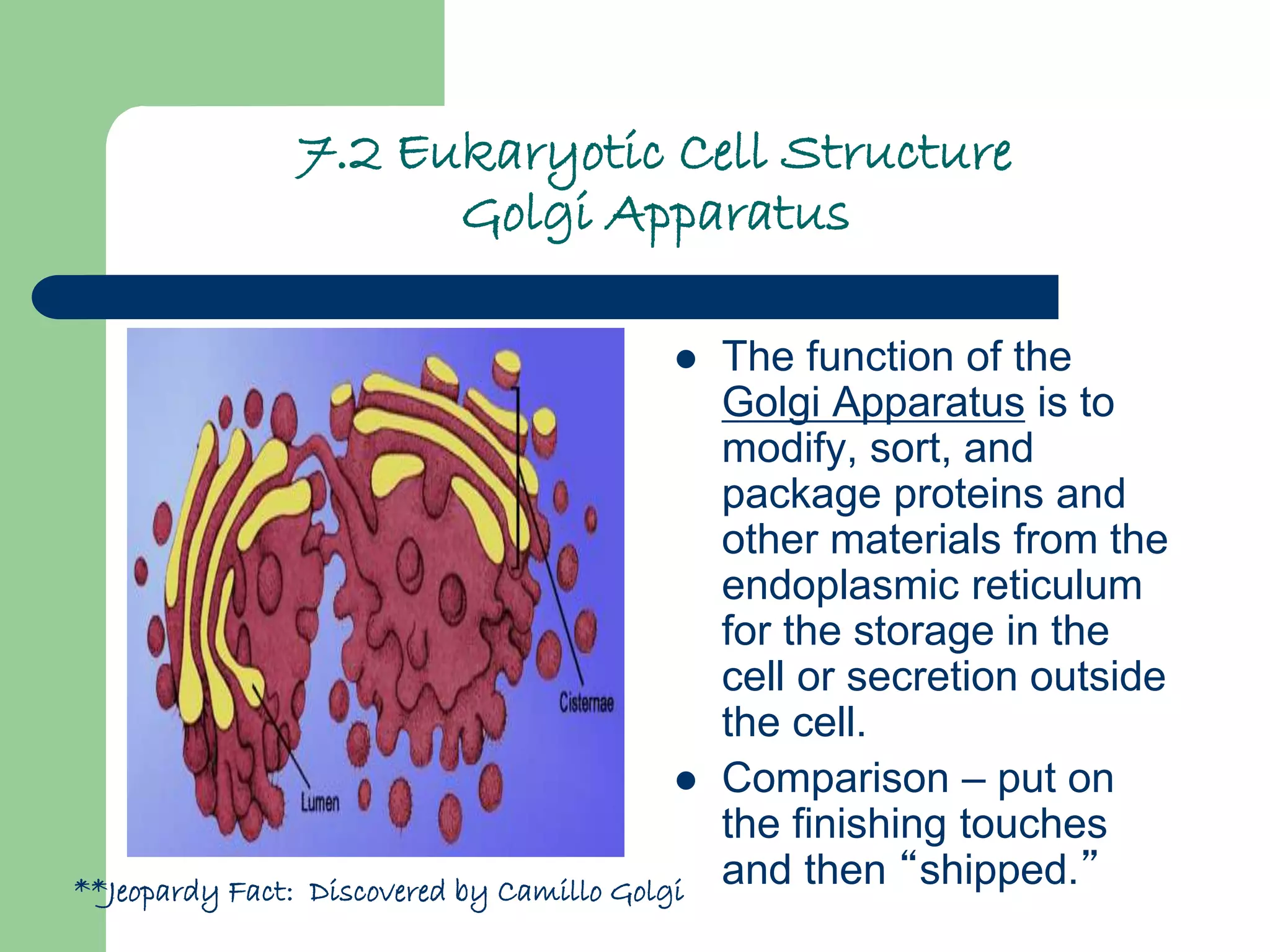
2. 7.1 Life is Cellular
The Discovery of the Cell
1665, Robert Hooke
used a microscope to
look at a thin slice of
cork.
Look like tiny, empty
chambers.
Hooke named the
chambers “cells”
Anton van
Leeuwenhoek observed
pond water, revealed
tiny living organisms.
3. 7.1 Life is Cellular
The Cell Theory
Cells are the basic unit of
life.
1838, Matthias Schleiden
concluded all plants are
made up of cells.
1839, Theodor Schwann
stated that all animals were
made up of cells
1855, Rudolf Virchow
concluded that new cells
could be produced only from
the division of existing cells.
4. 7.1 Life is Cellular
The Cell Theory
These discoveries are
summarized in the cell
theory:
1. All living things are
composed of cells.
2. Cells are the basic unit of
structure and function in
living things.
3. New cells are produced
from existing cells.
5. 7.1 Life is Cellular
Exploring the Cell
Fluorescent labels and light
microscopy
Confocal light microscopy
High-resolution video
technology
Scanning Electron
Microscope
Transmission Electron
Microscope
Scanning Probe
Microscopes
Let’s look at page 170…
6. 7.1 Life is Cellular
Prokaryotes and Eukaryotes
Cells are very diverse
Size ranges from 5 to 50
micrometers in diameter
Tiniest – mycoplasm, 0.2
micrometers
Larger – Chaos chaos
(Giant Amoeba), 1000
micrometers
All are surrounded by a
barrier called a cell
membrane and contain
DNA.
7.
8. 7.1 Life is Cellular
Prokaryotes and Eukaryotes
Two categories
depending on the
presence of a nucleus
Nucleus – a large
membrane-enclosed
structure that contains the
cell’s genetic material in
the form of DNA
Eukaryotes – cells that
contain nuclei.
Prokaryotes – cells that
do not contain nuclei.
Prokaryotic Cell
Cell membrane
Cell membrane
Cytoplasm
Cytoplasm
Nucleus
Organelles
Eukaryotic Cell
9. 7.1 Life is Cellular
Prokaryotes
Smaller and simpler
than eukaryotic cells.
Genetic material is not
contained in the
nucleus.
Carry out every activity
associated with living
things.
Ex. Bacteria
Can you name this bacteria?
10. 7.1 Life is Cellular
Eukaryotes
Larger and more complex
than prokaryotic cells.
Contain dozens of structures
and internal membranes.
Contain a nucleus in which
their genetic information is
separated from the rest of
the cell.
Can be single celled, or
multicellular.
Ex. Plants, animals, fungi,
and protists.
13. 7.2 Eukaryotic Cell Structure
Comparing the Cell to a Factory
Eukaryotic cells have
structures that can be
compared to
specialized organs
called organelles, “little
organs.”
Two major parts: the
nucleus and the
cytoplasm.
Cytoplasm – portion of
the cell outside the
nucleus.
14. 7.2 Eukaryotic Cell Structure
Nucleus
Nuclear envelope –
surrounds the nucleus,
contains thousands of
nuclear pores.
Chromatin – DNA bound to
protein (condenses when a
cell divides to form
chromosomes).
Chromosomes – contain
genetic information that is
passed from one generation
of cells to the next.
Nucleolus – small, dense
region where the assembly
of ribosomes begin.
The nucleus contains nearly all the
cell’s
DNA and with it the coded instructions
for making protein and other important
molecules.
15. 7.2 Eukaryotic Cell Structure
Ribosomes
Proteins are assembled on
ribosomes.
Produce proteins by
following coded instruction
that come from the nucleus.
Cell that are active in protein
synthesis are often packed
with ribosomes.
Can be free or fixed
16. 7.2 Eukaryotic Cell Structure
Endoplasmic Reticulum
Endoplasmic reticulum – the
site where lipid components of
the cell membrane are
assembled, along with
proteins and other materials
that are exported from the
cell.
Rough Endoplasmic
Reticulum (RER) – synthesis
of proteins where they can be
modified
Smooth Endoplasmic
Reticulum (SER) – synthesis
of membrane lipids and the
detoxification of drugs
17. 7.2 Eukaryotic Cell Structure
Golgi Apparatus
The function of the
Golgi Apparatus is to
modify, sort, and
package proteins and
other materials from the
endoplasmic reticulum
for the storage in the
cell or secretion outside
the cell.
Comparison – put on
the finishing touches
and then “shipped.”**Jeopardy Fact: Discovered by Camillo Golgi
18. 7.2 Eukaryotic Cell Structure
Lysosomes
“Clean up crew”
Small organelles filled
with enzymes that
digest lipids,
carbohydrates, and
proteins that can be
used by the rest of the
cell.
Also, remove “old”
organelles
19. 7.2 Eukaryotic Cell Structure
Vacuoles
Vacuoles – saclike
structure that store
“things.”
Store water, salts,
proteins, and
carbohydrates
Ex. Central vacuole
(plant) and contractile
vacuole (paramecium)
20. 7.2 Eukaryotic Cell Structure
Mitochondria
All living things need a
source of energy.
Mitochondria are organelles
that convert chemical energy
stored in food into
compounds that are more
convenient for the cell to
use.
“power house of the cell”
Mitochondria have two
membranes
Inherited from the female
21. 7.2 Eukaryotic Cell Structure
Chloroplasts
Chloroplasts –
organelles that capture
the energy from
sunlight and convert it
into chemical energy in
a process called
photosynthesis.
“Solar power plants”
Also contain a double
membrane.
Green pigment -
chlorophyll
22. 7.2 Eukaryotic Cell Structure
Organelle DNA
Mitochondria and
chloroplast have their
own DNA
Theory – descendants
of ancient prokaryotes
(developed a symbiotic
relationship)
Predation, Competition, Symbiosis, Mutalism, Commensalism, Parasitism
23. 7.2 Eukaryotic Cell Structure
Cytoskeleton
Cytoskeleton – network of
protein filaments that helps
the cell to maintain its
shape. The cytoskeleton is
involved in movement.
Microfilaments – threadlike,
made up of actin, helps cell
move
Microtubules – hollow, made
up of tubulin, maintain cell
shape, separate
chromosomes during cell
division (centrioles), help
build flagella and cilia
Cell membrane
Endoplasmic
reticulum
Microtubule
Microfilament
Ribosomes Mitochondrion
26. 7.3 Cell Boundaries
Cell Membrane
The cell membrane
regulates what enters
and leaves the cell and
also provides protection
and support
Called a “phospholipid
bilayer.”
Also contains proteins
(channels and pumps)
and carbohydrates
(identification), referred
to as a mosaic
28. 7.3 Cell Boundaries
Cell Wall
Present in many
organisms, including
plants, algae, and many
prokaryotes
Allows water, oxygen,
and carbon dioxide to
pass through
Main Function: provide
support and protection
Composed of mostly
cellulose.
29. 7.3 Cell Boundaries
Diffusion Through Cell Boundries
One of the most important
functions of the cell
membrane is to regulate
movement of dissolved
molecules from the liquid
side of the membrane to the
liquid on the other side
Measuring Concentration
Concentration – the mass of
solute in a given volume of
solution, or mass/volume
30. 7.3 Cell Boundaries
Diffusion
Diffusion – movement from
an area of more
concentration to an area of
less concentration
Equilibrium – when the
concentration of the solute is
the same throughout the
system
Why do we care?
This occurs on both sides of
the cell membrane.
Does NOT require energy
Particles are constantly
moving
http://www.indiana.edu/~phys215/lecture/lecnotes/lecgraphics/diffusion2.gif
31. 7.3 Cell Boundaries
Osmosis
Some substances cannot
pass through the cell
membrane – impermeable
Most biological membranes
are selectively permeable.
Water is permeable to
most membranes.
Osmosis – the diffusion of
water through a selectively
permeable membrane.
33. 7.3 Cell Boundaries
How Osmosis works
Movement of water
from an area of high
concentration to the
area of low
concentration – until
equilibrium is met
Isotonic – “same
strength”
Hypertonic – “above
strength”
Hypotonic – “below
strength”
34. 7.3 Cell Boundaries
Osmotic Pressure
Osmotic pressure can
cause serious problems
for a cell.
Cells are bathed in
fluids, such as blood
that are isotonic, to
prevent this
Plant and bacteria cells
are surrounded by
tough cell walls, to
prevent expansion
35. 7.3 Cell Boundaries
Facilitated Diffusion
Cell membranes have
protein channels that
make it easy for certain
molecules to cross the
membrane
The cell protein is said to
facilitate, or help, the
molecule. This is called
facilitated diffusion.
Fast and specific
Will only in occur with a
concentration gradient
Does NOT need energy
High
Concentration
Cell
Membrane
Low
Concentration
Protein
channel
Glucose
molecules
36. 7.3 Cell Boundaries
Active Transport
Sometimes, molecules
need to be moved
against the
concentration gradient
This is called active
transport, because it
requires energy.
Carried out by protein
channels or “pumps.”
Low
Concentration
Cell
Membrane
High
Concentration
Molecule
being carried
Low
Concentration
Cell
Membrane
High
Concentration
37. 7.3 Cell Boundaries
Molecular Transport
Many cells use
proteins to move
calcium, potassium,
and sodium ions
across cell
membranes
A considerable
amount of energy is
used by cells to keep
active transport
working
38. 7.3 Cell Boundaries
Endocytosis and Exocytosis
Endocytosis – the process of
taking material into the cell by
means of enfolding's, or
pockets, of the cell membrane
– Pinocytosis – process by which
a cell takes in liquid from the
surrounding environment
– Phagocytosis – process by
which extensions of cytoplasm
surround and engulf large
particles and take them into the
cell
Exocytosis – the membrane of
the vacuole surrounding the
material fuses with the cell
membrane, forcing the contents
out of the cell
39. 7.4The Diversity of Cellular Life
Unicellular Organisms
A single celled
organism is also called
a unicellular organism
They grow, respond to
the environment,
transform energy, and
reproduce
They dominate this
Earth!
Leptospira interrogans
40. 7.4The Diversity of Cellular Life
Multicellular Organisms
Organisms that are
made up of many cells
are called multicellular.
Cells throughout an
organism can develop
in different ways to
perform different tasks,
this is called cell
specialization.
41. 7.4The Diversity of Cellular Life
Specialized Animal Cells
Ex. Red blood cells
carry oxygen,
pancreatic cells
produce proteins for
digestion, muscle cells
to help us move
Can you identify these specialized
Animal cells???
42. 7.4The Diversity of Cellular Life
Specialized Plant Cells
Ex. Guard cells monitor
the plants internal
conditions,
communicates with the
stomata
What’s going on here???
43. 7.4The Diversity of Cellular Life
Levels of Organization
The levels of organization in a multicellular organism
are individual cells, tissues, organs, and organ
systems.
Muscle cell Smooth muscle tissue Stomach Digestive system
44. 7.4The Diversity of Cellular Life
Tissues
Tissue – group of
similar cells that
perform a particular
funtion.
Ex. Pancreatic tissues
that make enzymes
Animal tissue: muscle,
epithelial, nervous, and
connective
Do you know what type of tissue this is?
45. 7.4The Diversity of Cellular Life
Organs
Organ – many tissues
working together
Ex. Within a muscle
there is more than one
type of tissue…nerve,
connective, etc.
Jeopardy question…What is OUR
Largest organ???
46. 7.4The Diversity of Cellular Life
Organ Systems
Organ system – a
group of organs that
work together to
perform a specific
function.
We rely on
Interdependence