This document provides information about cell structure and organization. It discusses the key components and features of prokaryotic and eukaryotic cells. Some of the main points covered include:
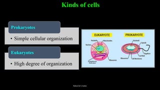
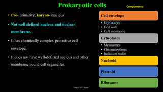
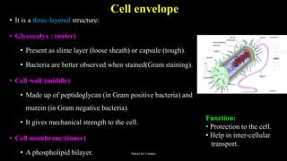
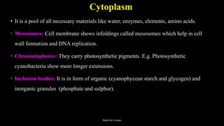
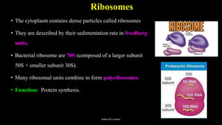
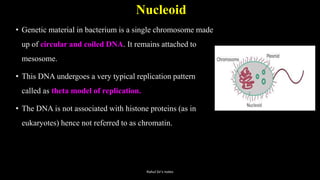
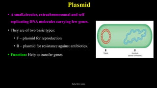
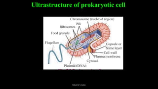
- Prokaryotic cells like bacteria have no nucleus or membrane-bound organelles. They have a cell envelope, cytoplasm, ribosomes, and a nucleoid containing DNA.
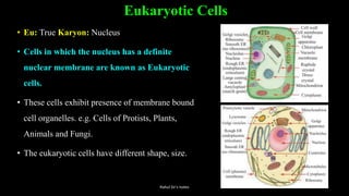
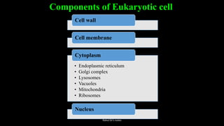
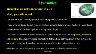
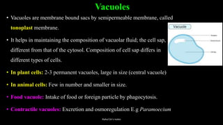
- Eukaryotic cells contain membrane-bound organelles like the nucleus, mitochondria, endoplasmic reticulum, Golgi bodies, lysosomes, and vacuoles. Their genetic material is contained within the nucleus.
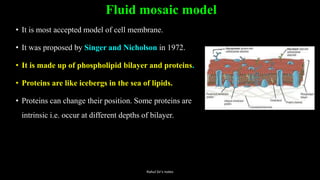
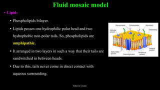
- Cell membranes are semipermeable and made of a phospholipid bilayer. They regulate the passage of materials


![Cell structure function[1]](https://cdn.slidesharecdn.com/ss_thumbnails/cellstructurefunction1-101211104112-phpapp01-thumbnail.jpg?width=640&height=640&fit=bounds)


