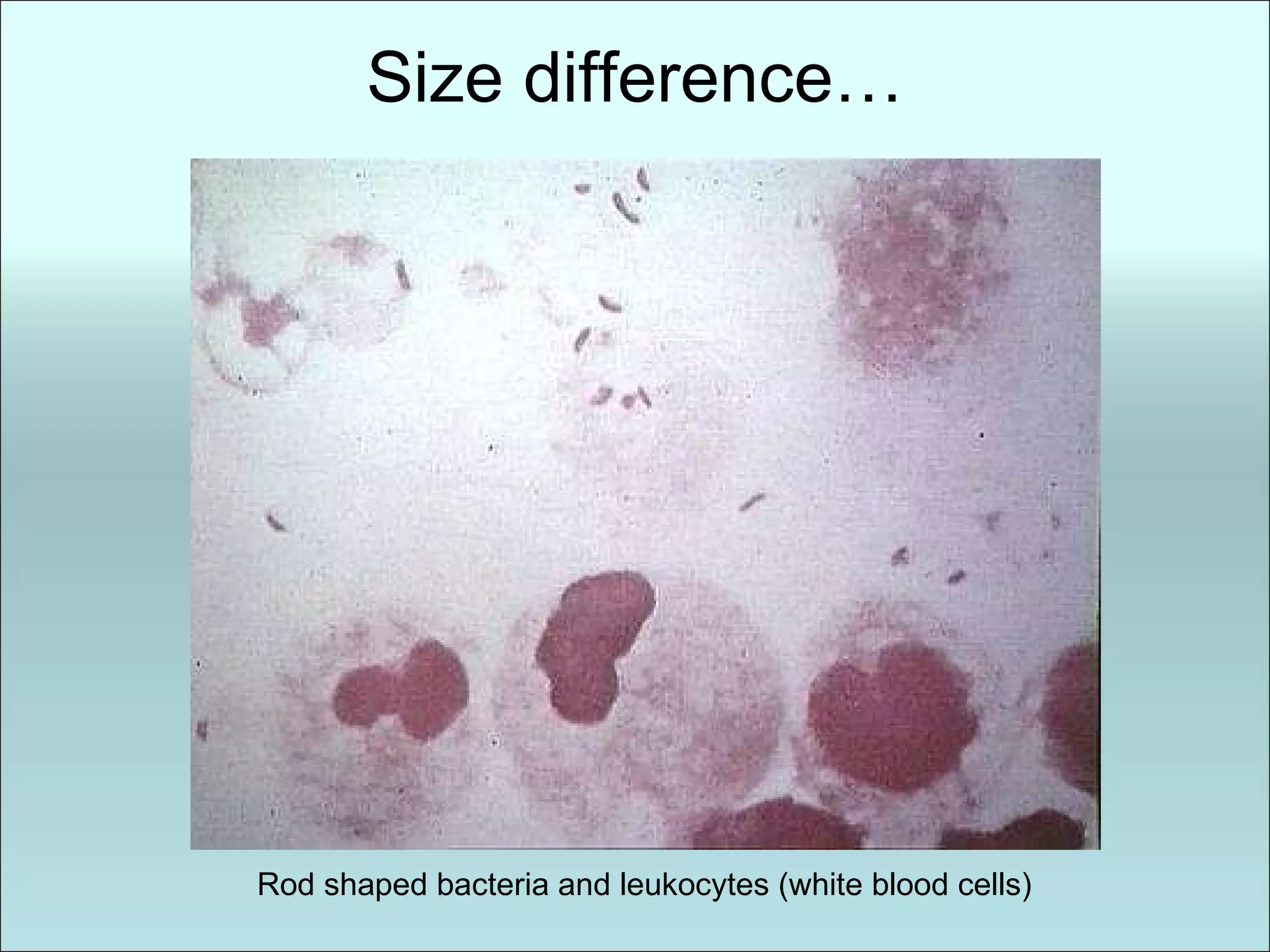
- Prokaryotic cells like bacteria are smaller and less complex than eukaryotic cells, lacking membrane-bound organelles. They have no nucleus, and their DNA is located in the nucleoid region.
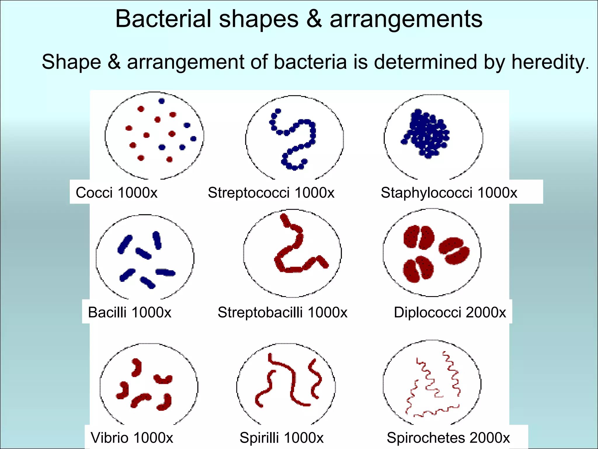
- Bacteria come in different shapes determined by heredity, including coccus, bacillus, spirillum, and spirochetes. They can be motile with flagella or non-motile.
- The bacterial cell contains a cell wall, plasma membrane, cytoplasm, and nucleoid region with circular DNA. Transport across the membrane includes diffusion, facilitated diffusion and active transport.
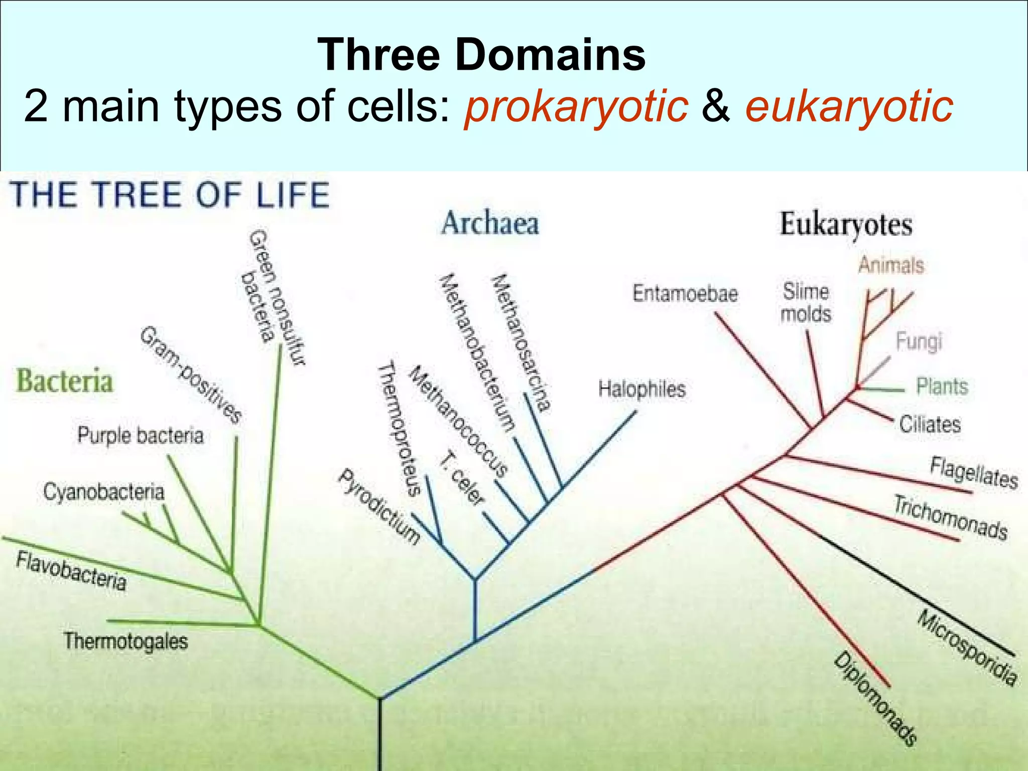
Three Domains 2 main types of cells: prokaryotic & eukaryotic
3.
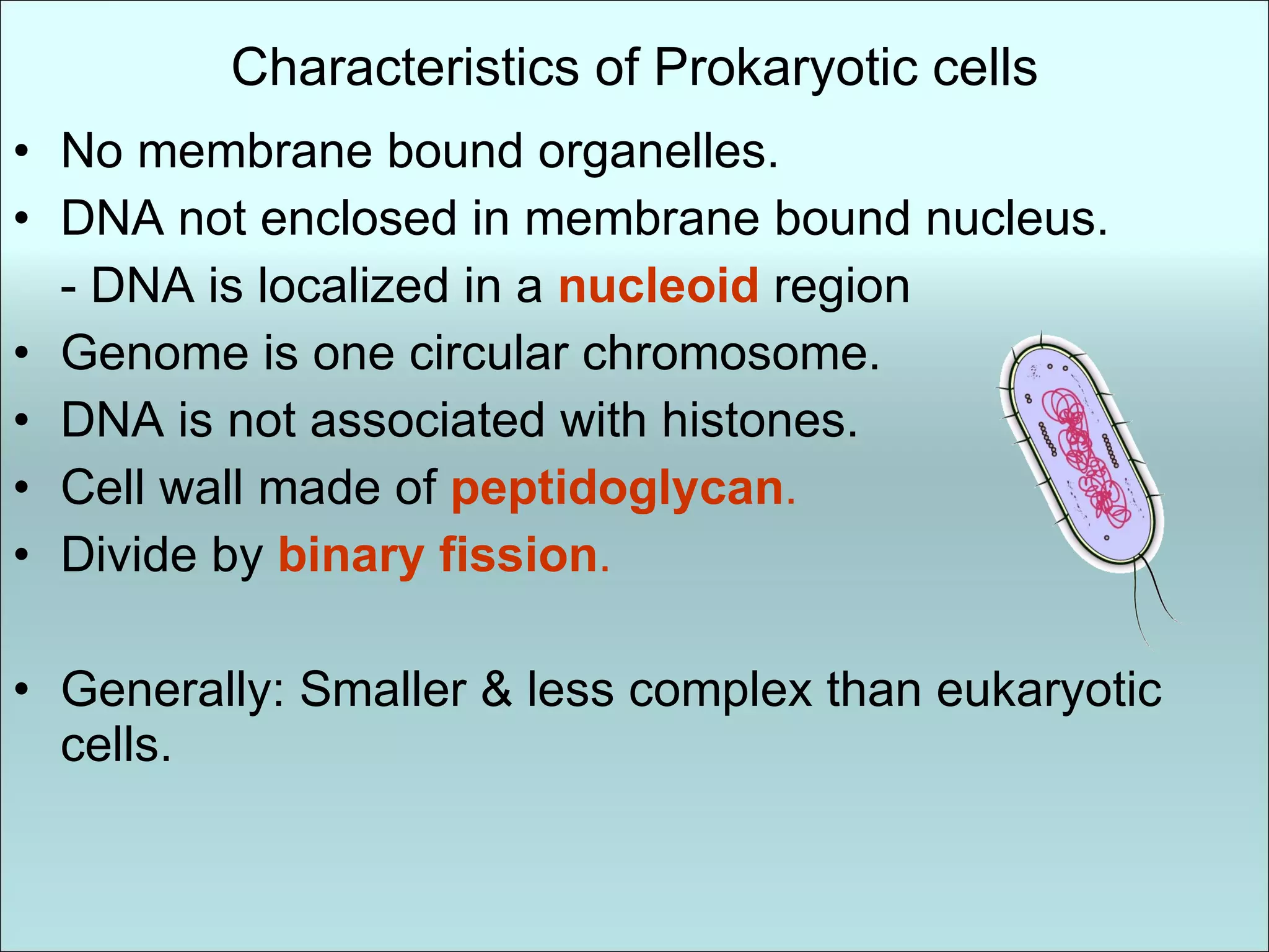
Characteristics of Prokaryoticcells No membrane bound organelles. DNA not enclosed in membrane bound nucleus. - DNA is localized in a nucleoid region Genome is one circular chromosome. DNA is not associated with histones. Cell wall made of peptidoglycan . Divide by binary fission . Generally: Smaller & less complex than eukaryotic cells.
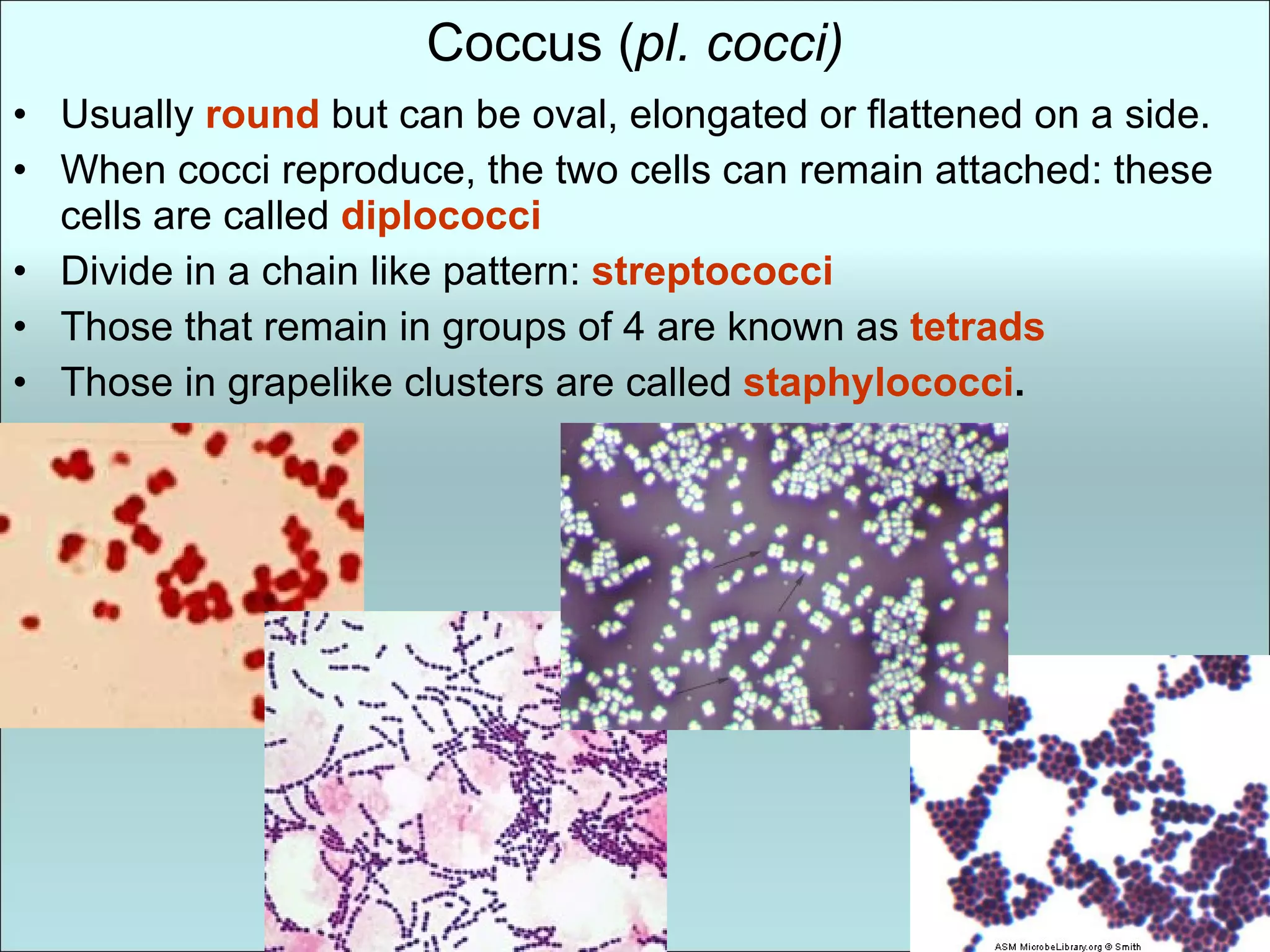
Coccus ( pl.cocci) Usually round but can be oval, elongated or flattened on a side. When cocci reproduce, the two cells can remain attached: these cells are called diplococci Divide in a chain like pattern: streptococci Those that remain in groups of 4 are known as tetrads Those in grapelike clusters are called staphylococci .
7.
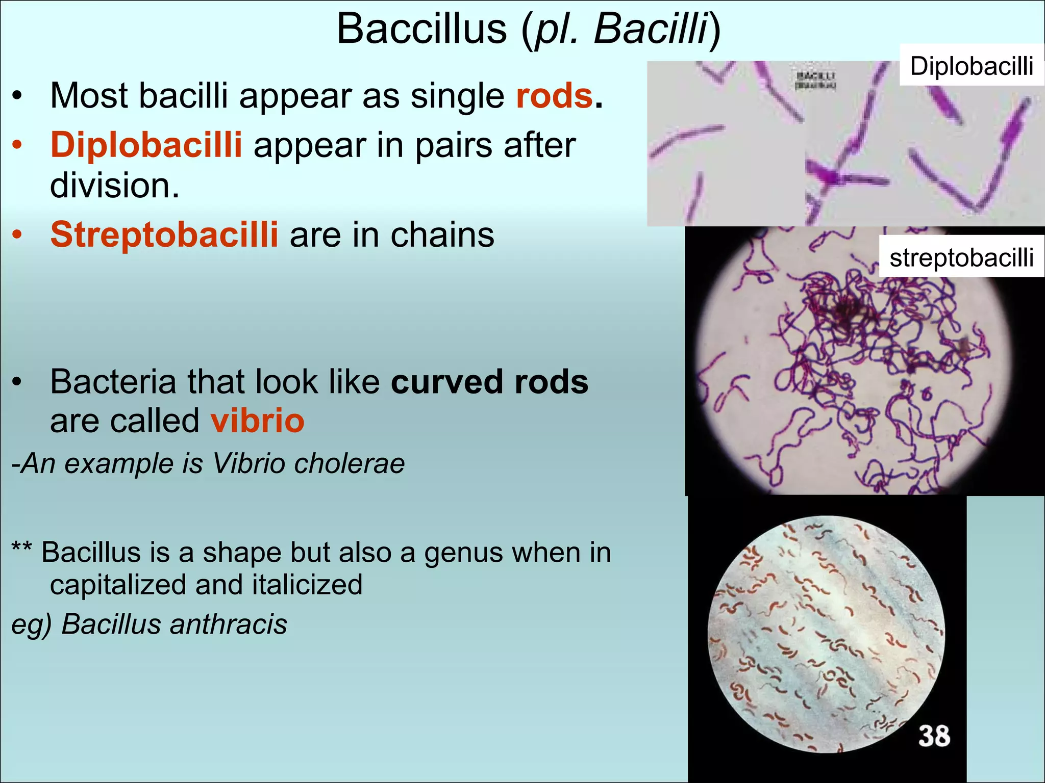
Baccillus ( pl.Bacilli ) Most bacilli appear as single rods . Diplobacilli appear in pairs after division. Streptobacilli are in chains Bacteria that look like curved rods are called vibrio -An example is Vibrio cholerae ** Bacillus is a shape but also a genus when in capitalized and italicized eg) Bacillus anthracis Diplobacilli streptobacilli
8.
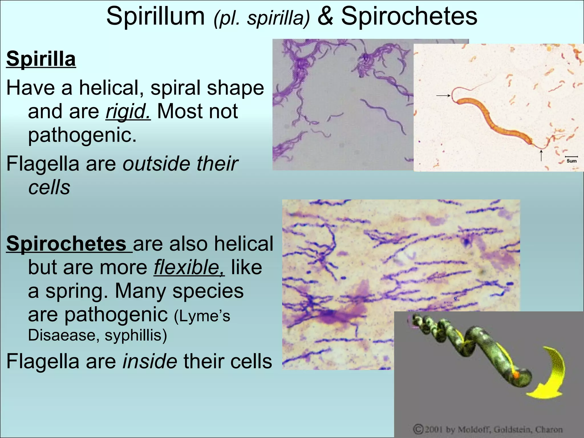
Spirillum (pl.spirilla) & Spirochetes Spirilla Have a helical, spiral shape and are rigid. Most not pathogenic. Flagella are outside their cells Spirochetes are also helical but are more flexible, like a spring. Many species are pathogenic (Lyme’s Disaease, syphillis) Flagella are inside their cells
9.
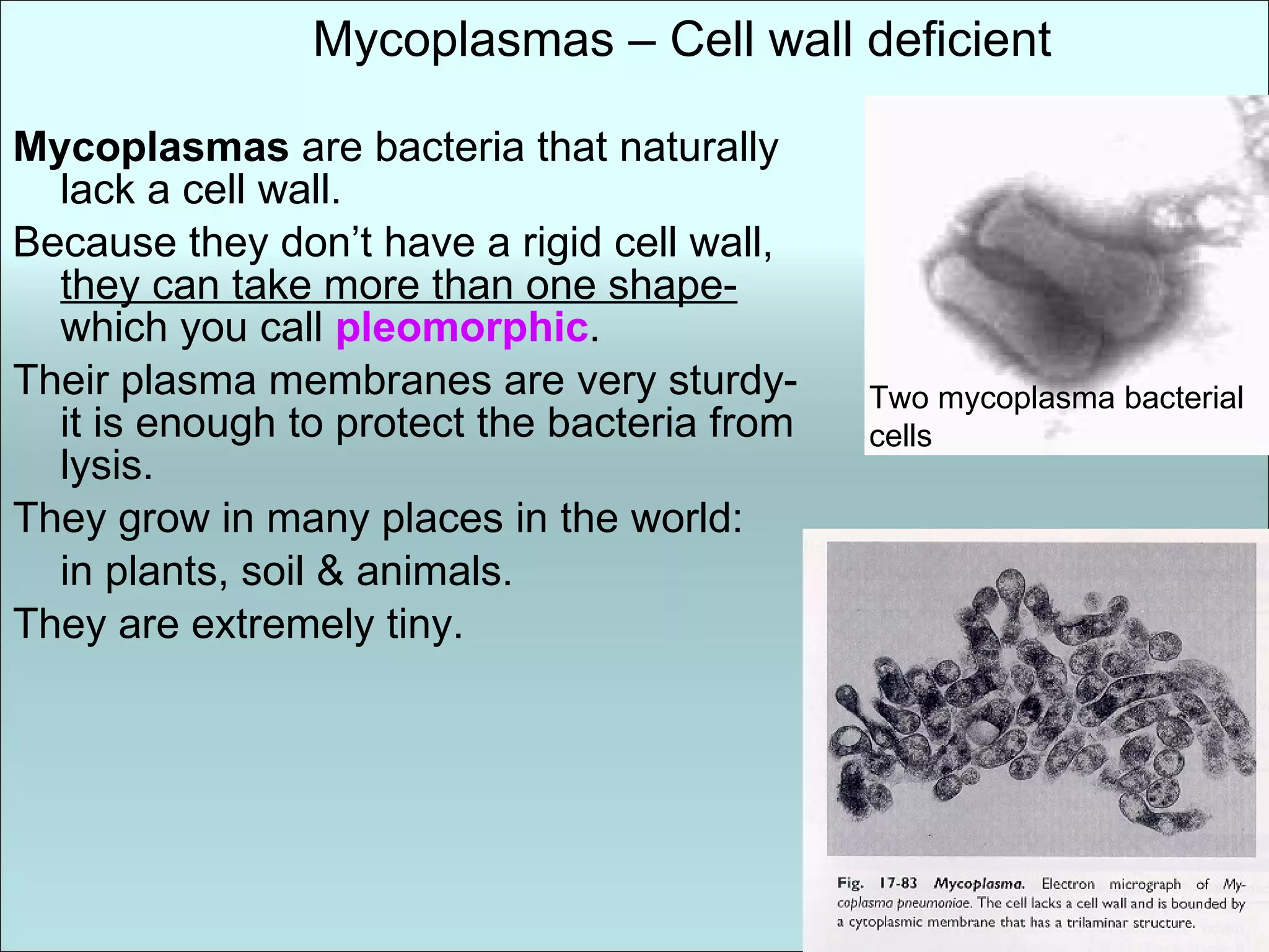
Mycoplasmas arebacteria that naturally lack a cell wall. Because they don’t have a rigid cell wall, they can take more than one shape- which you call pleomorphic . Their plasma membranes are very sturdy- it is enough to protect the bacteria from lysis. They grow in many places in the world: in plants, soil & animals. They are extremely tiny. Mycoplasmas – Cell wall deficient Two mycoplasma bacterial cells
10.
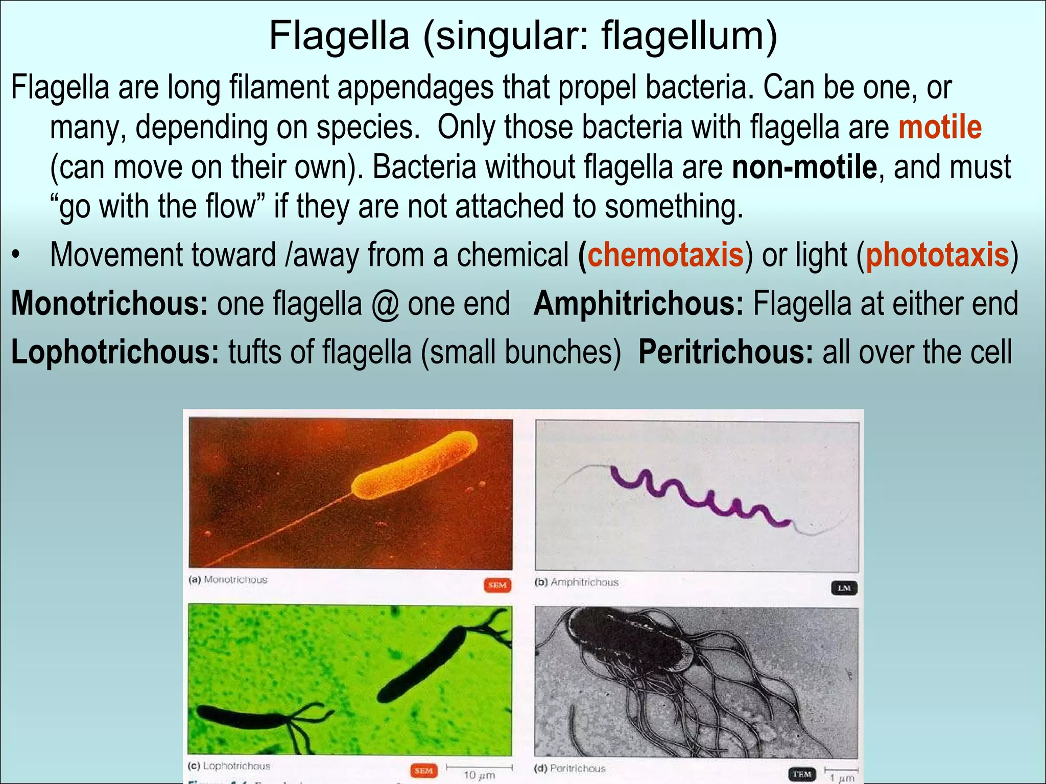
Flagella (singular: flagellum)Flagella are long filament appendages that propel bacteria. Can be one, or many, depending on species. Only those bacteria with flagella are motile (can move on their own). Bacteria without flagella are non-motile , and must “go with the flow” if they are not attached to something. Movement toward /away from a chemical ( chemotaxis ) or light ( phototaxis ) Monotrichous: one flagella @ one end Amphitrichous: Flagella at either end Lophotrichous: tufts of flagella (small bunches) Peritrichous: all over the cell
11.
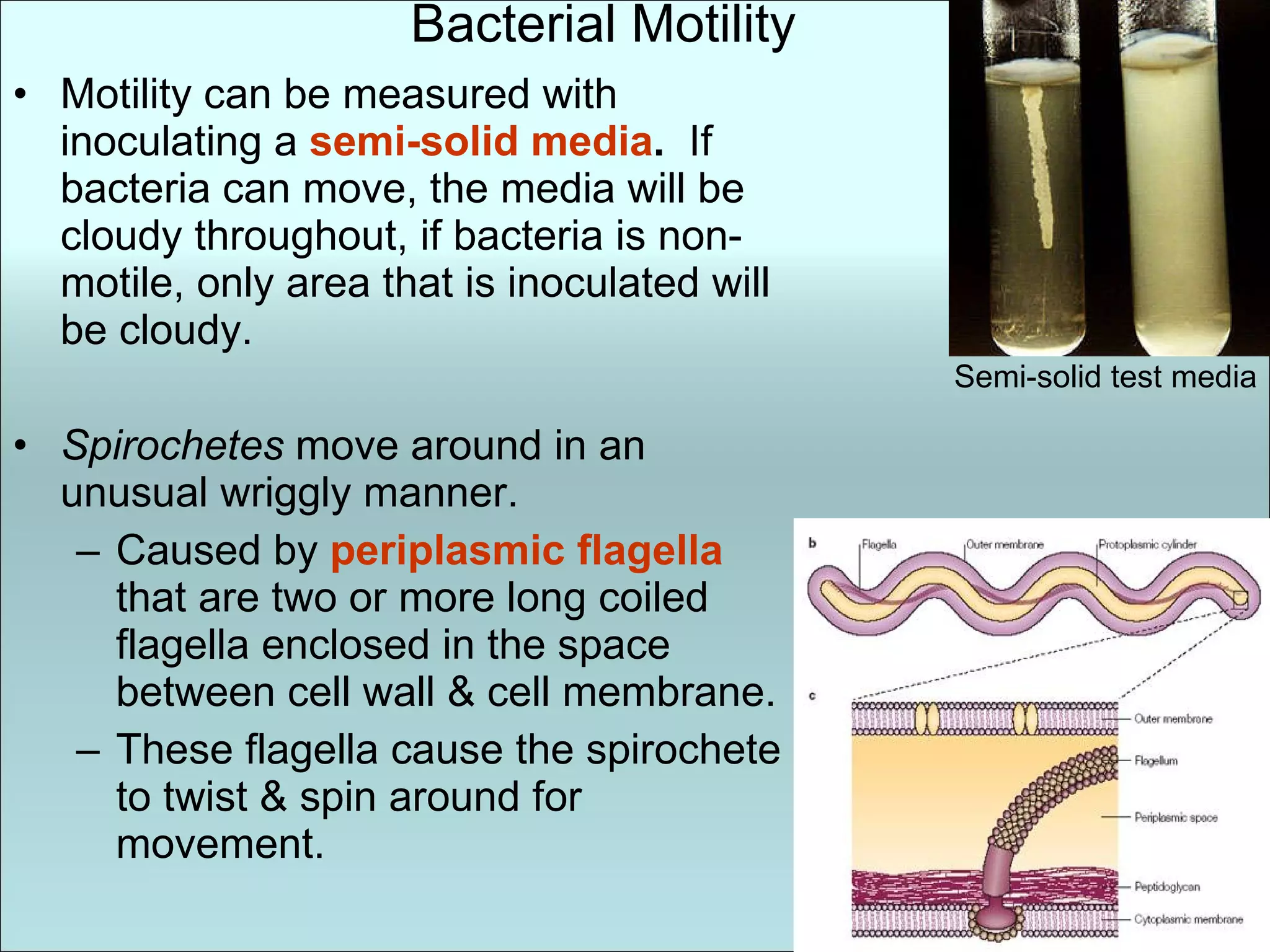
Bacterial Motility Motilitycan be measured with inoculating a semi-solid media . If bacteria can move, the media will be cloudy throughout, if bacteria is non-motile, only area that is inoculated will be cloudy. Spirochetes move around in an unusual wriggly manner. Caused by periplasmic flagella that are two or more long coiled flagella enclosed in the space between cell wall & cell membrane. These flagella cause the spirochete to twist & spin around for movement. Semi-solid test media
12.
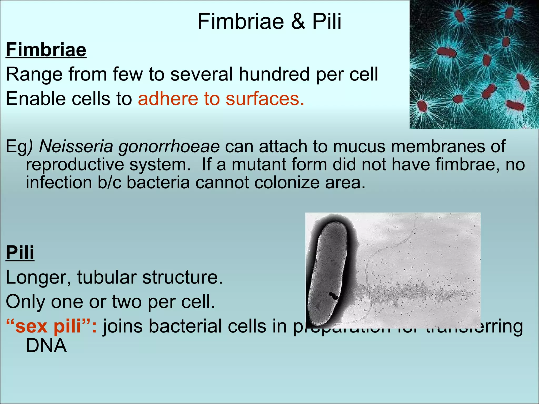
Fimbriae & PiliFimbriae Range from few to several hundred per cell Enable cells to adhere to surfaces. Eg ) Neisseria gonorrhoeae can attach to mucus membranes of reproductive system. If a mutant form did not have fimbrae, no infection b/c bacteria cannot colonize area. Pili Longer, tubular structure. Only one or two per cell. “ sex pili”: joins bacterial cells in preparation for transferring DNA
13.
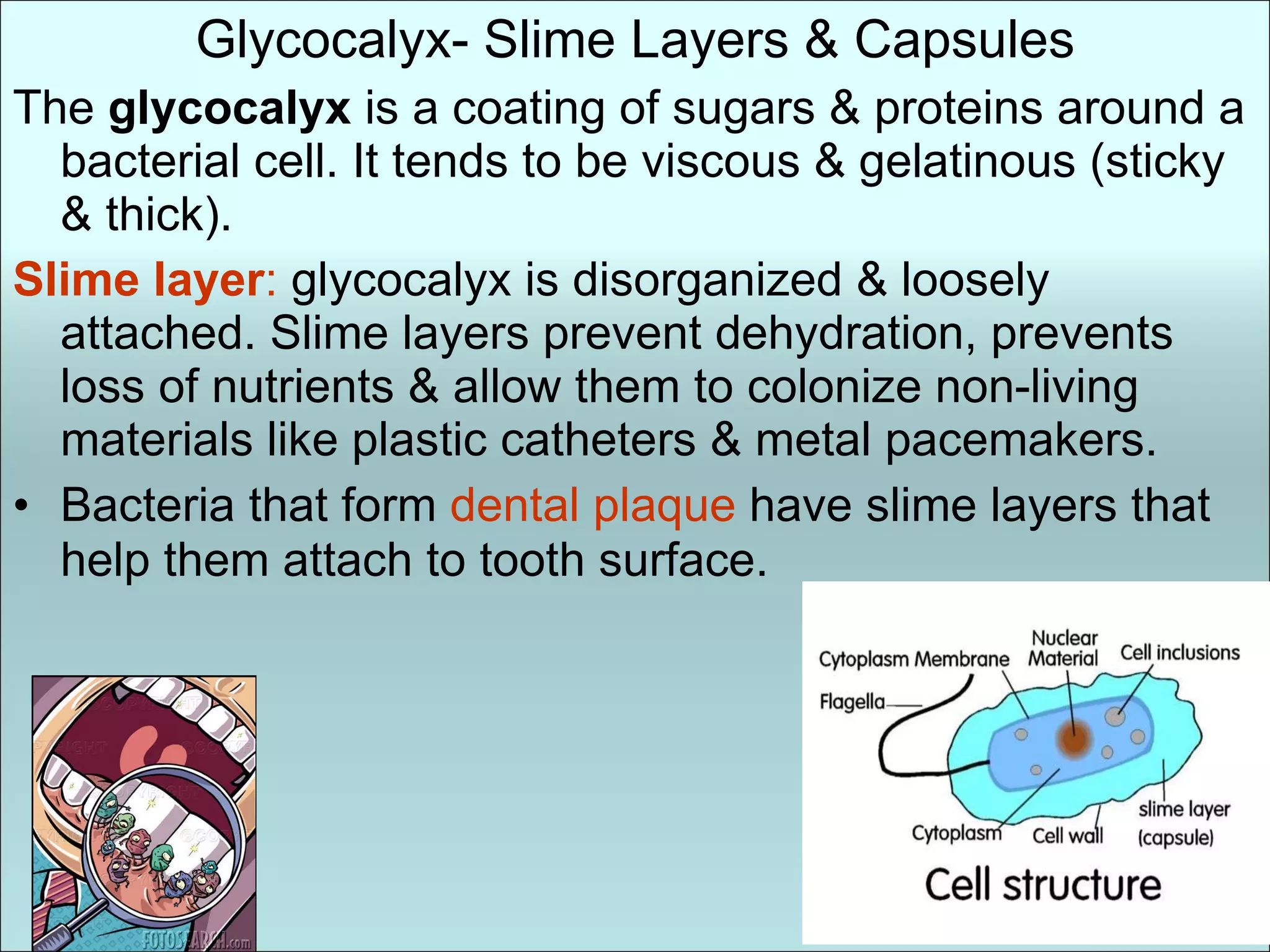
Glycocalyx- Slime Layers& Capsules The glycocalyx is a coating of sugars & proteins around a bacterial cell. It tends to be viscous & gelatinous (sticky & thick). Slime layer : glycocalyx is disorganized & loosely attached. Slime layers prevent dehydration, prevents loss of nutrients & allow them to colonize non-living materials like plastic catheters & metal pacemakers. Bacteria that form dental plaque have slime layers that help them attach to tooth surface .
14.
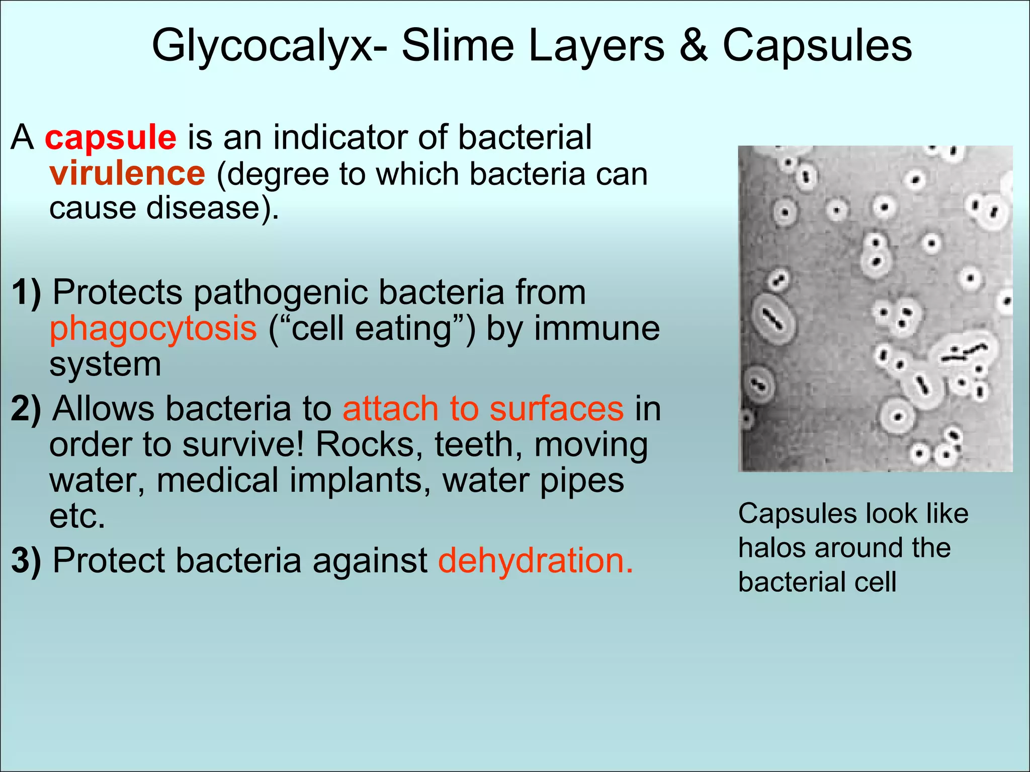
Glycocalyx- Slime Layers& Capsules A capsule is an indicator of bacterial virulence (degree to which bacteria can cause disease). 1) Protects pathogenic bacteria from phagocytosis (“cell eating”) by immune system 2) Allows bacteria to attach to surfaces in order to survive! Rocks, teeth, moving water, medical implants, water pipes etc. 3) Protect bacteria against dehydration. Capsules look like halos around the bacterial cell
15.
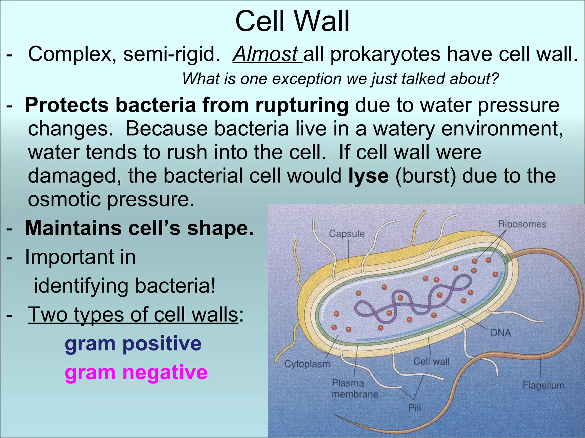
Cell Wall Complex,semi-rigid. Almost all prokaryotes have cell wall. What is one exception we just talked about? - Protects bacteria from rupturing due to water pressure changes. Because bacteria live in a watery environment, water tends to rush into the cell. If cell wall were damaged, the bacterial cell would lyse (burst) due to the osmotic pressure. - Maintains cell’s shape. - Important in identifying bacteria! Two types of cell walls : gram positive gram negative
16.
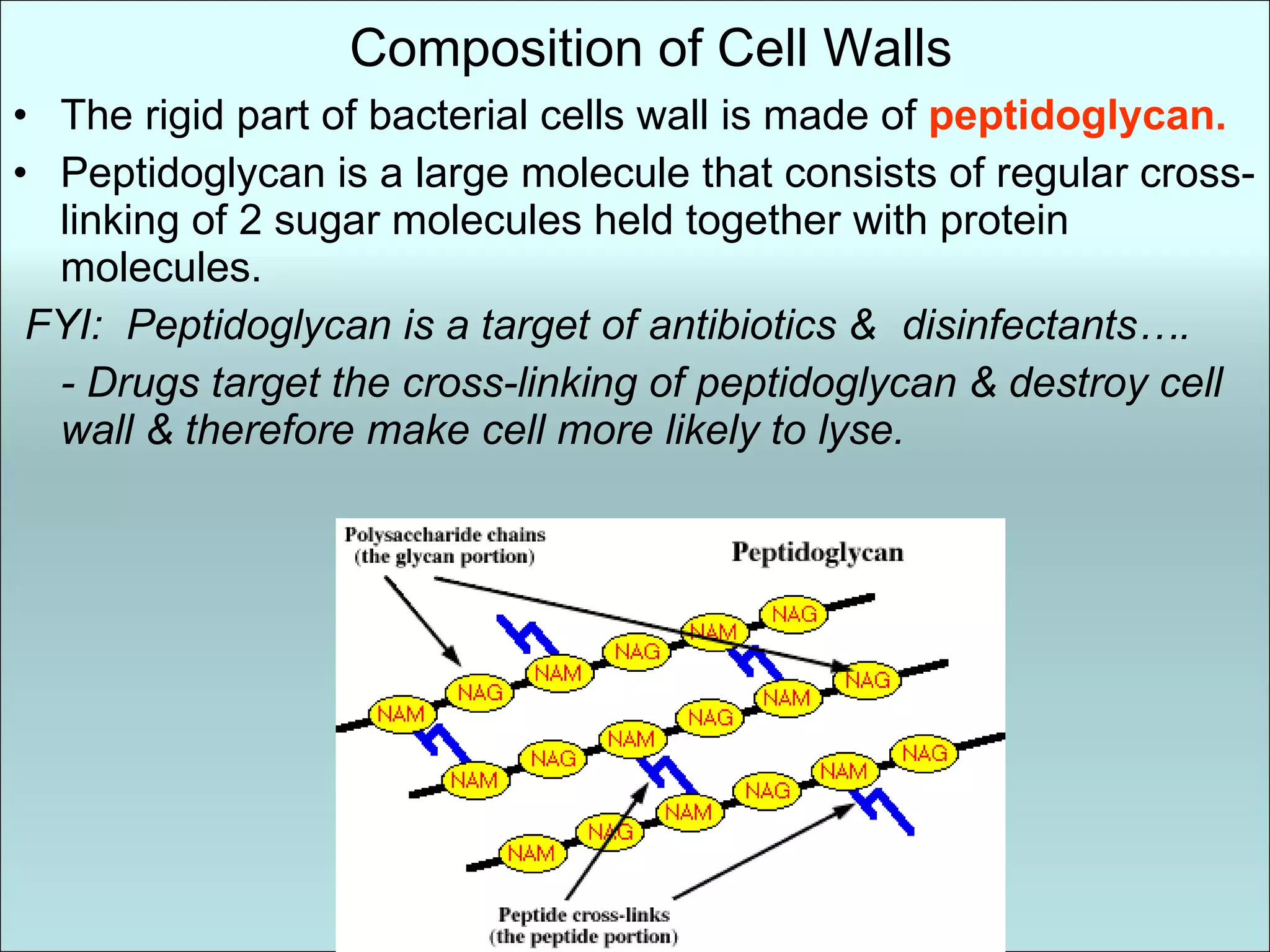
Composition of CellWalls The rigid part of bacterial cells wall is made of peptidoglycan. Peptidoglycan is a large molecule that consists of regular cross-linking of 2 sugar molecules held together with protein molecules. FYI: Peptidoglycan is a target of antibiotics & disinfectants…. - Drugs target the cross-linking of peptidoglycan & destroy cell wall & therefore make cell more likely to lyse.
17.
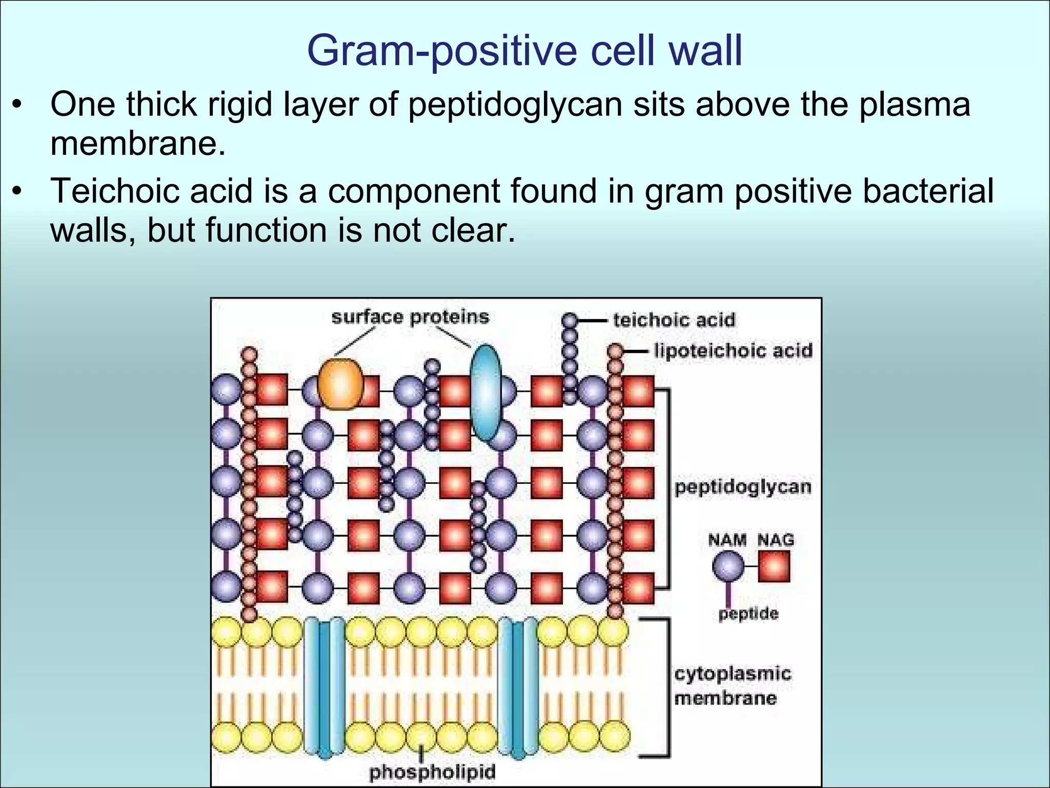
Gram-positive cell wallOne thick rigid layer of peptidoglycan sits above the plasma membrane. Teichoic acid is a component found in gram positive bacterial walls, but function is not clear.
18.
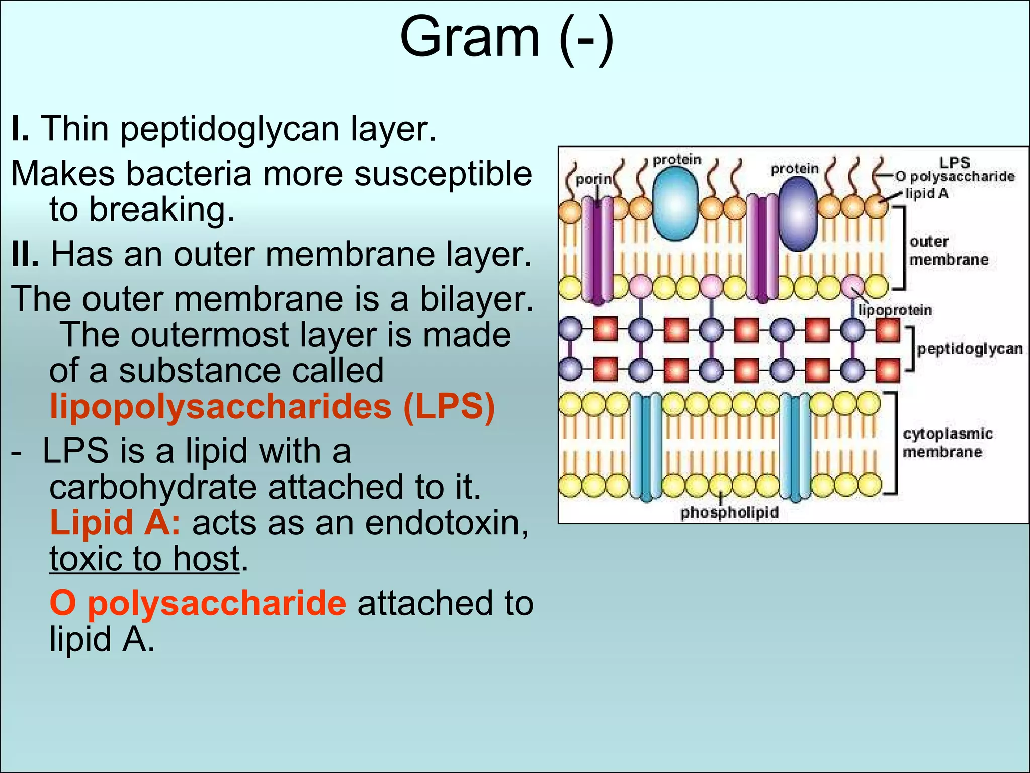
Gram (-) I. Thin peptidoglycan layer. Makes bacteria more susceptible to breaking. II. Has an outer membrane layer. The outer membrane is a bilayer. The outermost layer is made of a substance called lipopolysaccharides (LPS) - LPS is a lipid with a carbohydrate attached to it. Lipid A: acts as an endotoxin, toxic to host . O polysaccharide attached to lipid A.
19.
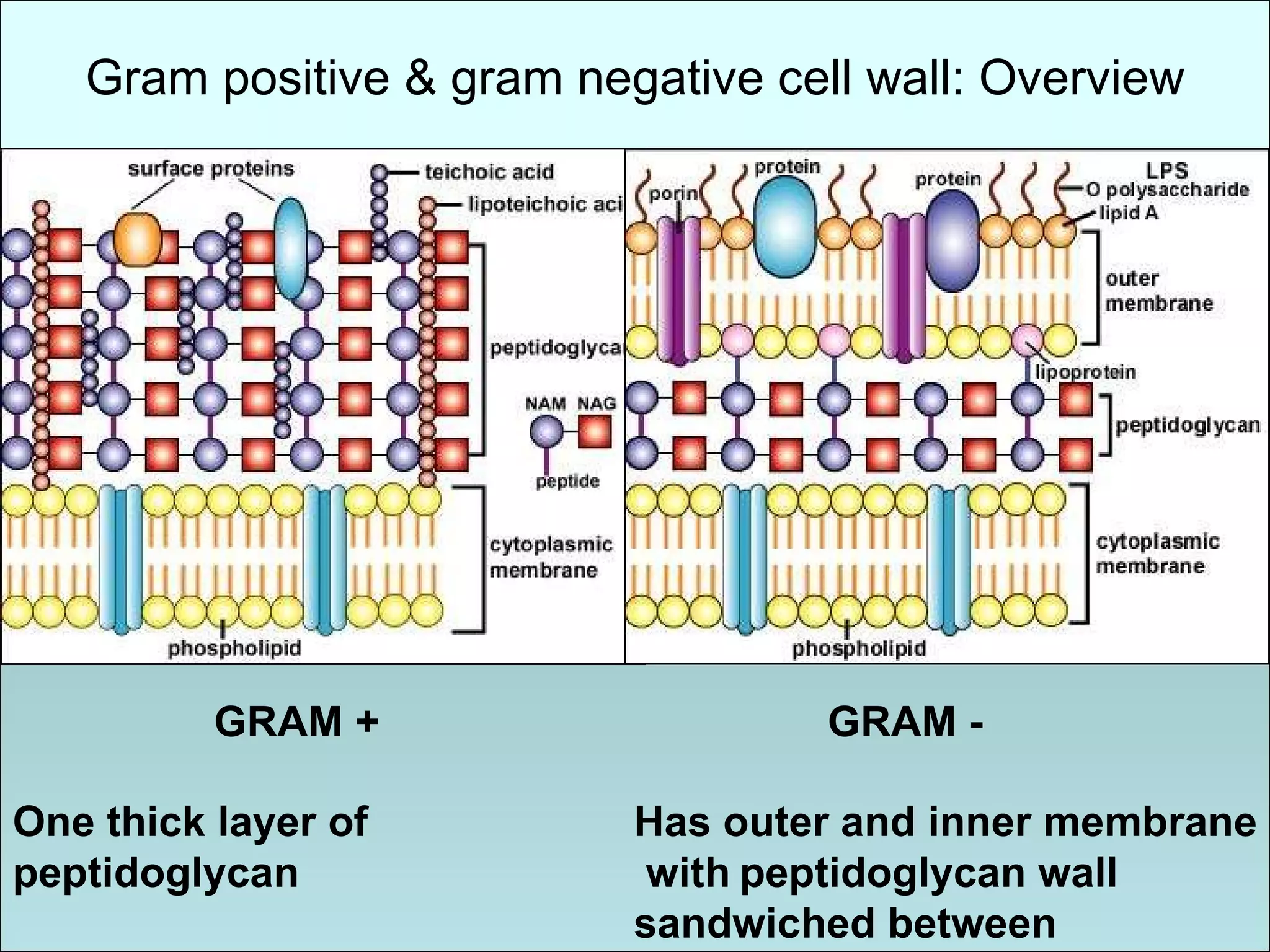
Gram positive &gram negative cell wall: Overview One thick layer of peptidoglycan Has outer and inner membrane with peptidoglycan wall sandwiched between GRAM + GRAM -
20.
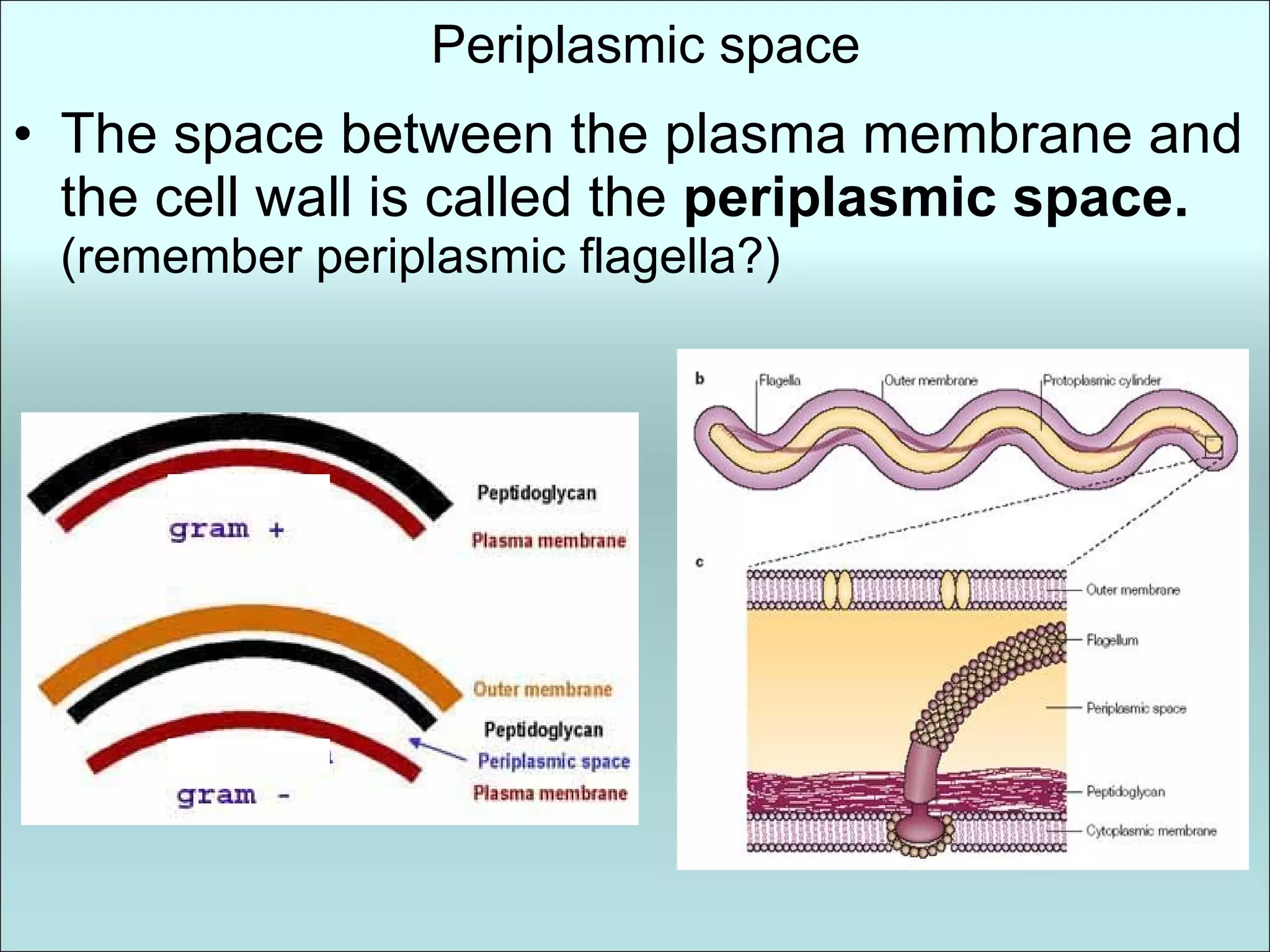
Periplasmic space Thespace between the plasma membrane and the cell wall is called the periplasmic space. (remember periplasmic flagella?)
21.
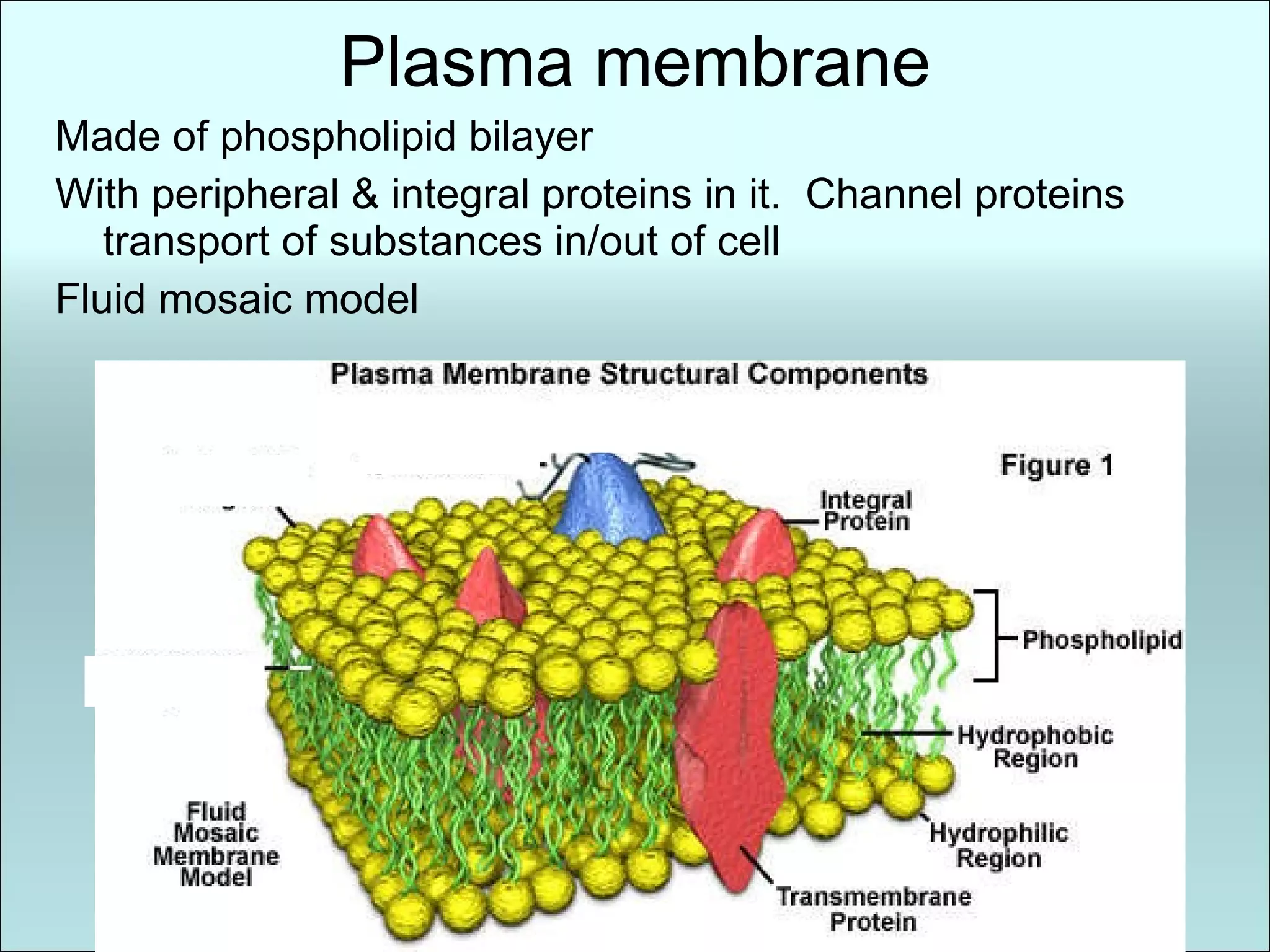
Plasma membrane Madeof phospholipid bilayer With peripheral & integral proteins in it. Channel proteins transport of substances in/out of cell Fluid mosaic model
22.
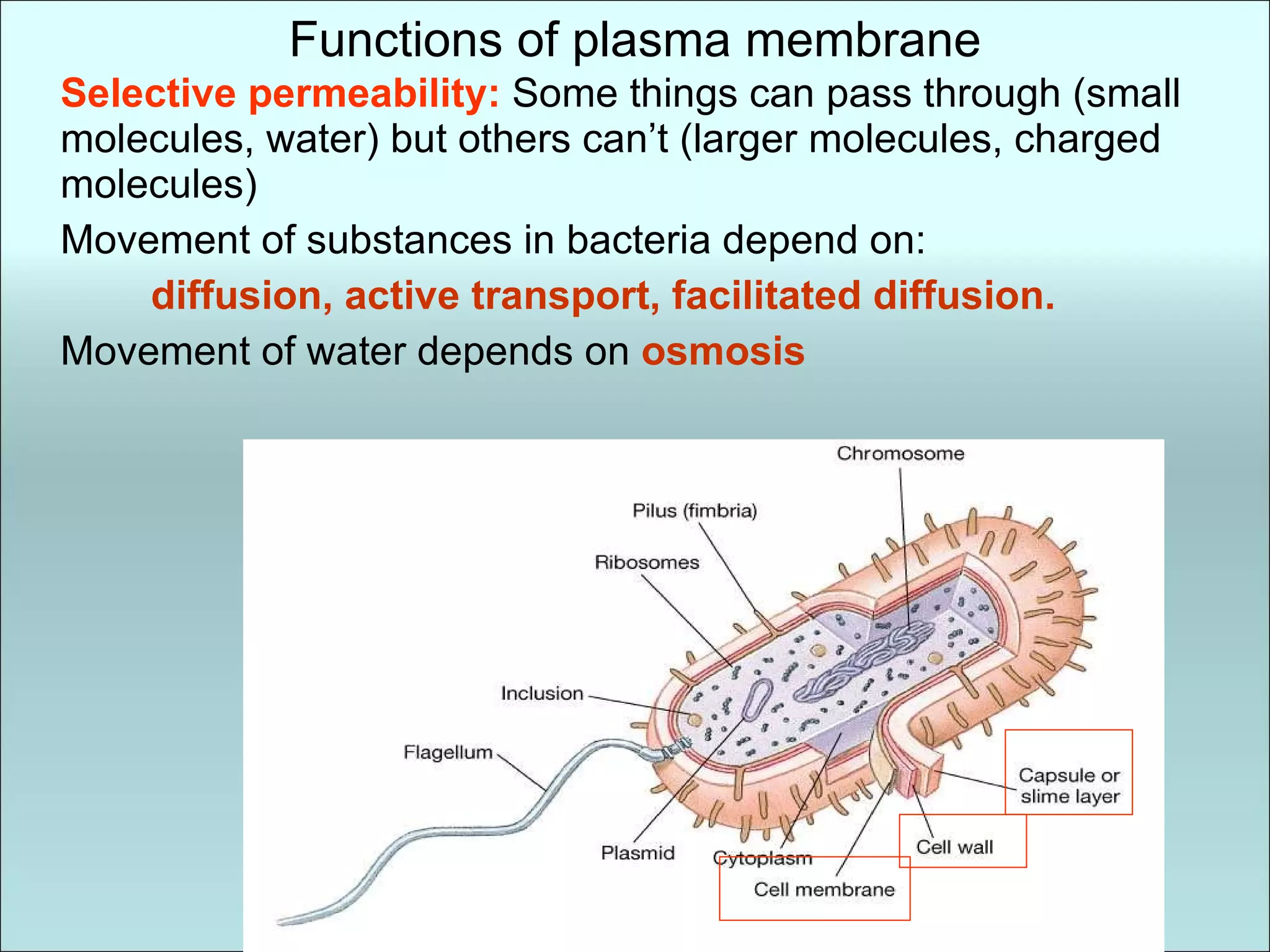
Functions of plasmamembrane Selective permeability: Some things can pass through (small molecules, water) but others can’t (larger molecules, charged molecules) Movement of substances in bacteria depend on: diffusion, active transport, facilitated diffusion. Movement of water depends on osmosis
23.
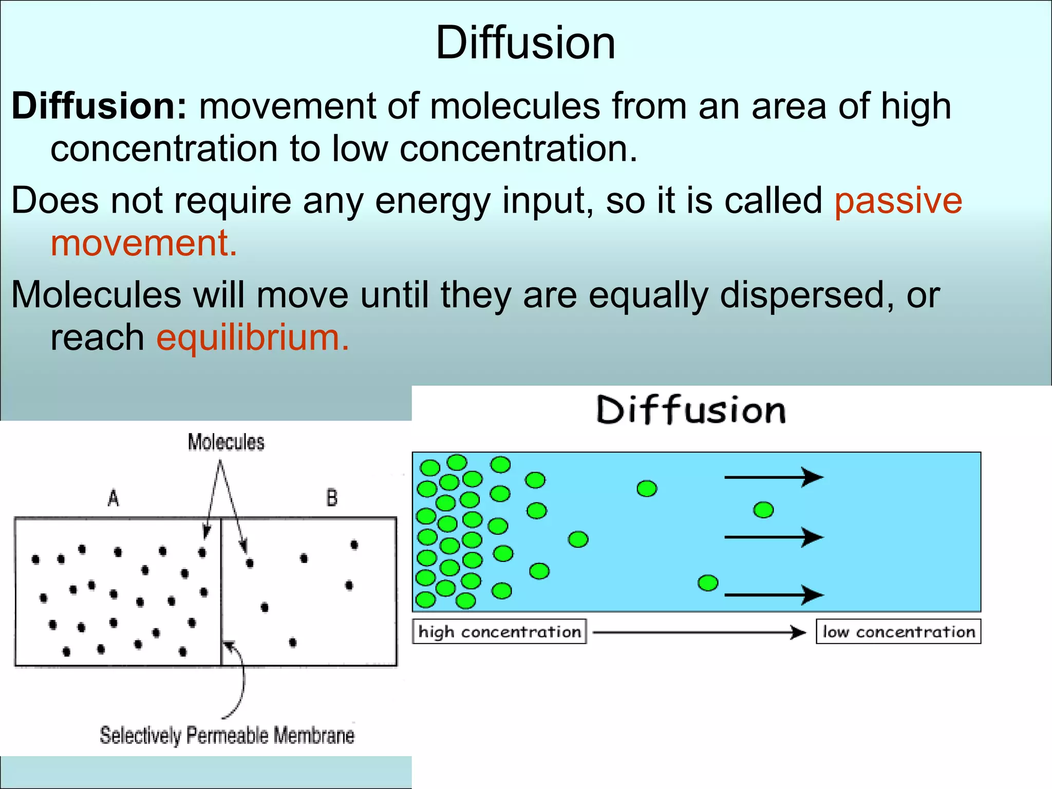
Diffusion Diffusion: movement of molecules from an area of high concentration to low concentration. Does not require any energy input, so it is called passive movement. Molecules will move until they are equally dispersed, or reach equilibrium.
24.
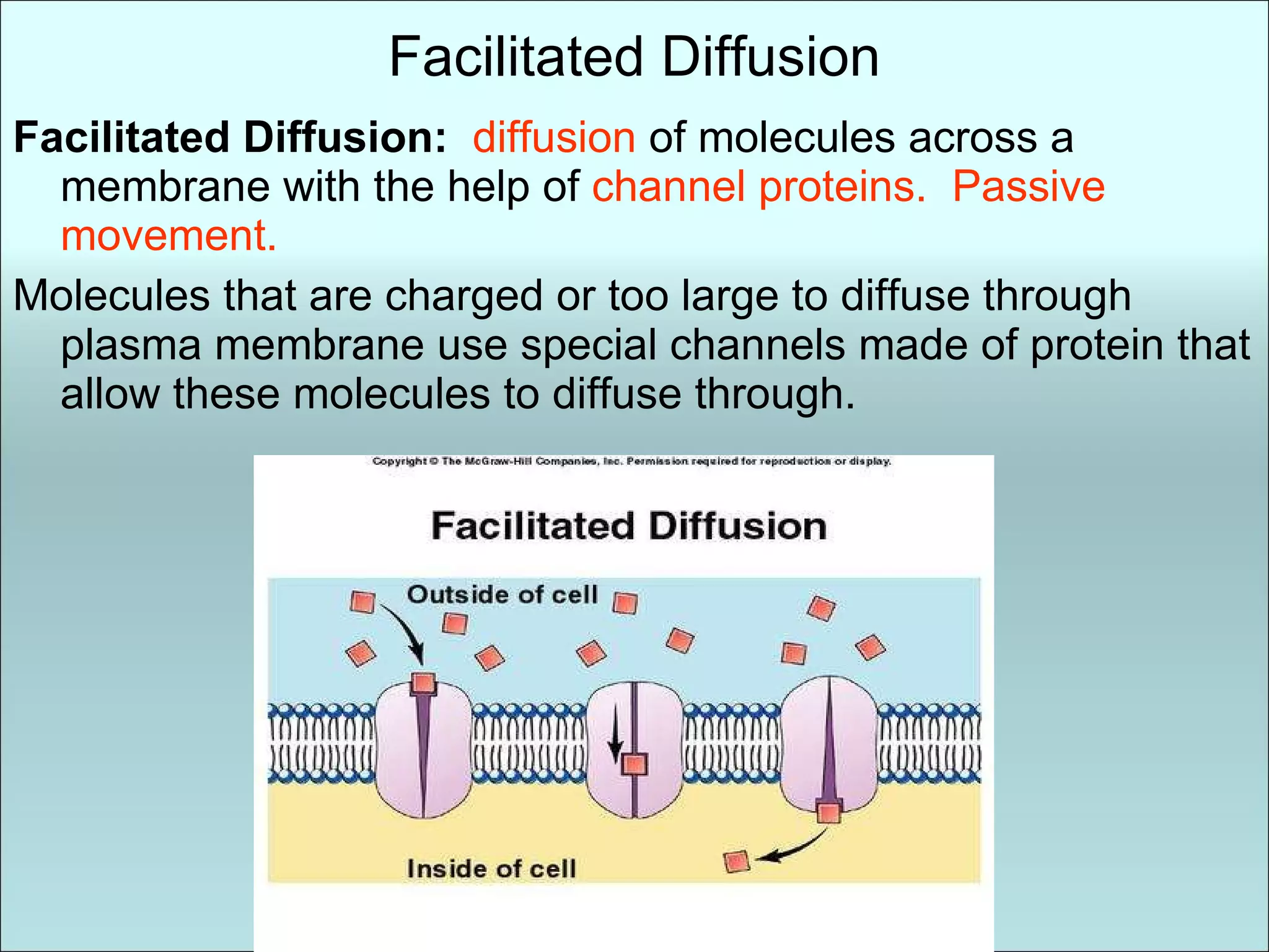
Facilitated Diffusion FacilitatedDiffusion: diffusion of molecules across a membrane with the help of channel proteins. Passive movement. Molecules that are charged or too large to diffuse through plasma membrane use special channels made of protein that allow these molecules to diffuse through.
25.
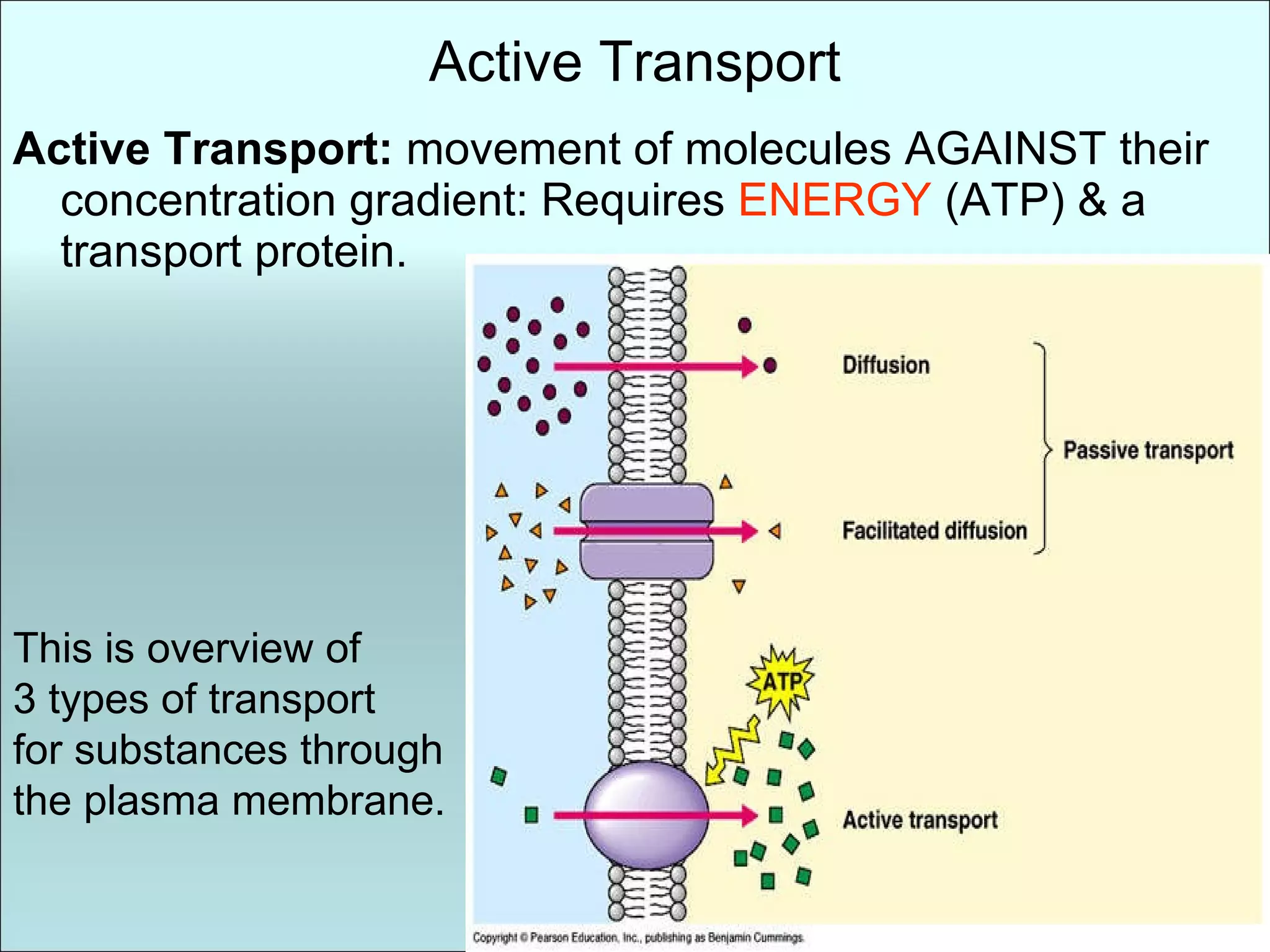
Active Transport ActiveTransport: movement of molecules AGAINST their concentration gradient: Requires ENERGY (ATP) & a transport protein. This is overview of 3 types of transport for substances through the plasma membrane.
26.
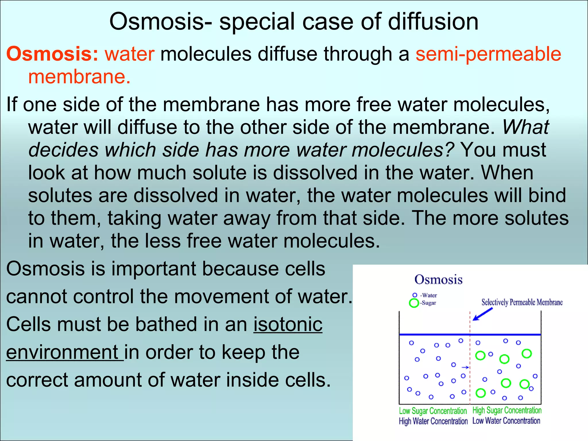
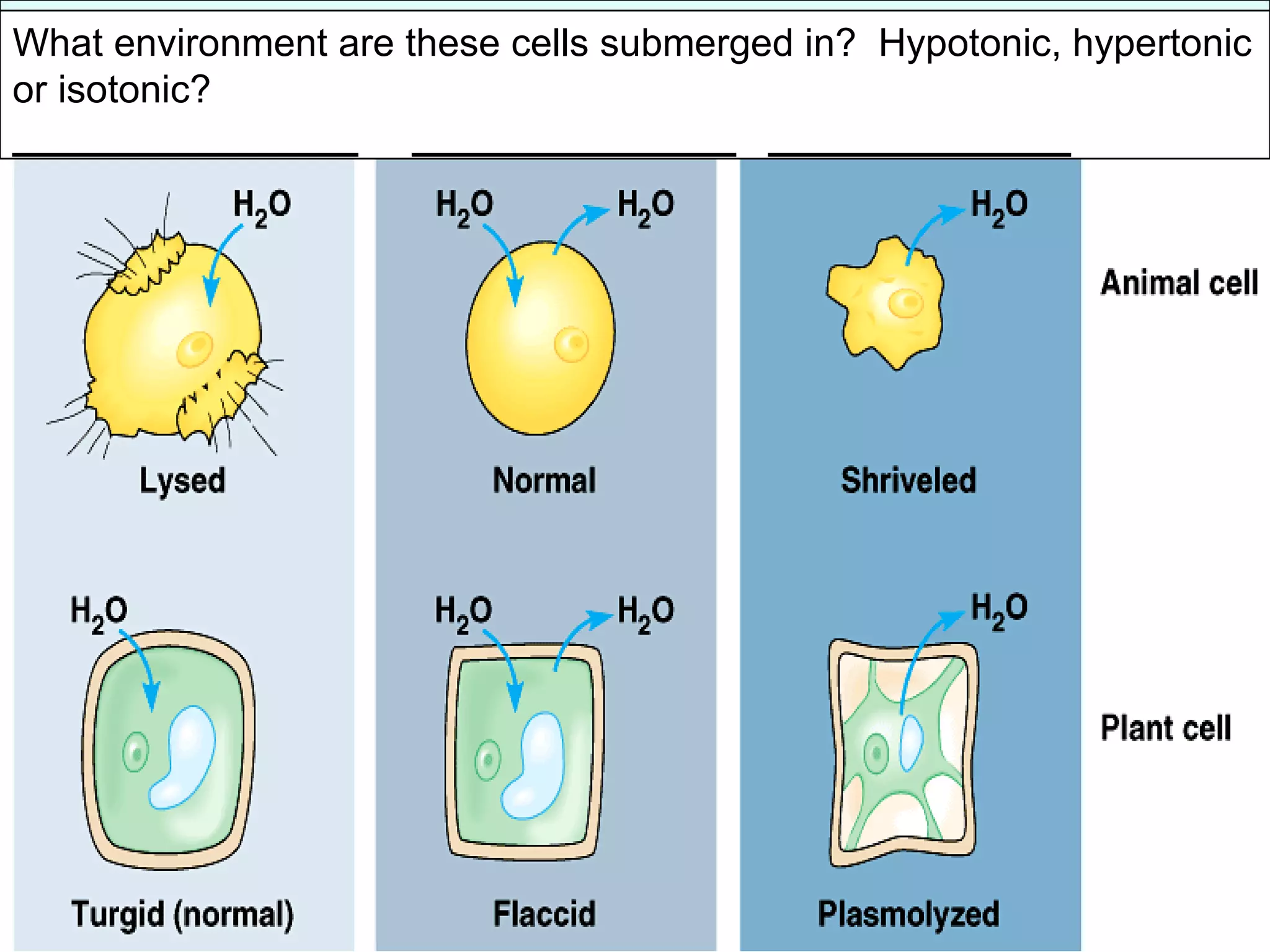
Osmosis- special caseof diffusion Osmosis: water molecules diffuse through a semi-permeable membrane. If one side of the membrane has more free water molecules, water will diffuse to the other side of the membrane. What decides which side has more water molecules? You must look at how much solute is dissolved in the water. When solutes are dissolved in water, the water molecules will bind to them, taking water away from that side. The more solutes in water, the less free water molecules. Osmosis is important because cells cannot control the movement of water. Cells must be bathed in an isotonic environment in order to keep the correct amount of water inside cells.
27.
When comparing twosolutions of water: The solution with the higher concentration of solutes is hypertonic to the other. The solution with the lower concentration of solutes is hypotonic to the other. Solution : homogenous mixture of solute & solvent Solute: What is being dissolved, the solid substance. eg) Salt Solvent: What is doing the dissolving. The liquid. eg) water Osmosis Animated tutorial Another tutorial with quiz
28.
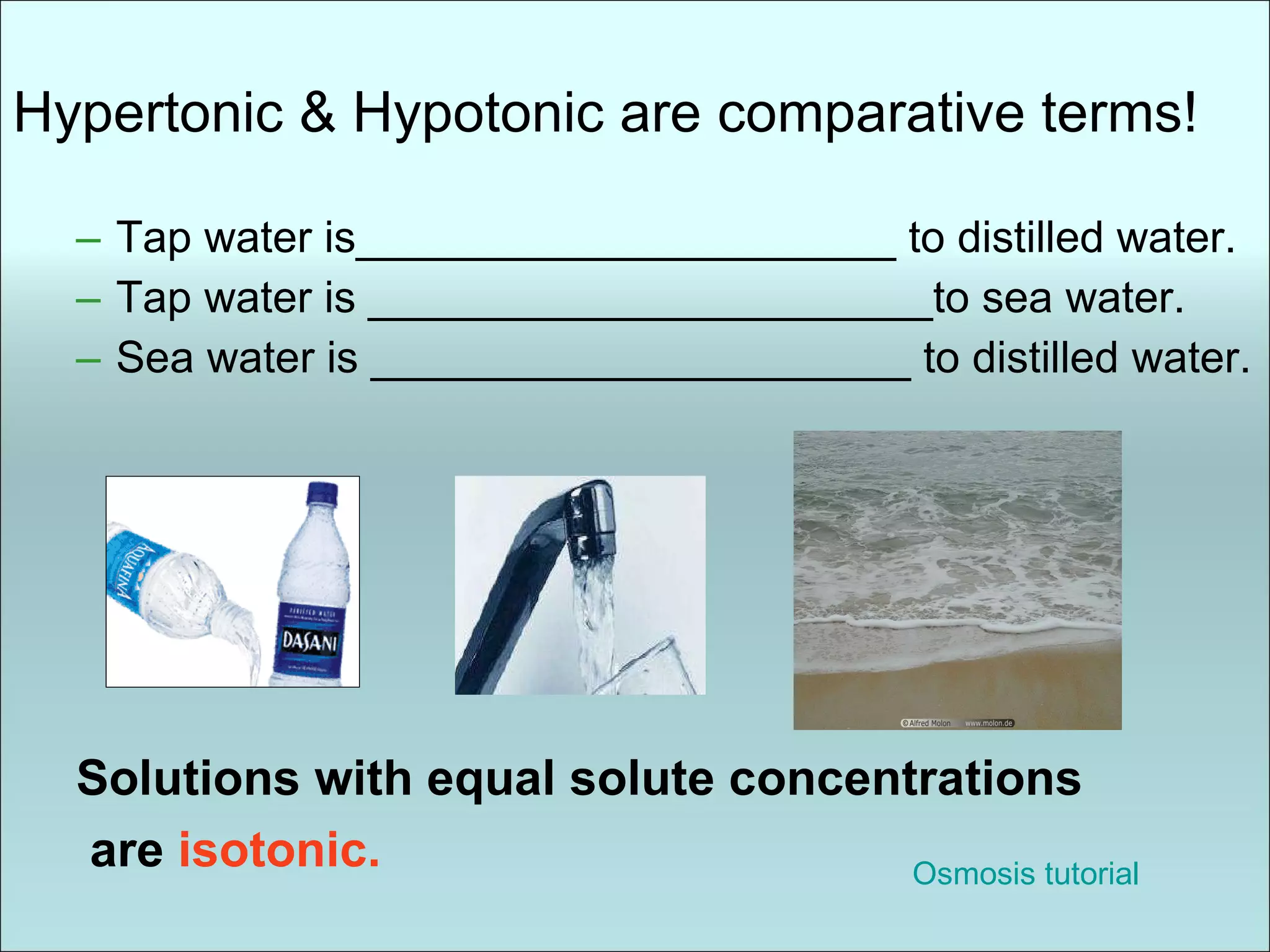
Hypertonic & Hypotonicare comparative terms! Tap water is______________________ to distilled water. Tap water is _______________________to sea water. Sea water is ______________________ to distilled water. Solutions with equal solute concentrations are isotonic. Osmosis tutorial
29.
What environment arethese cells submerged in? Hypotonic, hypertonic or isotonic? ________________ _______________ ______________
30.
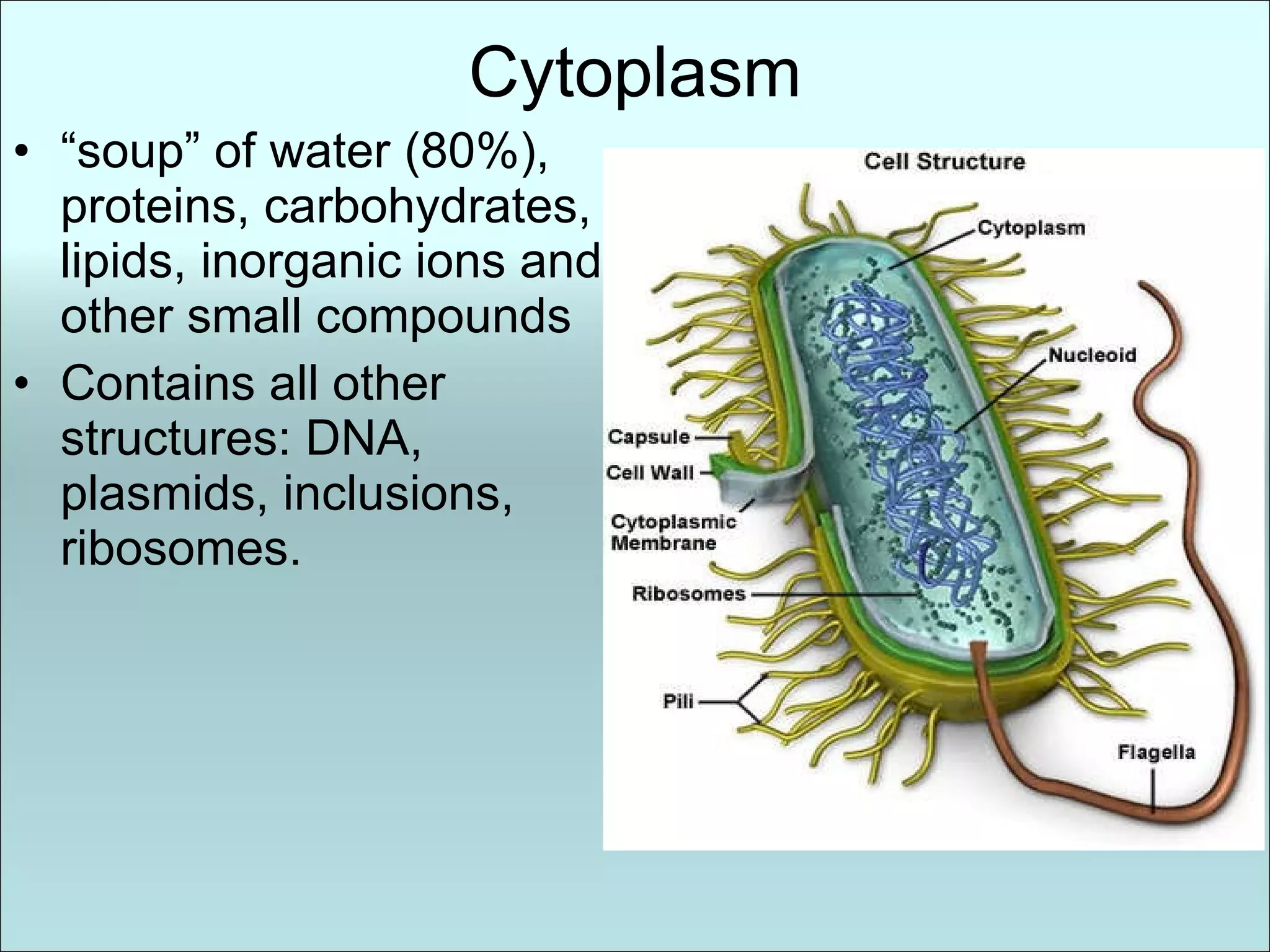
Cytoplasm “ soup”of water (80%), proteins, carbohydrates, lipids, inorganic ions and other small compounds Contains all other structures: DNA, plasmids, inclusions, ribosomes.
31.
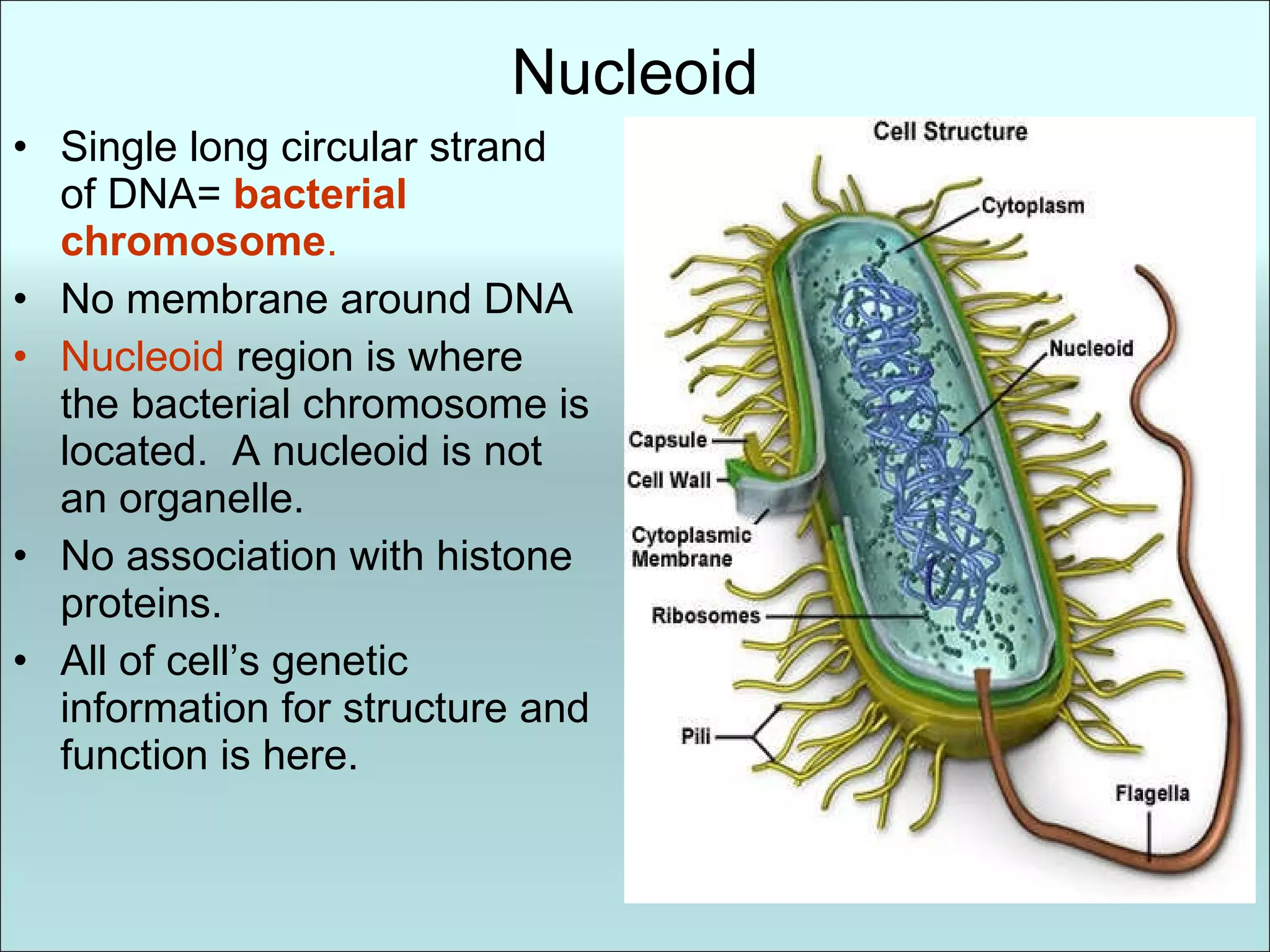
Nucleoid Single longcircular strand of DNA= bacterial chromosome . No membrane around DNA Nucleoid region is where the bacterial chromosome is located. A nucleoid is not an organelle. No association with histone proteins. All of cell’s genetic information for structure and function is here.
32.
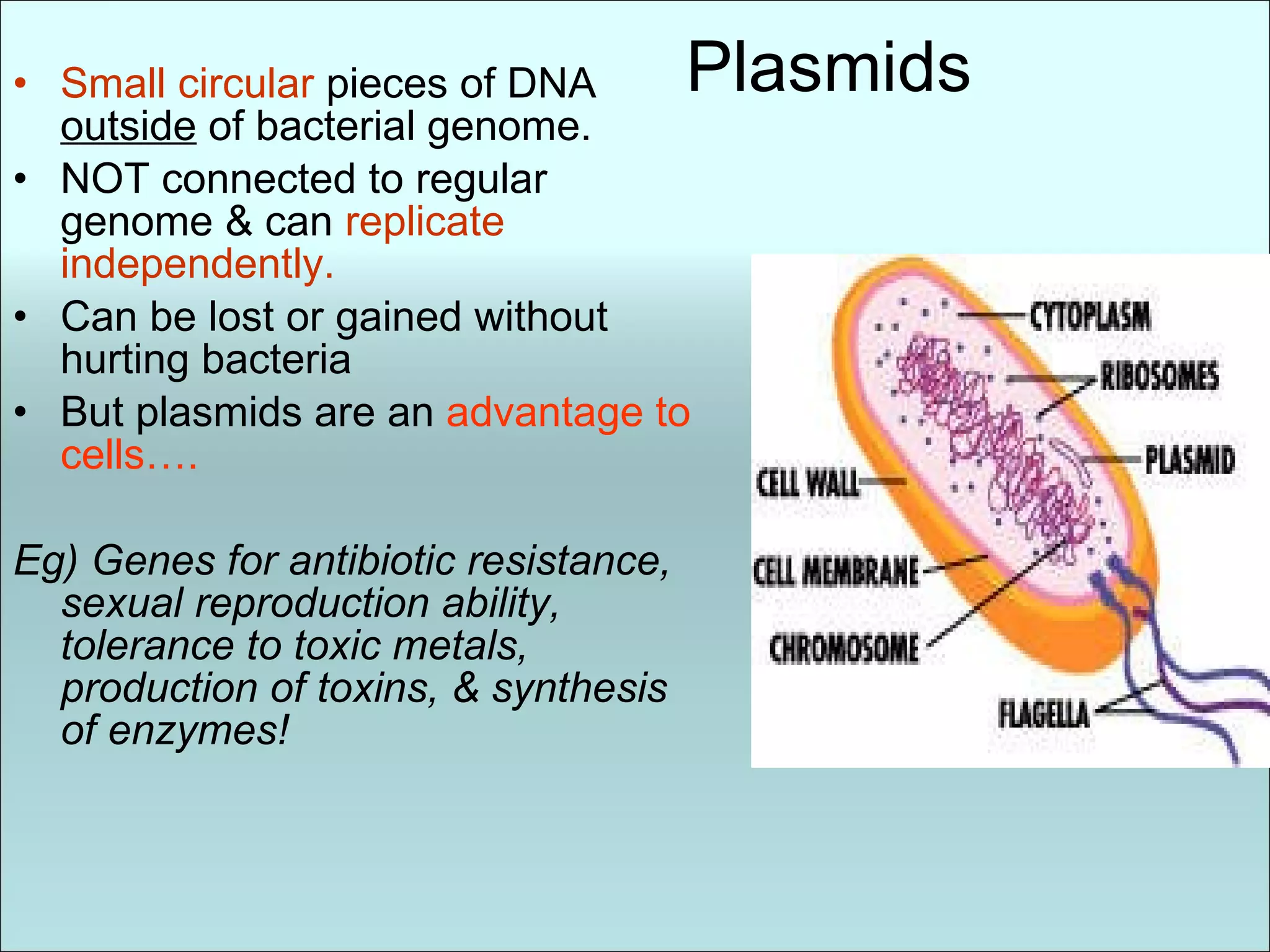
Plasmids Small circular pieces of DNA outside of bacterial genome. NOT connected to regular genome & can replicate independently. Can be lost or gained without hurting bacteria But plasmids are an advantage to cells…. Eg) Genes for antibiotic resistance, sexual reproduction ability, tolerance to toxic metals, production of toxins, & synthesis of enzymes!
33.
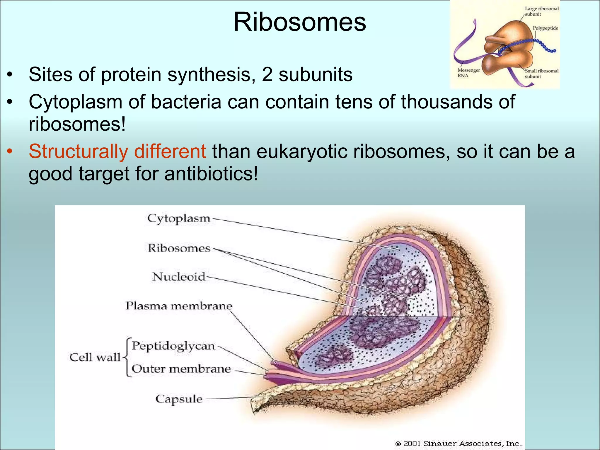
Ribosomes Sites ofprotein synthesis, 2 subunits Cytoplasm of bacteria can contain tens of thousands of ribosomes! Structurally different than eukaryotic ribosomes, so it can be a good target for antibiotics!
34.
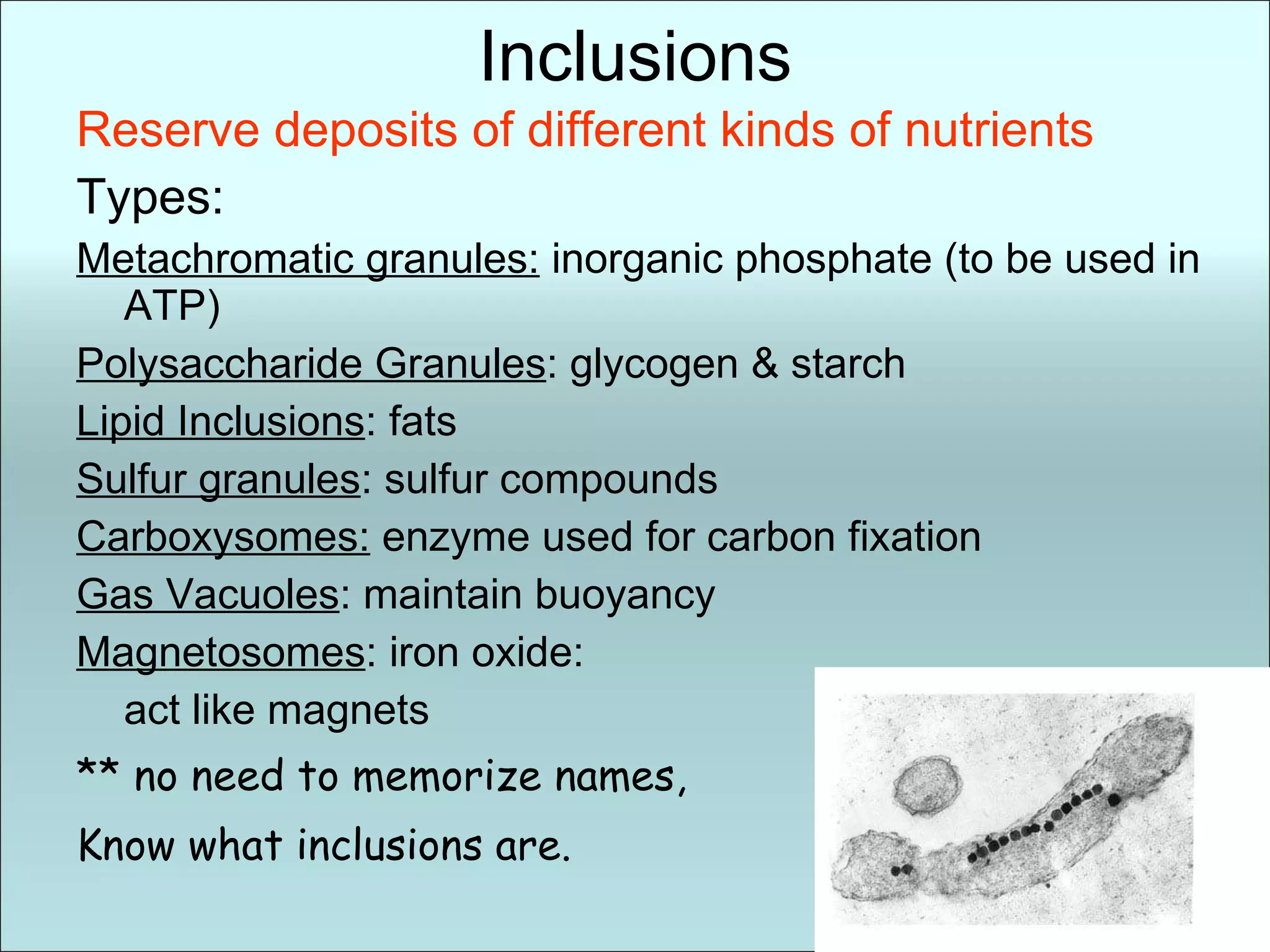
Inclusions Reserve depositsof different kinds of nutrients Types: Metachromatic granules: inorganic phosphate (to be used in ATP) Polysaccharide Granules : glycogen & starch Lipid Inclusions : fats Sulfur granules : sulfur compounds Carboxysomes: enzyme used for carbon fixation Gas Vacuoles : maintain buoyancy Magnetosomes : iron oxide: act like magnets ** no need to memorize names, Know what inclusions are.
35.
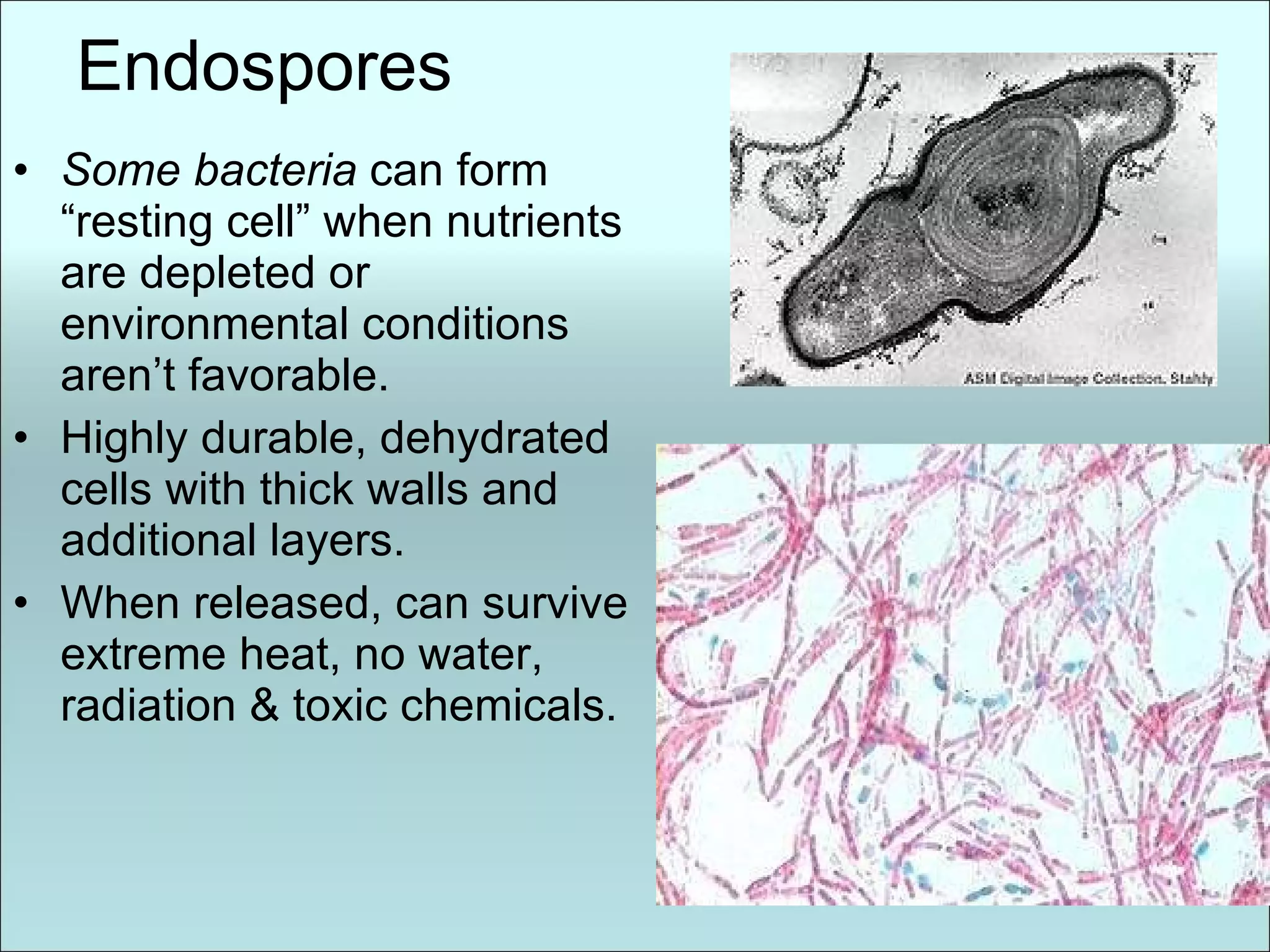
Endospores Some bacteria can form “resting cell” when nutrients are depleted or environmental conditions aren’t favorable. Highly durable, dehydrated cells with thick walls and additional layers. When released, can survive extreme heat, no water, radiation & toxic chemicals.
36.
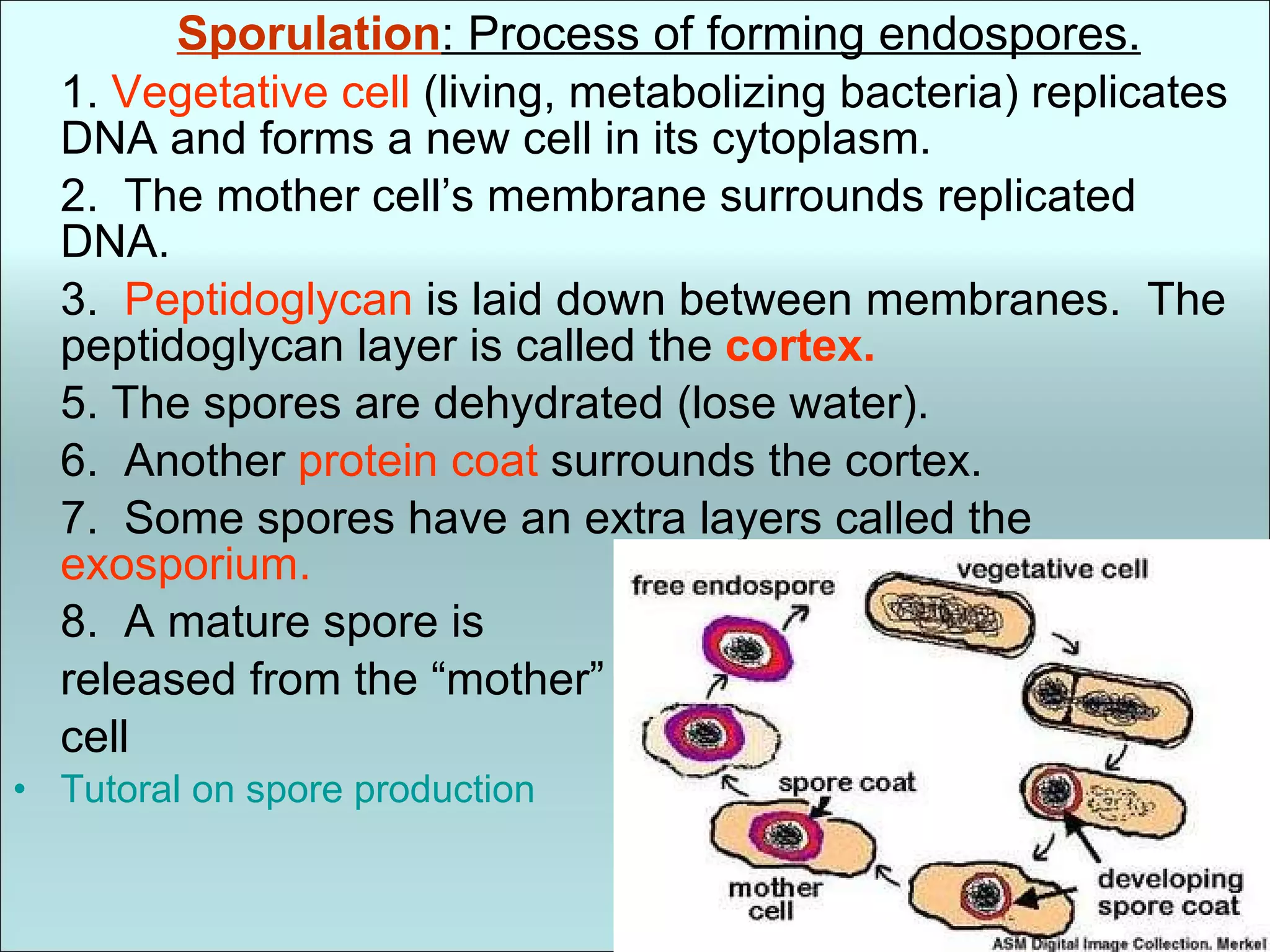
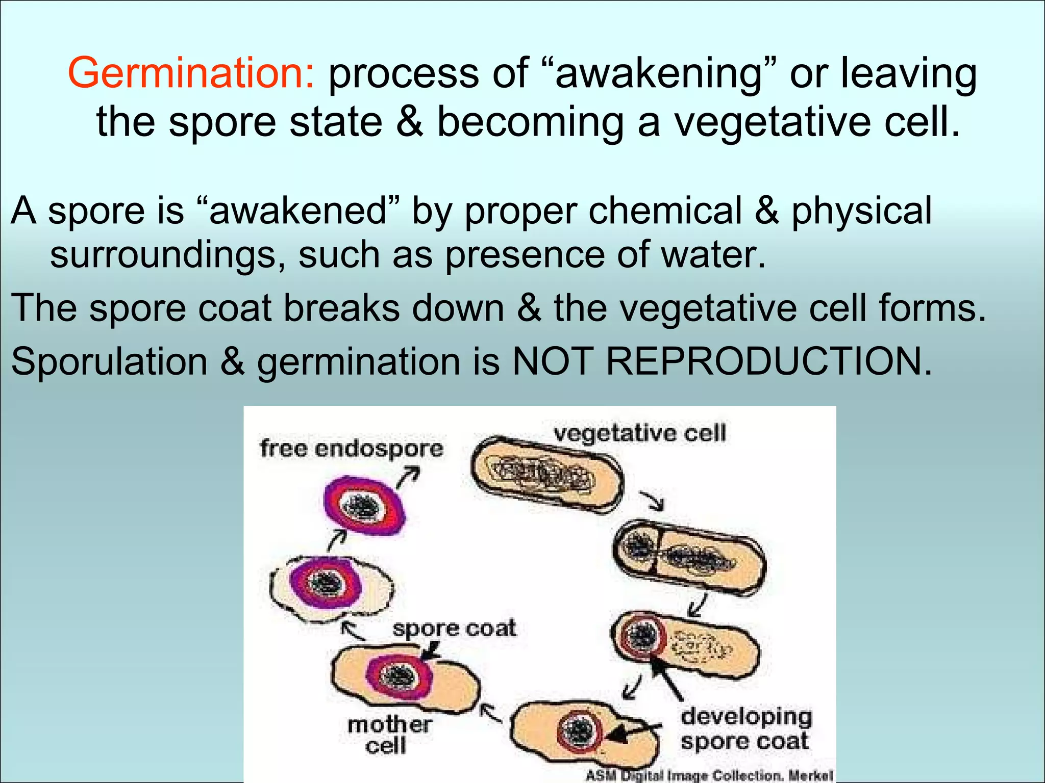
Sporulation : Processof forming endospores. 1. Vegetative cell (living, metabolizing bacteria) replicates DNA and forms a new cell in its cytoplasm. 2. The mother cell’s membrane surrounds replicated DNA. 3. Peptidoglycan is laid down between membranes. The peptidoglycan layer is called the cortex. 5. The spores are dehydrated (lose water). 6. Another protein coat surrounds the cortex. 7. Some spores have an extra layers called the exosporium. 8. A mature spore is released from the “mother” cell Tutoral on spore production
37.
Germination: processof “awakening” or leaving the spore state & becoming a vegetative cell. A spore is “awakened” by proper chemical & physical surroundings, such as presence of water. The spore coat breaks down & the vegetative cell forms. Sporulation & germination is NOT REPRODUCTION.
38.
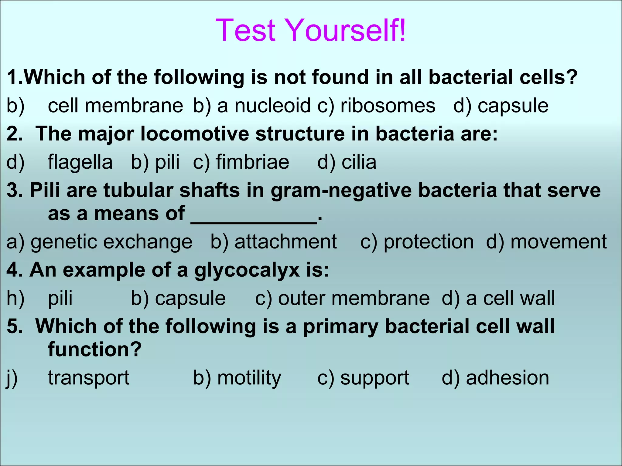
Test Yourself! 1.Whichof the following is not found in all bacterial cells? cell membrane b) a nucleoid c) ribosomes d) capsule 2. The major locomotive structure in bacteria are: flagella b) pili c) fimbriae d) cilia 3. Pili are tubular shafts in gram-negative bacteria that serve as a means of ___________. a) genetic exchange b) attachment c) protection d) movement 4. An example of a glycocalyx is: pili b) capsule c) outer membrane d) a cell wall 5. Which of the following is a primary bacterial cell wall function? transport b) motility c) support d) adhesion
39.
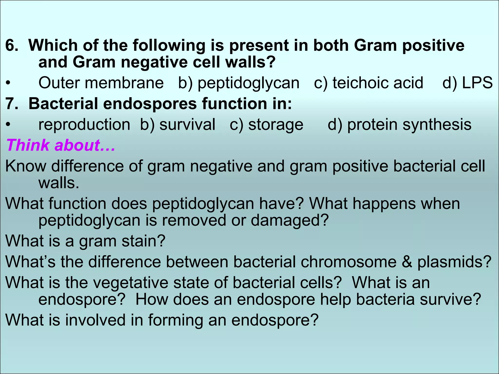
6. Whichof the following is present in both Gram positive and Gram negative cell walls? Outer membrane b) peptidoglycan c) teichoic acid d) LPS 7. Bacterial endospores function in: reproduction b) survival c) storage d) protein synthesis Think about… Know difference of gram negative and gram positive bacterial cell walls. What function does peptidoglycan have? What happens when peptidoglycan is removed or damaged? What is a gram stain? What’s the difference between bacterial chromosome & plasmids? What is the vegetative state of bacterial cells? What is an endospore? How does an endospore help bacteria survive? What is involved in forming an endospore?


![04 [chapter 4 the tissue level of organization][11e]](https://cdn.slidesharecdn.com/ss_thumbnails/04chapter4thetissueleveloforganization11e-170828035609-thumbnail.jpg?width=640&height=640&fit=bounds)


![Ch 32 intro to animal diversity 10-11 [compatibility mode]](https://cdn.slidesharecdn.com/ss_thumbnails/ch32-introtoanimaldiversity10-11compatibilitymode-100928195423-phpapp02-thumbnail.jpg?width=640&height=640&fit=bounds)


![BACTERIA STRUCTURE AND FUNCTION [Autosaved].pptx](https://cdn.slidesharecdn.com/ss_thumbnails/bacteriastructureandfunctionautosaved-230206013850-80c9a713-thumbnail.jpg?width=640&height=640&fit=bounds)


