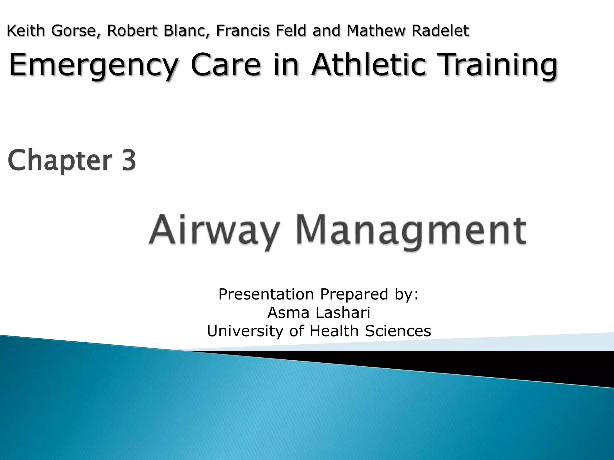
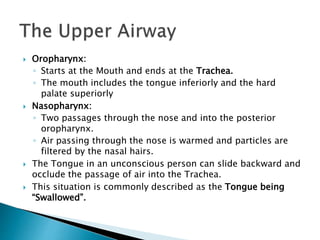
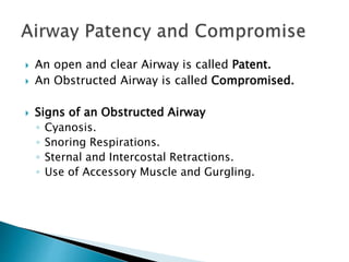
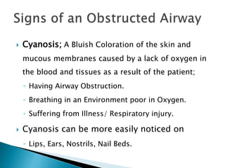
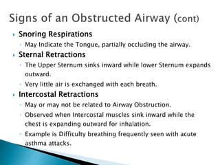
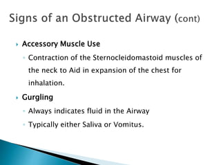
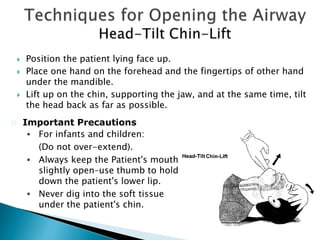
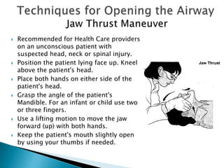
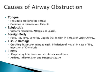
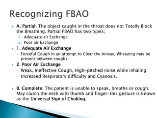
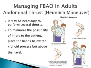
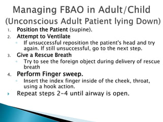
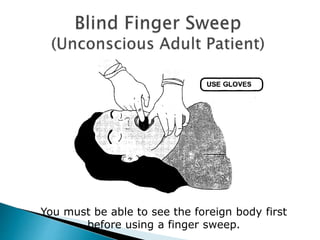
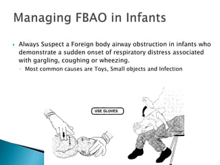
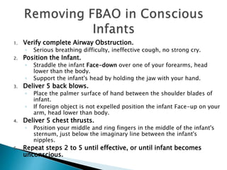
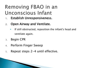
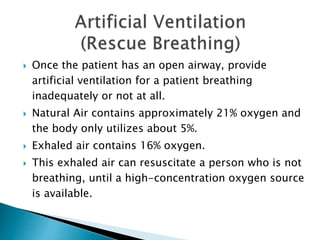
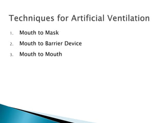
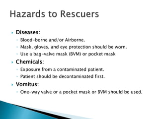
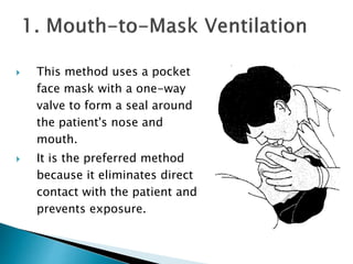
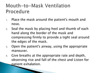
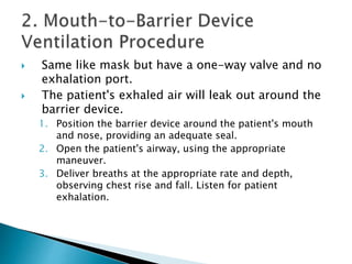
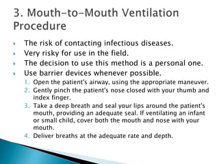
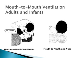
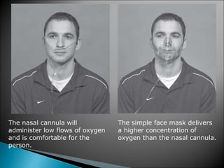
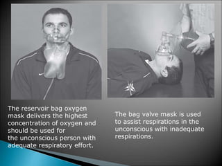
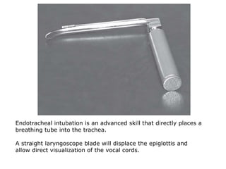
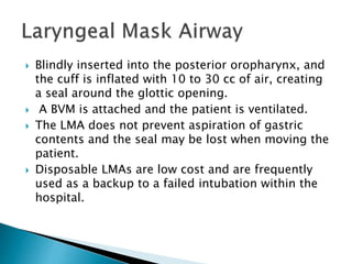
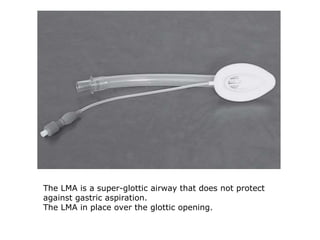
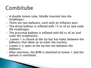
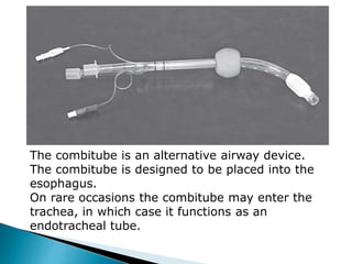
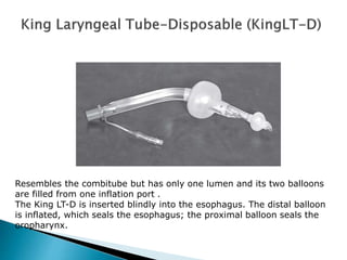
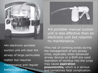
Emergency Care in Athletic Training
Chapter 3
Keith Gorse, Robert Blanc, Francis Feld and Mathew Radelet
Presentation Prepared by:
Asma Lashari
University of Health Sciences
 The Airway can be divided into two parts:
◦ Upper Airway
◦ Lower Airway
 The Upper Airway:
◦ Oropharynx
◦ Nasopharynx
 Oropharynx:
◦ Starts at the Mouth and ends at the Trachea.
◦ The mouth includes the tongue inferiorly and the hard
palate superiorly
 Nasopharynx:
◦ Two passages through the nose and into the posterior
oropharynx.
◦ Air passing through the nose is warmed and particles are
filtered by the nasal hairs.
 The Tongue in an unconscious person can slide backward and
occlude the passage of air into the Trachea.
 This situation is commonly described as the Tongue being
“Swallowed”.
 Epiglottis
◦ A Flap that covers the opening to the Trachea (called glottis)
when food or fluid passes into the Esophagus.
 Larynx
◦ Composed of Nine Cartilages and Muscles.
◦ Located anterior to the Forth, Fifth, and Sixth Cervical
vertebrae in adults.
◦ The larynx is also known as the Adam’s Apple.
◦ Protects the glottis while also allowing phonation.
 An open and clear Airway is called Patent.
 An Obstructed Airway is called Compromised.
 Signs of an Obstructed Airway
◦ Cyanosis.
◦ Snoring Respirations.
◦ Sternal and Intercostal Retractions.
◦ Use of Accessory Muscle and Gurgling.
 Cyanosis; A Bluish Coloration of the skin and
mucous membranes caused by a lack of oxygen in
the blood and tissues as a result of the patient;
◦ Having Airway Obstruction.
◦ Breathing in an Environment poor in Oxygen.
◦ Suffering from Illness/ Respiratory injury.
 Cyanosis can be more easily noticed on
◦ Lips, Ears, Nostrils, Nail Beds.
 Snoring Respirations
◦ May Indicate the Tongue, partially occluding the airway.
 Sternal Retractions
◦ The Upper Sternum sinks inward while lower Sternum expands
outward.
◦ Very little air is exchanged with each breath.
 Intercostal Retractions
◦ May or may not be related to Airway Obstruction.
◦ Observed when Intercostal muscles sink inward while the
chest is expanding outward for inhalation.
◦ Example is Difficulty breathing frequently seen with acute
asthma attacks.
 Accessory Muscle Use
◦ Contraction of the Sternocleidomastoid muscles of
the neck to Aid in expansion of the chest for
inhalation.
 Gurgling
◦ Always indicates fluid in the Airway
◦ Typically either Saliva or Vomitus.
 Position the patient lying face up.
 Place one hand on the forehead and the fingertips of other hand
under the mandible.
 Lift up on the chin, supporting the jaw, and at the same time, tilt
the head back as far as possible.
 Important Precautions
 For infants and children:
(Do not over-extend).
 Always keep the Patient's mouth
slightly open–use thumb to hold
down the patient's lower lip.
 Never dig into the soft tissue
under the patient's chin.
 Recommended for Health Care providers
on an unconscious patient with
suspected head, neck or spinal injury.
 Position the patient lying face up. Kneel
above the patient's head.
 Place both hands on either side of the
patient's head.
 Grasp the angle of the patient's
Mandible. For an infant or child use two
or three fingers.
 Use a lifting motion to move the jaw
forward (up) with both hands.
 Keep the patient's mouth slightly open
by using your thumbs if needed.
Foreign Body Airway
Obstruction (FBAO)
 Upper Airway Obstruction
◦ Anything that blocks the back of the Mouth or Throat,
or the Nasal Passages.
 Lower Airway Obstruction
◦ Breathing in, a Foreign Body
◦ Severe Spasm of the Bronchial passages, such as
Asthma.
 The most common Airway Obstruction in a Responsive
patient is FOOD, and in Unresponsive patient is the
TONGUE.
 Tongue
◦ Falls back blocking the Throat
◦ Common in Unconscious Patients.
 Epiglottitis
◦ Valsalva maneuver, Allergies or Spasm.
 Foreign Body
◦ Food, Ice, Toys, Vomitus, Liquids that remain in Throat or Upper Airway.
 Tissue Damage
◦ Crushing Trauma or Injury to neck, Inhalation of Hot air in case of fire,
Ingestion of Chemicals
 Illness
◦ Respiratory Infections, certain chronic conditions
◦ Asthma, Inflammation and Muscular Spasm
 A. Partial: The object caught in the throat does not Totally Block
the Breathing. Partial FBAO has two types;
1. Adequate air Exchange
2. Poor air Exchange
 1. Adequate Air Exchange
◦ Forceful Cough in an attempt to Clear the Airway, Wheezing may be
present between coughs.
 2. Poor Air Exchange
◦ Weak, Ineffective Cough, High-pitched noise while inhaling
◦ Increased Respiratory difficulty and Cyanosis.
 B. Complete: The patient is unable to speak, breathe or cough.
May clutch the neck with thumb and finger-this gesture is known
as the Universal Sign of Choking.
 It may be necessary to
perform several thrusts.
 To minimize the possibility
of injury to the patient,
place the hands below the
xiphoid process but above
the navel.
1. Position the Patient (supine).
2. Attempt to Ventilate
◦ If unsuccessful reposition the patient's head and try
again. If still unsuccessful, go to the next step.
3. Give a Rescue Breath
◦ Try to see the foreign object during delivery of rescue
breath
4. Perform Finger sweep.
◦ Insert the index finger inside of the cheek, throat,
using a hook action.
 Repeat steps 2-4 until airway is open.
You must be able to see the foreign body first
before using a finger sweep.
 Always Suspect a Foreign body airway obstruction in infants who
demonstrate a sudden onset of respiratory distress associated
with gargling, coughing or wheezing.
◦ Most common causes are Toys, Small objects and Infection
1. Verify complete Airway Obstruction.
◦ Serious breathing difficulty, ineffective cough, no strong cry.
2. Position the Infant.
◦ Straddle the infant Face-down over one of your forearms, head
lower than the body.
◦ Support the infant's head by holding the jaw with your hand.
3. Deliver 5 back blows.
◦ Place the palmer surface of hand between the shoulder blades of
infant.
◦ If foreign object is not expelled position the infant Face-up on your
arm, head lower than body.
4. Deliver 5 chest thrusts.
◦ Position your middle and ring fingers in the middle of the infant's
sternum, just below the imaginary line between the infant's
nipples.
5. Repeat steps 2 to 5 until effective, or until infant becomes
unconscious.
1. Establish Unresponsiveness.
2. Open Airway and Ventilate.
 If still obstructed, reposition the infant's head and
ventilate again.
3. Begin CPR
4. Perform Finger Sweep
5. Repeat steps 2-4 until effective.
 Once the patient has an open airway, provide
artificial ventilation for a patient breathing
inadequately or not at all.
 Natural Air contains approximately 21% oxygen and
the body only utilizes about 5%.
 Exhaled air contains 16% oxygen.
 This exhaled air can resuscitate a person who is not
breathing, until a high-concentration oxygen source
is available.
1. Mouth to Mask
2. Mouth to Barrier Device
3. Mouth to Mouth
 Diseases:
◦ Blood-borne and/or Airborne.
◦ Mask, gloves, and eye protection should be worn.
◦ Use a bag-valve mask (BVM) or pocket mask
 Chemicals:
◦ Exposure from a contaminated patient.
◦ Patient should be decontaminated first.
 Vomitus:
◦ One-way valve or a pocket mask or BVM should be used.
 This method uses a pocket
face mask with a one-way
valve to form a seal around
the patient's nose and
mouth.
 It is the preferred method
because it eliminates direct
contact with the patient and
prevents exposure.
1. Place the mask around the patient's mouth and
nose.
2. Seal the mask by placing heel and thumb of each
hand along the border of the mask and
compressing firmly to provide a tight seal around
the edges of the mask.
3. Open the patient's airway, using the appropriate
maneuver.
4. Give breaths at the appropriate rate and depth,
observing rise and fall of the chest and Listen for
patient exhalation.
 Same like mask but have a one-way valve and no
exhalation port.
 The patient's exhaled air will leak out around the
barrier device.
1. Position the barrier device around the patient's mouth
and nose, providing an adequate seal.
2. Open the patient's airway, using the appropriate
maneuver.
3. Deliver breaths at the appropriate rate and depth,
observing chest rise and fall. Listen for patient
exhalation.
 The risk of contacting infectious diseases.
 Very risky for use in the field.
 The decision to use this method is a personal one.
 Use barrier devices whenever possible.
1. Open the patient's airway, using the appropriate maneuver.
2. Gently pinch the patient's nose closed with your thumb and
index finger.
3. Take a deep breath and seal your lips around the patient's
mouth, providing an adequate seal. If ventilating an infant
or small child, cover both the mouth and nose with your
mouth.
4. Deliver breaths at the adequate rate and depth.
Mouth-to-Mouth Ventilation Mouth to Mouth and Nose
Oxygen Therapy
 Airway management is not complete without administration of
supplemental oxygen.
 High-flow oxygen to any patient with difficulty breathing in an
emergency situation is recommended no matter what past
medical history exists.
 Oxygen administration over a long period (hours) may lead to
Hypoventilation or even Apnea.
 For this reason there is a common misconception among health
care providers that patients with COPD (emphysema,bronchitis,
asthma, and black lung disease) should never receive oxygen
by any means other than a Nasal Cannula at low flow rates.
Various-sized Oral and Nasal Pharyngeal Airways
Device FiO2(%) Flow Rate (L/m)
 Nasal Cannula 25–40 1–6
 Simple Face mask 40–60 6–10
 Reservoir Bag Face mask 60–90 10–15
 The Bag Valve mask (BVM) 100 10–15
The nasal cannula will
administer low flows of oxygen
and is comfortable for the
person.
The simple face mask delivers
a higher concentration of
oxygen than the nasal cannula.
The reservoir bag oxygen
mask delivers the highest
concentration of oxygen and
should be used for
the unconscious person with
adequate respiratory effort.
The bag valve mask is used
to assist respirations in the
unconscious with inadequate
respirations.
Advanced Airway
Devices
 Effective Ventilation with a BVM is possible for a
short time, eventually the airway must be secured
by an advanced airway device.
 The gold standard is Endotracheal Intubation.
 This Technique involves using a laryngoscope to
directly visualize the vocal cords at the glottic
opening and passing a cuffed endotracheal tube
into the trachea.
 Once the tube is inserted, the cuff is inflated, and
trachea is sealed.
Endotracheal intubation is an advanced skill that directly places a
breathing tube into the trachea.
A straight laryngoscope blade will displace the epiglottis and
allow direct visualization of the vocal cords.
 Blindly inserted into the posterior oropharynx, and
the cuff is inflated with 10 to 30 cc of air, creating
a seal around the glottic opening.
 A BVM is attached and the patient is ventilated.
 The LMA does not prevent aspiration of gastric
contents and the seal may be lost when moving the
patient.
 Disposable LMAs are low cost and are frequently
used as a backup to a failed intubation within the
hospital.
The LMA is a super-glottic airway that does not protect
against gastric aspiration.
The LMA in place over the glottic opening.
 A double lumen tube, blindly inserted into the
esophagus .
 There are two balloons, each with an inflation port.
 The distal balloon is inflated with 15 cc of air and seals
the esophagus.
 The proximal balloon is inflated with 60 cc of air and
seals the oropharynx.
 Lumen 1 is closed at the tip but has holes between the
balloons that allow air to enter the trachea.
 Lumen 2 is open at the tip but not between the
balloons.
 After insertion, the BVM is attached to lumen 1 and the
patient is ventilated .
The combitube is an alternative airway device.
The combitube is designed to be placed into the
esophagus.
On rare occasions the combitube may enter the
trachea, in which case it functions as an
endotracheal tube.
Resembles the combitube but has only one lumen and its two balloons
are filled from one inflation port .
The King LT-D is inserted blindly into the esophagus. The distal balloon
is inflated, which seals the esophagus; the proximal balloon seals the
oropharynx.
An electronic portable
suction unit will clear the
airway of large particulate
matter but requires
maintenance and regular
battery charging.
A portable manual suction
unit is less effective than an
electronic unit but requires
no maintenance.
The risk of vomiting exists during
the management of any airway
crisis, especially when advanced
airway devices or a BVM are used.
Aspiration of vomitus into the lungs
may cause aspiration
pneumonitis, which is a serious
and sometimes fatal complication.
Thank you 

Chapter 3 Airway Management.

  • 1.
    Emergency Care inAthletic Training Chapter 3 Keith Gorse, Robert Blanc, Francis Feld and Mathew Radelet Presentation Prepared by: Asma Lashari University of Health Sciences
  • 2.
     The Airwaycan be divided into two parts: ◦ Upper Airway ◦ Lower Airway  The Upper Airway: ◦ Oropharynx ◦ Nasopharynx
  • 3.
     Oropharynx: ◦ Startsat the Mouth and ends at the Trachea. ◦ The mouth includes the tongue inferiorly and the hard palate superiorly  Nasopharynx: ◦ Two passages through the nose and into the posterior oropharynx. ◦ Air passing through the nose is warmed and particles are filtered by the nasal hairs.  The Tongue in an unconscious person can slide backward and occlude the passage of air into the Trachea.  This situation is commonly described as the Tongue being “Swallowed”.
  • 4.
     Epiglottis ◦ AFlap that covers the opening to the Trachea (called glottis) when food or fluid passes into the Esophagus.  Larynx ◦ Composed of Nine Cartilages and Muscles. ◦ Located anterior to the Forth, Fifth, and Sixth Cervical vertebrae in adults. ◦ The larynx is also known as the Adam’s Apple. ◦ Protects the glottis while also allowing phonation.
  • 6.
     An openand clear Airway is called Patent.  An Obstructed Airway is called Compromised.  Signs of an Obstructed Airway ◦ Cyanosis. ◦ Snoring Respirations. ◦ Sternal and Intercostal Retractions. ◦ Use of Accessory Muscle and Gurgling.
  • 7.
     Cyanosis; ABluish Coloration of the skin and mucous membranes caused by a lack of oxygen in the blood and tissues as a result of the patient; ◦ Having Airway Obstruction. ◦ Breathing in an Environment poor in Oxygen. ◦ Suffering from Illness/ Respiratory injury.  Cyanosis can be more easily noticed on ◦ Lips, Ears, Nostrils, Nail Beds.
  • 8.
     Snoring Respirations ◦May Indicate the Tongue, partially occluding the airway.  Sternal Retractions ◦ The Upper Sternum sinks inward while lower Sternum expands outward. ◦ Very little air is exchanged with each breath.  Intercostal Retractions ◦ May or may not be related to Airway Obstruction. ◦ Observed when Intercostal muscles sink inward while the chest is expanding outward for inhalation. ◦ Example is Difficulty breathing frequently seen with acute asthma attacks.
  • 9.
     Accessory MuscleUse ◦ Contraction of the Sternocleidomastoid muscles of the neck to Aid in expansion of the chest for inhalation.  Gurgling ◦ Always indicates fluid in the Airway ◦ Typically either Saliva or Vomitus.
  • 10.
     Position thepatient lying face up.  Place one hand on the forehead and the fingertips of other hand under the mandible.  Lift up on the chin, supporting the jaw, and at the same time, tilt the head back as far as possible.  Important Precautions  For infants and children: (Do not over-extend).  Always keep the Patient's mouth slightly open–use thumb to hold down the patient's lower lip.  Never dig into the soft tissue under the patient's chin.
  • 11.
     Recommended forHealth Care providers on an unconscious patient with suspected head, neck or spinal injury.  Position the patient lying face up. Kneel above the patient's head.  Place both hands on either side of the patient's head.  Grasp the angle of the patient's Mandible. For an infant or child use two or three fingers.  Use a lifting motion to move the jaw forward (up) with both hands.  Keep the patient's mouth slightly open by using your thumbs if needed.
  • 12.
  • 13.
     Upper AirwayObstruction ◦ Anything that blocks the back of the Mouth or Throat, or the Nasal Passages.  Lower Airway Obstruction ◦ Breathing in, a Foreign Body ◦ Severe Spasm of the Bronchial passages, such as Asthma.  The most common Airway Obstruction in a Responsive patient is FOOD, and in Unresponsive patient is the TONGUE.
  • 14.
     Tongue ◦ Fallsback blocking the Throat ◦ Common in Unconscious Patients.  Epiglottitis ◦ Valsalva maneuver, Allergies or Spasm.  Foreign Body ◦ Food, Ice, Toys, Vomitus, Liquids that remain in Throat or Upper Airway.  Tissue Damage ◦ Crushing Trauma or Injury to neck, Inhalation of Hot air in case of fire, Ingestion of Chemicals  Illness ◦ Respiratory Infections, certain chronic conditions ◦ Asthma, Inflammation and Muscular Spasm
  • 15.
     A. Partial:The object caught in the throat does not Totally Block the Breathing. Partial FBAO has two types; 1. Adequate air Exchange 2. Poor air Exchange  1. Adequate Air Exchange ◦ Forceful Cough in an attempt to Clear the Airway, Wheezing may be present between coughs.  2. Poor Air Exchange ◦ Weak, Ineffective Cough, High-pitched noise while inhaling ◦ Increased Respiratory difficulty and Cyanosis.  B. Complete: The patient is unable to speak, breathe or cough. May clutch the neck with thumb and finger-this gesture is known as the Universal Sign of Choking.
  • 16.
     It maybe necessary to perform several thrusts.  To minimize the possibility of injury to the patient, place the hands below the xiphoid process but above the navel.
  • 17.
    1. Position thePatient (supine). 2. Attempt to Ventilate ◦ If unsuccessful reposition the patient's head and try again. If still unsuccessful, go to the next step. 3. Give a Rescue Breath ◦ Try to see the foreign object during delivery of rescue breath 4. Perform Finger sweep. ◦ Insert the index finger inside of the cheek, throat, using a hook action.  Repeat steps 2-4 until airway is open.
  • 18.
    You must beable to see the foreign body first before using a finger sweep.
  • 19.
     Always Suspecta Foreign body airway obstruction in infants who demonstrate a sudden onset of respiratory distress associated with gargling, coughing or wheezing. ◦ Most common causes are Toys, Small objects and Infection
  • 20.
    1. Verify completeAirway Obstruction. ◦ Serious breathing difficulty, ineffective cough, no strong cry. 2. Position the Infant. ◦ Straddle the infant Face-down over one of your forearms, head lower than the body. ◦ Support the infant's head by holding the jaw with your hand. 3. Deliver 5 back blows. ◦ Place the palmer surface of hand between the shoulder blades of infant. ◦ If foreign object is not expelled position the infant Face-up on your arm, head lower than body. 4. Deliver 5 chest thrusts. ◦ Position your middle and ring fingers in the middle of the infant's sternum, just below the imaginary line between the infant's nipples. 5. Repeat steps 2 to 5 until effective, or until infant becomes unconscious.
  • 21.
    1. Establish Unresponsiveness. 2.Open Airway and Ventilate.  If still obstructed, reposition the infant's head and ventilate again. 3. Begin CPR 4. Perform Finger Sweep 5. Repeat steps 2-4 until effective.
  • 22.
     Once thepatient has an open airway, provide artificial ventilation for a patient breathing inadequately or not at all.  Natural Air contains approximately 21% oxygen and the body only utilizes about 5%.  Exhaled air contains 16% oxygen.  This exhaled air can resuscitate a person who is not breathing, until a high-concentration oxygen source is available.
  • 23.
    1. Mouth toMask 2. Mouth to Barrier Device 3. Mouth to Mouth
  • 24.
     Diseases: ◦ Blood-borneand/or Airborne. ◦ Mask, gloves, and eye protection should be worn. ◦ Use a bag-valve mask (BVM) or pocket mask  Chemicals: ◦ Exposure from a contaminated patient. ◦ Patient should be decontaminated first.  Vomitus: ◦ One-way valve or a pocket mask or BVM should be used.
  • 25.
     This methoduses a pocket face mask with a one-way valve to form a seal around the patient's nose and mouth.  It is the preferred method because it eliminates direct contact with the patient and prevents exposure.
  • 26.
    1. Place themask around the patient's mouth and nose. 2. Seal the mask by placing heel and thumb of each hand along the border of the mask and compressing firmly to provide a tight seal around the edges of the mask. 3. Open the patient's airway, using the appropriate maneuver. 4. Give breaths at the appropriate rate and depth, observing rise and fall of the chest and Listen for patient exhalation.
  • 27.
     Same likemask but have a one-way valve and no exhalation port.  The patient's exhaled air will leak out around the barrier device. 1. Position the barrier device around the patient's mouth and nose, providing an adequate seal. 2. Open the patient's airway, using the appropriate maneuver. 3. Deliver breaths at the appropriate rate and depth, observing chest rise and fall. Listen for patient exhalation.
  • 28.
     The riskof contacting infectious diseases.  Very risky for use in the field.  The decision to use this method is a personal one.  Use barrier devices whenever possible. 1. Open the patient's airway, using the appropriate maneuver. 2. Gently pinch the patient's nose closed with your thumb and index finger. 3. Take a deep breath and seal your lips around the patient's mouth, providing an adequate seal. If ventilating an infant or small child, cover both the mouth and nose with your mouth. 4. Deliver breaths at the adequate rate and depth.
  • 29.
  • 30.
  • 31.
     Airway managementis not complete without administration of supplemental oxygen.  High-flow oxygen to any patient with difficulty breathing in an emergency situation is recommended no matter what past medical history exists.  Oxygen administration over a long period (hours) may lead to Hypoventilation or even Apnea.  For this reason there is a common misconception among health care providers that patients with COPD (emphysema,bronchitis, asthma, and black lung disease) should never receive oxygen by any means other than a Nasal Cannula at low flow rates.
  • 32.
    Various-sized Oral andNasal Pharyngeal Airways
  • 33.
    Device FiO2(%) FlowRate (L/m)  Nasal Cannula 25–40 1–6  Simple Face mask 40–60 6–10  Reservoir Bag Face mask 60–90 10–15  The Bag Valve mask (BVM) 100 10–15
  • 34.
    The nasal cannulawill administer low flows of oxygen and is comfortable for the person. The simple face mask delivers a higher concentration of oxygen than the nasal cannula.
  • 35.
    The reservoir bagoxygen mask delivers the highest concentration of oxygen and should be used for the unconscious person with adequate respiratory effort. The bag valve mask is used to assist respirations in the unconscious with inadequate respirations.
  • 36.
  • 37.
     Effective Ventilationwith a BVM is possible for a short time, eventually the airway must be secured by an advanced airway device.  The gold standard is Endotracheal Intubation.  This Technique involves using a laryngoscope to directly visualize the vocal cords at the glottic opening and passing a cuffed endotracheal tube into the trachea.  Once the tube is inserted, the cuff is inflated, and trachea is sealed.
  • 38.
    Endotracheal intubation isan advanced skill that directly places a breathing tube into the trachea. A straight laryngoscope blade will displace the epiglottis and allow direct visualization of the vocal cords.
  • 39.
     Blindly insertedinto the posterior oropharynx, and the cuff is inflated with 10 to 30 cc of air, creating a seal around the glottic opening.  A BVM is attached and the patient is ventilated.  The LMA does not prevent aspiration of gastric contents and the seal may be lost when moving the patient.  Disposable LMAs are low cost and are frequently used as a backup to a failed intubation within the hospital.
  • 40.
    The LMA isa super-glottic airway that does not protect against gastric aspiration. The LMA in place over the glottic opening.
  • 41.
     A doublelumen tube, blindly inserted into the esophagus .  There are two balloons, each with an inflation port.  The distal balloon is inflated with 15 cc of air and seals the esophagus.  The proximal balloon is inflated with 60 cc of air and seals the oropharynx.  Lumen 1 is closed at the tip but has holes between the balloons that allow air to enter the trachea.  Lumen 2 is open at the tip but not between the balloons.  After insertion, the BVM is attached to lumen 1 and the patient is ventilated .
  • 42.
    The combitube isan alternative airway device. The combitube is designed to be placed into the esophagus. On rare occasions the combitube may enter the trachea, in which case it functions as an endotracheal tube.
  • 43.
    Resembles the combitubebut has only one lumen and its two balloons are filled from one inflation port . The King LT-D is inserted blindly into the esophagus. The distal balloon is inflated, which seals the esophagus; the proximal balloon seals the oropharynx.
  • 44.
    An electronic portable suctionunit will clear the airway of large particulate matter but requires maintenance and regular battery charging. A portable manual suction unit is less effective than an electronic unit but requires no maintenance. The risk of vomiting exists during the management of any airway crisis, especially when advanced airway devices or a BVM are used. Aspiration of vomitus into the lungs may cause aspiration pneumonitis, which is a serious and sometimes fatal complication.
  • 45.