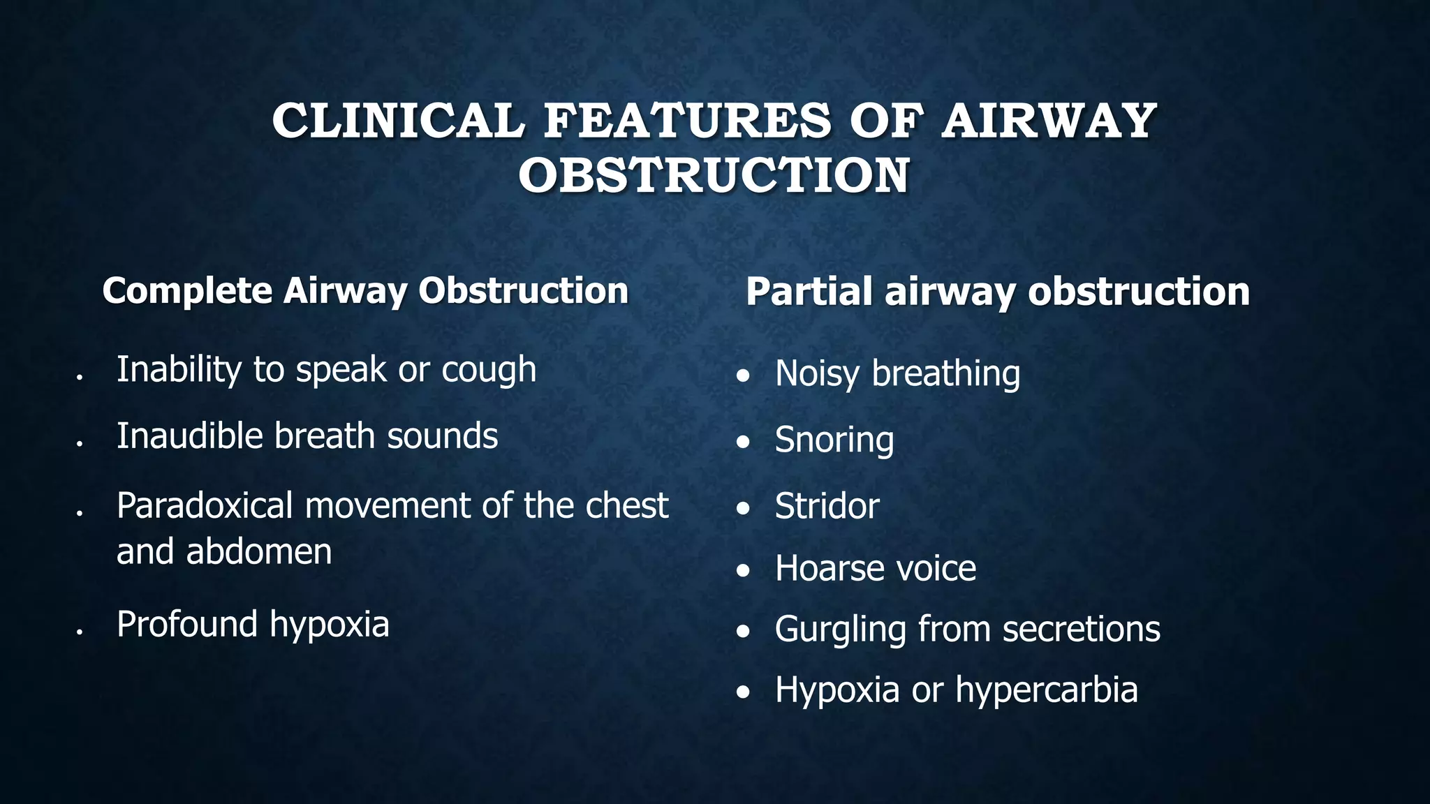
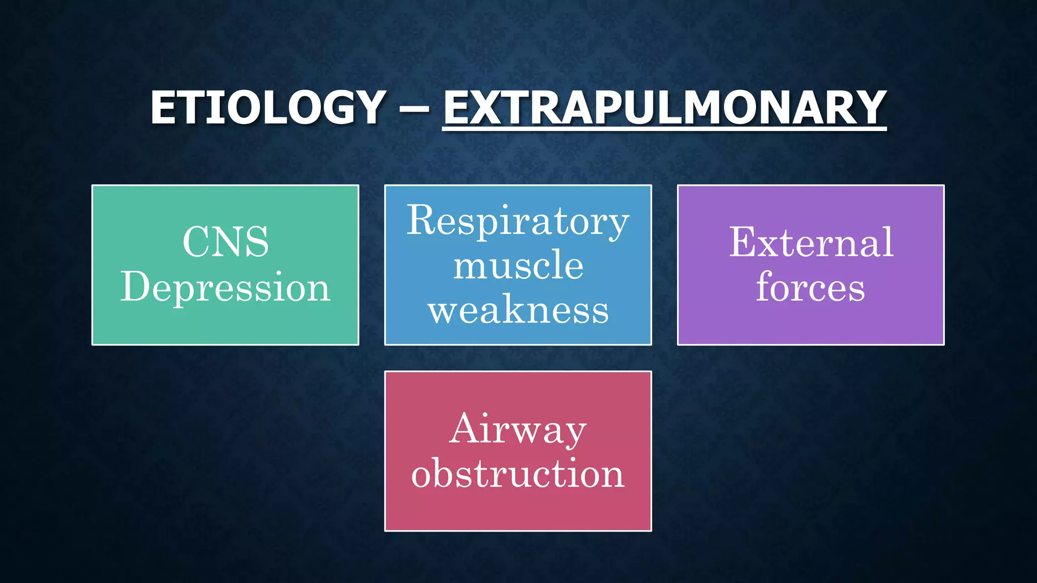
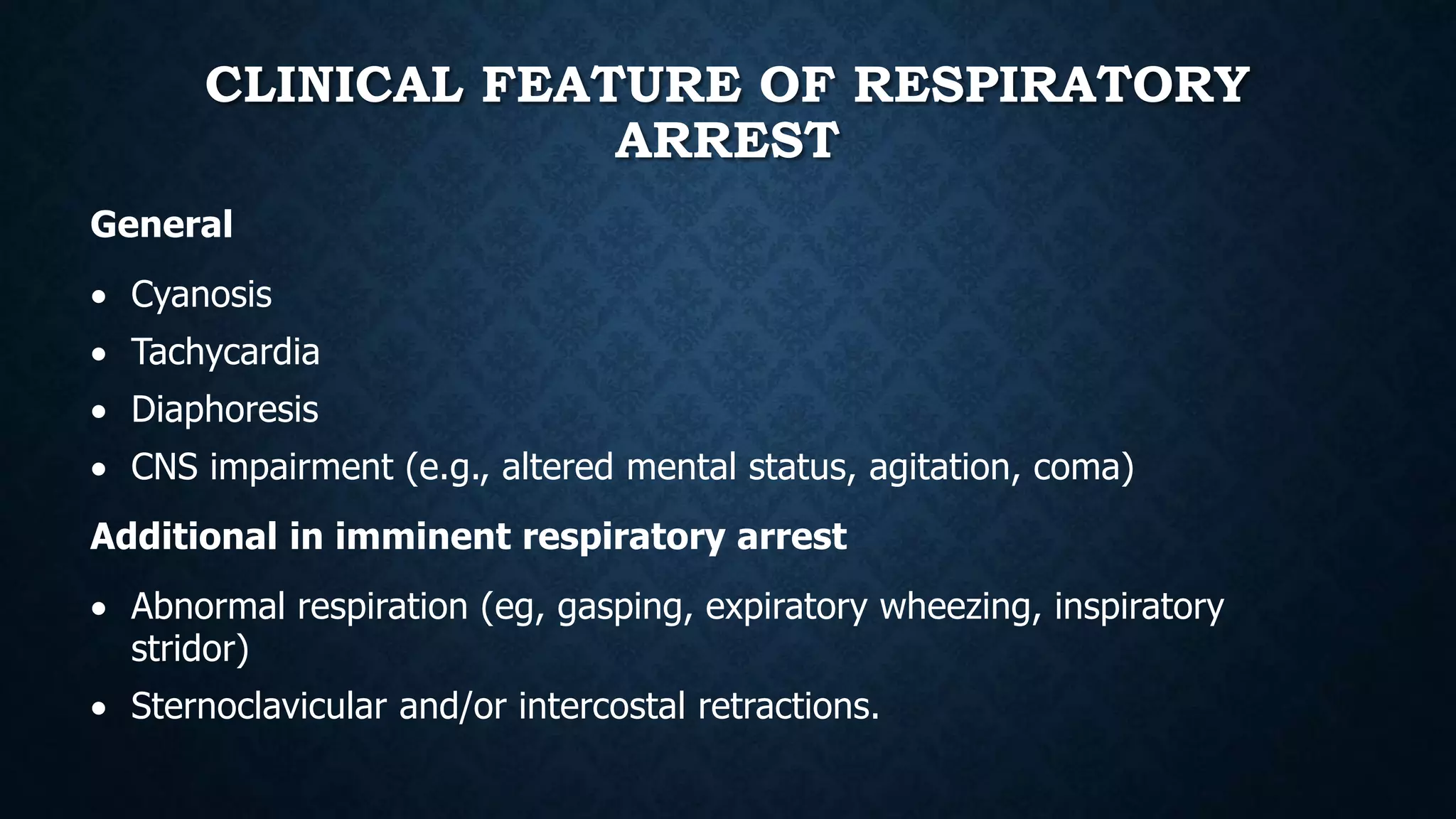
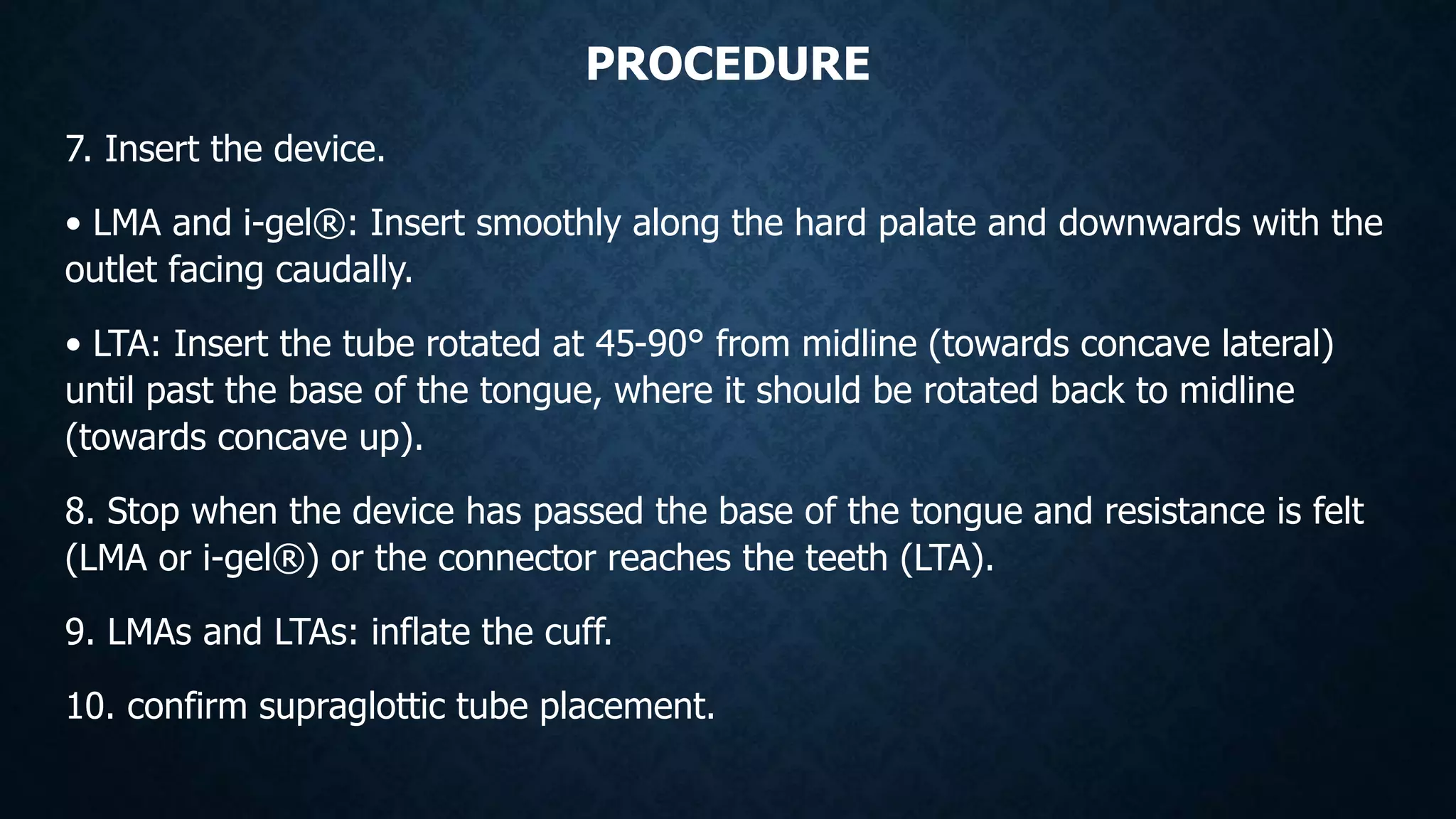
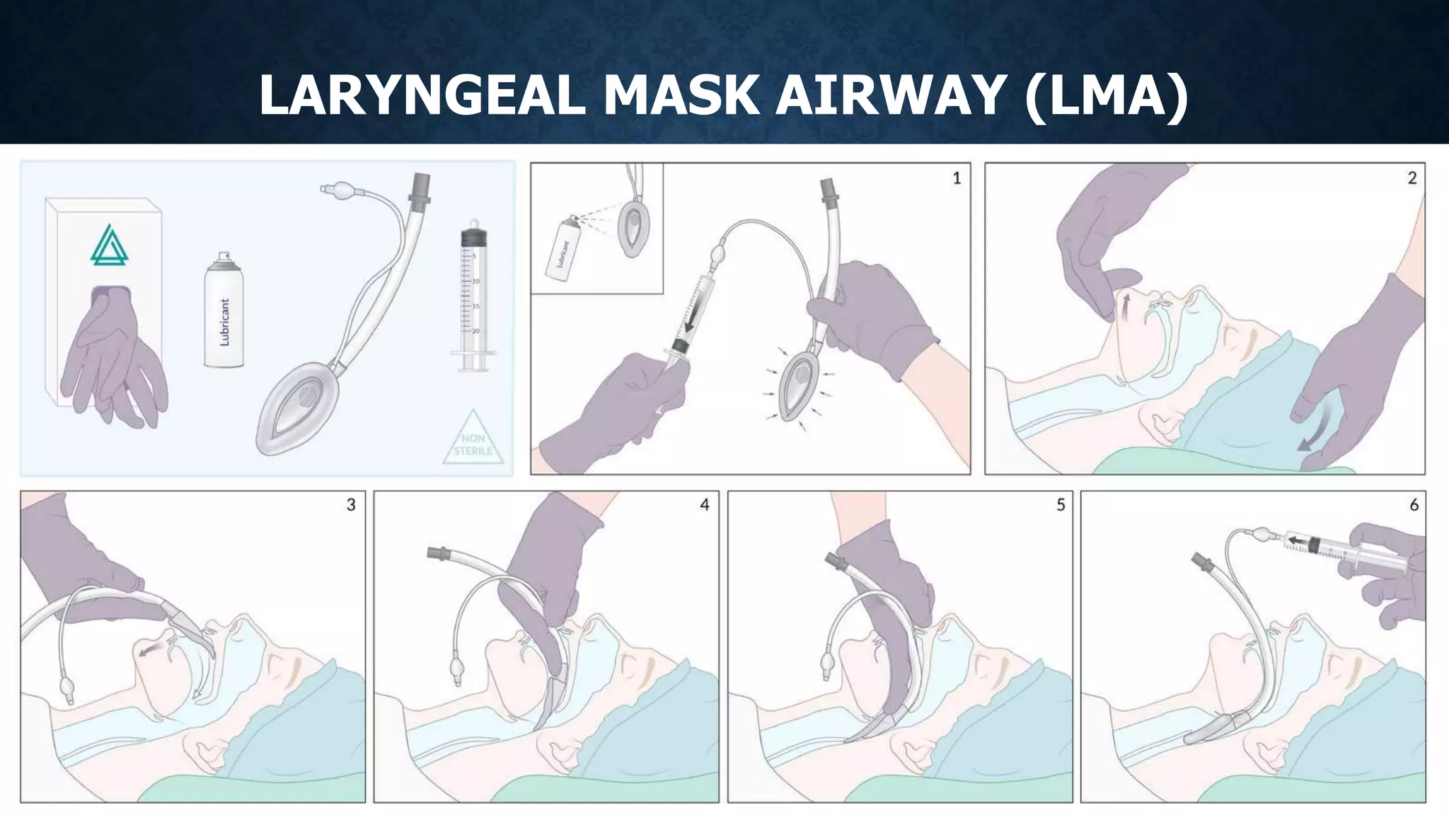
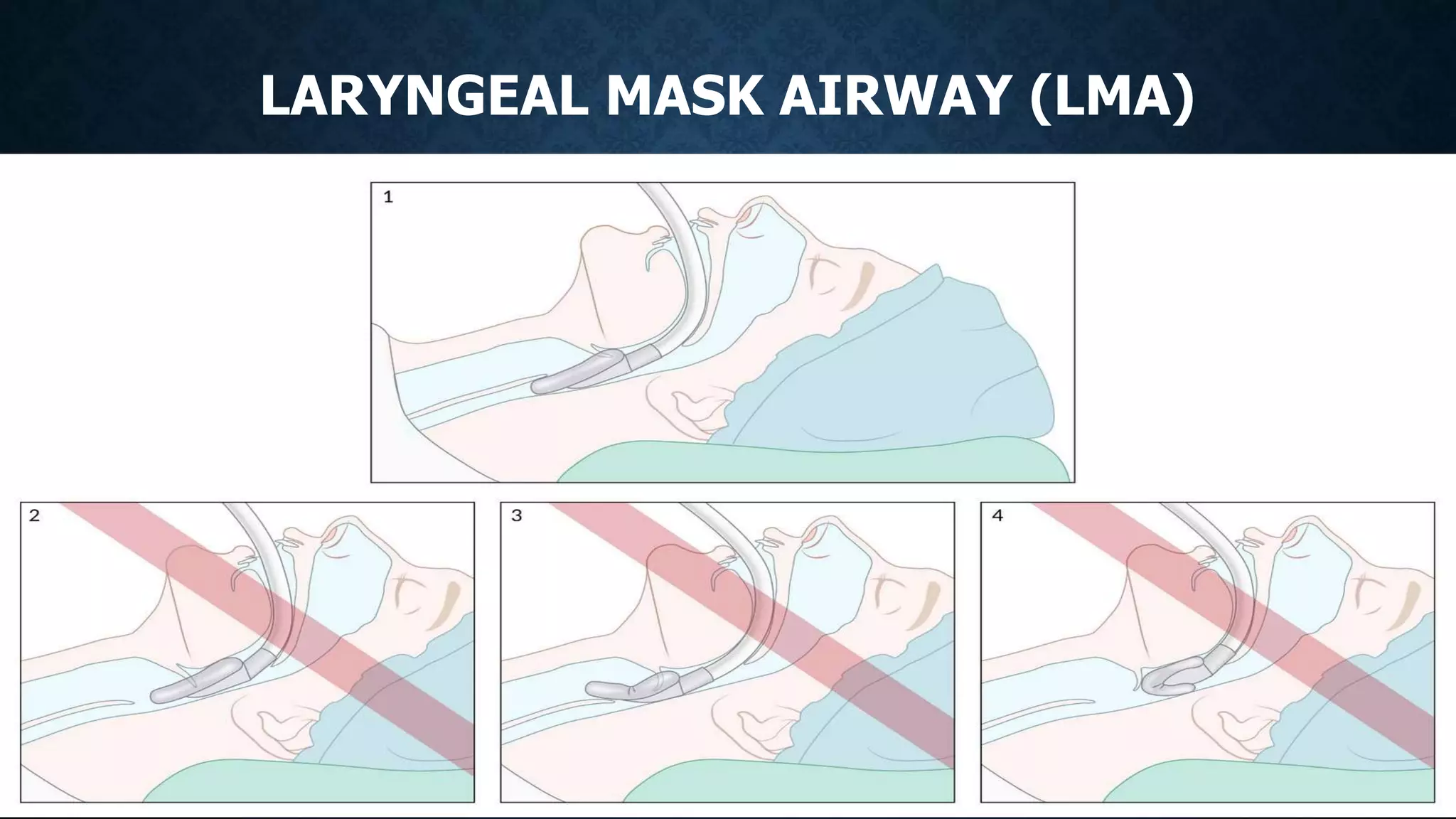
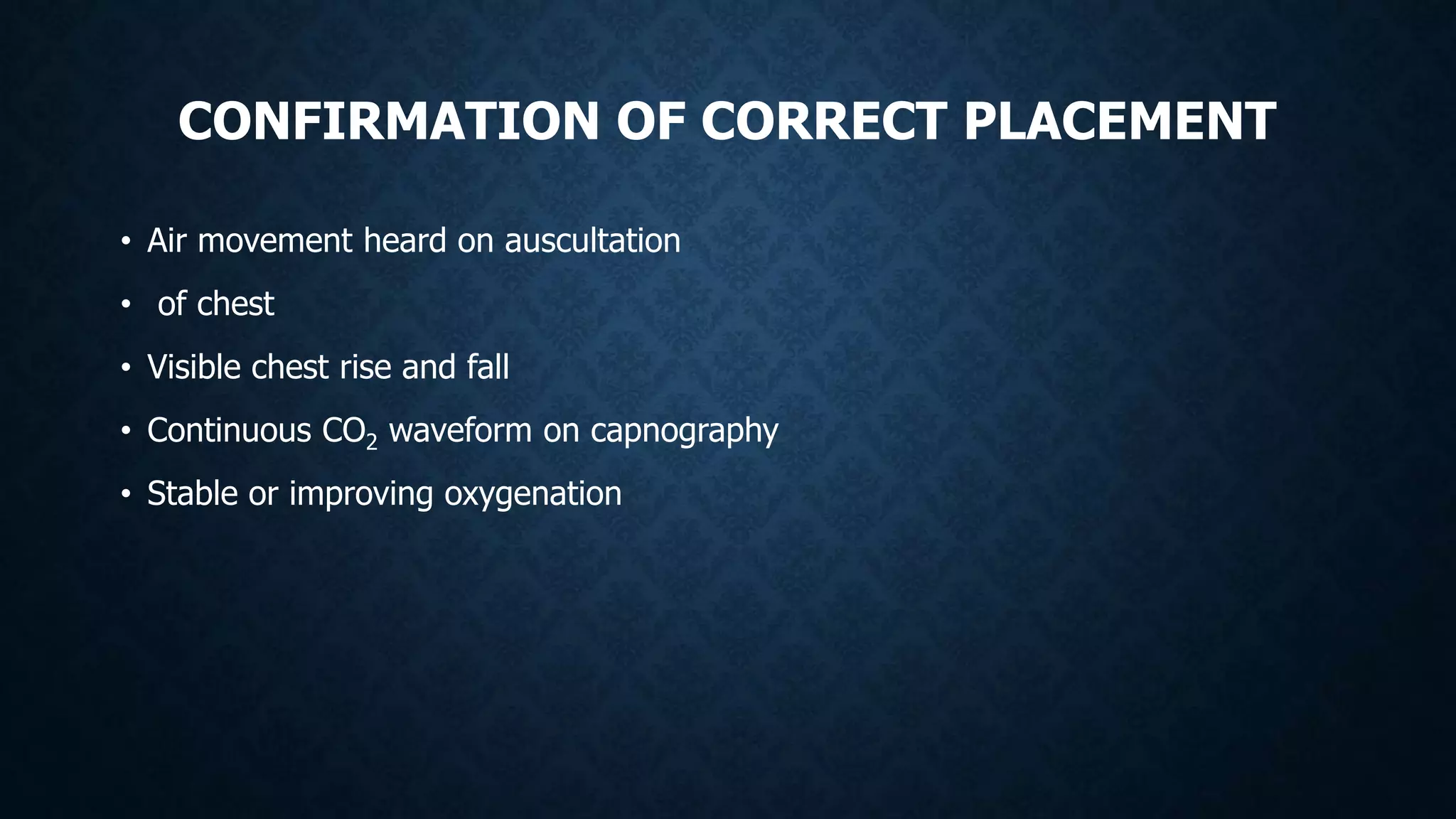
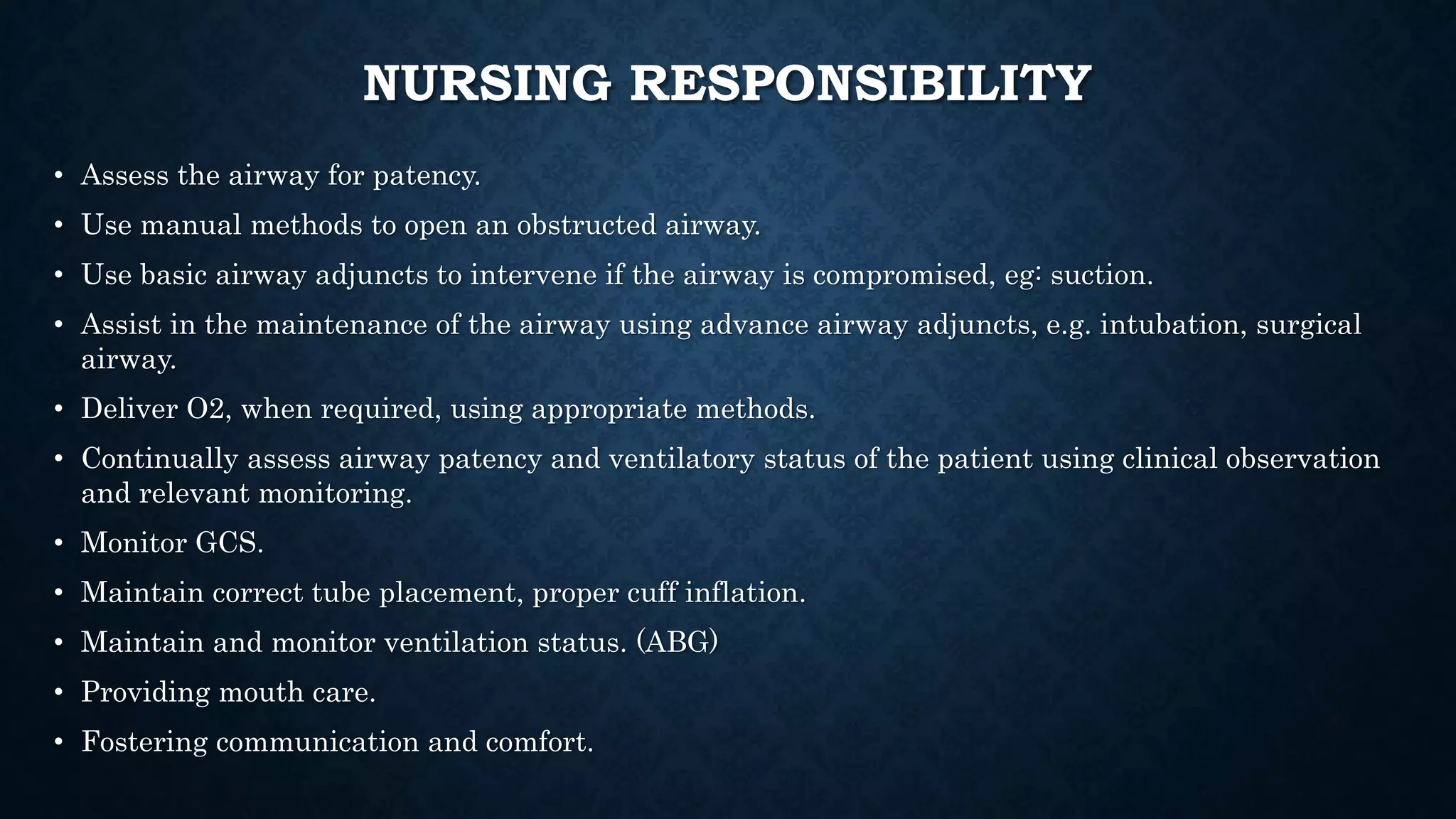
SlideShare a Scribd company logo
DEMONSTRATION
ON
AIRWAY MANAGEMENT
Presenter: Asha Prerna Tigga
OBJECTIVES:
• At the end of the class the student will be:
1. To define Airway management.
2. To enlist the clinical features of airway obstruction.
3. To discuss the respiration arrest.
4. To demonstrate the Basic airway maneuvers.
5. To demonstrate the basic air adjuncts.
6. To demonstrate the advance air device.
7. To explain procedure of the Intubation.
8. To elaborate the Adjuncts for intubation.
9. To discuss about the Surgical Airway management.
INTRODUCTION
• Patients in any healthcare setting can quickly become acutely unwell, and
assessment and management of the airway is always the priority in any
clinical situation (Resuscitation Council UK, 2021). When patients are
critically unwell, there is a high risk of respiratory deterioration, and many
patients require an artificial airway to facilitate their treatment. Knowing how
to assess and manage the airway is a key skill for the nurse working in
critical care.
INTRODUCTION
• Airway management is the practice of evaluating, planning and
using a wide array of medical procedures and device for the
purpose of maintaining or restoring a safe, effective pathway for
oxygenation and ventilation. These procedures are indicated in
patient with airway obstruction, respiratory failure, or a need for
airway protection.
DEFINITION OF AIRWAY MANAGEMENT
• Airway management involves ensuring that the patient has a patent
airway through which effective ventilation can take place.
Obstruction of the airway may be partial or complete and may
occur at any level from the nose to the trachea.
CLINICAL FEATURES OF AIRWAY
OBSTRUCTION
Complete Airway Obstruction
 Inability to speak or cough
 Inaudible breath sounds
 Paradoxical movement of the chest
and abdomen
 Profound hypoxia
Partial airway obstruction
 Noisy breathing
 Snoring
 Stridor
 Hoarse voice
 Gurgling from secretions
 Hypoxia or hypercarbia
CLINICAL FEATURES OF AIRWAY
OBSTRUCTION
Loss of airway protective
reflexes
 Reduced GCS (traditionally ≤ 8)
 Ability to comfortably tolerate an
oral airway
 Inability to swallow secretions
 Procedural sedation or general
anaesthesia.
Airway obstruction
• Facial trauma
• Burn injury and/or inhalational injury
• Progressive angioedema
• Known or suspected foreign body
aspiration
• Known laryngeal or pharyngeal
cancer
RESPIRATION ARREST
• Definition
“Respiratory arrest is the
complete cessation of
breathing in patients with
a pulse.”
ETIOLOGY – EXTRAPULMONARY
CNS
Depression
Respiratory
muscle
weakness
External
forces
Airway
obstruction
ETIOLOGY – PULMONARY
Airway
Obstruction
Impaired
alveolar
diffusion
CLINICAL FEATURE OF RESPIRATORY
ARREST
General
 Cyanosis
 Tachycardia
 Diaphoresis
 CNS impairment (e.g., altered mental status, agitation, coma)
Additional in imminent respiratory arrest
 Abnormal respiration (eg, gasping, expiratory wheezing, inspiratory
stridor)
 Sternoclavicular and/or intercostal retractions.
RESPIRATORY ARREST
Diagnostics
 Clinical evaluation (ABCDE approach)
 ABG: decreased PaO and increased PaCO
 Pulse oximetry
Management
 Intubation
 Mechanical ventilation
 In case of obstruction, airway opening maneuvers
 Respiratory and cardiac monitoring
 Treatment of underlying conditions
RESPIRATORY ARREST
• Complications
 Hypoxic organ
damage (eg., Hypoxic
brain injury)
• Cardiac arrest
•BASIC AIRWAY MANEUVERS
HEAD-TILT/CHIN-LIFT MANEUVER
“A method of opening the airway that involves head
and neck repositioning. It should be avoided if there
is concern for C-spine injury.”
• • Technique
• 1. Tilt the head of the patient posteriorly to 15-
30°.
• 2. Lift the chin with the fingers to pull the tongue
and oropharyngeal soft tissue anteriorly.
• 3. Use the fingers of the same hand to apply
pressure below the lip, slightly opening the mouth.
• 4. Maintain this "sniffing position" to align the
oral, pharyngeal, and laryngeal axes.
JAW-THRUST MANEUVER
“The mandible is moved anteriorly to open the
airway. May be used in conjunction with head-
tilt/chin-lift or alone in patients with suspected
C-spine injury.”
• • Technique
• 1. With the patient supine, place fingers
behind the angles of the lower jaw.
• 2. Move the jaw anteriorly to pull the base of
the tongue and soft tissues away from the
airway.
• 3. Use the thumbs to open the mouth
slightly.
https://www.youtube.com/watch?v=5iwogajl-n8
THE RECOVERY POSITION
“ positioning of the patient in
a lateral decubitus position
with slight neck extension “
• Aim
1. Prevention of airway occlusion
by the tongue and soft tissues
2. Reduction in the risk of
aspiration if patients
regurgitate
THE RECOVERY POSITION
• Indications
1. Prehospital settings
2. Temporal airway compromise
that can be managed by
positioning alone (e.g procedural
sedation, alcohol intoxication)
• Contraindications
1. C-Spine immobilization
2. Anticipated worsening of airway
compromise
3. Transportation outside of a
monitored environment
BAG-MASK VENTILATION
“Delivery of oxygen and provider-assisted breaths using a bag-valve-mask unit to
patients with inadequate ventilation.”
• Indications
 Bridge to intubation
• - Patients with acute respiratory failure
• - Preoxygenation prior to general anaesthesia
 Rescue ventilation: Use after failed intubation attempt (e.g., when safe apnea
time has been exceeded) or accidental oversedation.
 CPR
PROCEDURE OF BMV
Create a mask seal
EC-clamp technique (one-person
technique): commonly used in elective
perioperative situations when the provider
is alone.
• 1. With the patient supine, lift the jaw
towards the mask using the 3rd, 4th, and
5th fingers of one hand, forming an E-
shape
• 2. Squeeze the mask onto the face with
the thumb and index finger of the same
hand, forming a C-shape.
• 3. Deliver breaths with the second hand.
PROCEDURE OF BMV
Two-person bag-mask-ventilation technique:
used in emergency settings in which the patient is
deteriorating or ventilation is difficult, since it is more
effective.
• 1. One provider makes a seal and opens the airway
with both hands:
• - Squeeze the mask to the face using the eminences
of both hands, placed along the sides of the mask
with the thumbs pointing inferior
• - Perform a jaw-thrust maneuver using all 4 fingers
of both hands.
• - A two-handed EC-clamp technique may also be
used.
• 2. The second provider delivers breaths.
https://www.youtube.com/watch?v=zUGw90iL0Qw
PROCEDURE OF BMV
Provide breaths: Set minute ventilation
• Aim: Deliver 500-600 mL (6-7 mL/kg) volume at 10-12 breaths/minute.
• Procedure: Squeeze the bag slowly and gently over approx. 1 second before allowing it to fully reinflate. Repeat
every 5 seconds.
• Adjust based on the clinical situation: E.g., follow compression-to-breath ratio in patients undergoing CPR without
an advanced airway (e.g., 30:2).
Confirm adequacy of BMV
• Clinical: No leaks around mask, Bilateral chest rise, Air entry on auscultation of bilateral lung fields
• Monitoring
- Oxygen saturations in target range
- Normal capnometry (waveform and EtCO2 value)
ANTICIPATION AND COMPLICATION OF BMV
COMPLICATION
Poor mask seal or difficulty opening airway:
• Recommendation:
 Switch to 2 person BMV technique
 If this fails, insert a supraglottic device (eg, LMA) for ventilation.
Poor chest rise:
 Reposition, Check for obstruction, Use basic airway adjuncts.
Inadvertent hyperventilation
 Commonly occurs in stressful resuscitation scenarios, can lead to: stomach hyperinflation, vomiting, and aspiration, increase
intrathoracic pressure, decease cardiac output.
 Prevention: maintain steady pressure and depth of bag compression.
Hypoxia during apneic period
 Ensure adequate preoxygenation.
BASIC AIRWAY ADJUNCTS
• These devices may be used alongside bag-mask ventilation or airway opening
maneuvers to improve airway patency.
OROPHARYNGEAL AIRWAY (OPA)
Description: a rigid curved device placed in the mouth to prevent the tongue from occluding the
airway
Indications
• Unconscious patients with a large tongue, obstructed nasal passages, or copious nasal secretions
• Typically used as a bridge to intubation
Contraindications: conscious patient with intact gag reflex
Sizing rule: from the incisors to the angle of the mandible, or corner of the mouth (oral commissure)
to the earlobe
Insertion technique: Adults: Insert concave up or concave lateral until past the tongue and then
rotate until concave down. Ensure OPA has bypassed the tongue and is not pushing it backwards.
Further management: Toleration of an oropharyngeal airway indicates an at-risk airway;
preparations should be made for intubation.
Procedure Additional information/rationale
1. Select appropriate size.
If too short the airway would not separate the soft palate from
the posterior wall of the pharynx; too long may displace the
epiglottis.
2. Where no risk of cervical spine injury exists, hyperextend the
head and neck. Grasp the patient’s jaw and lift anteriorly.
Stretches the anterior neck structures to relieve obstruction of
the soft palate and epiglottis.
3. Using other hand, hold the OPA at its proximal end and insert it
into the patients mouth with the tip pointing towards the roof of
the mouth.
Avoids unnecessary trauma to the delicate tissues in the mouth
and inadvertent blocking of the airway by pushing the tongue
back.
4. Once the tip reaches the level of the soft palate, gently rotate
the airway 180° until it comes to rest over the tongue.
Brings the OPA into the alignment required for use.
5. The flattened, reinforced section of a correctly sized OPA should
lie between the patient’s teeth/dentures or gums. The lips should
not be pulled over the flange of the OPA as this may cause damage
to the labial frenulum.
Acts as a bite block.
6. Verify appropriate position by listening for clear breath sounds
and looking for chest rise and fall.
Ensures correct placement.
SIZE OF OPA
• https://www.youtube.com/watch?v=Hzc_T4QBp4E
NASOPHARYNGEAL AIRWAY (NPA)
• Description: a long flexible tube inserted into the nostril and down into the nasopharynx to prevent the
tongue from occluding the airway
• Indications: conscious or unconscious patients with current or potential oropharyngeal obstruction
• Contraindications: facial fractures, basilar skull fractures
• Sizing rule: nostril to the ipsilateral tragus
• Equipment required
 Range of nasopharyngeal airways
 Sterile gloves
 Sanitizer
 Sterile tray
 Water-based lubricant
Procedure Additional information/rationale
1. Select appropriate size, 7.0 as a starting point for an average adult male
and 6.0 for an average adult female.
2. Once the selection has been made, measure from the tip of the nose to
the tragus of the ear. Insert safety pin to mark the maximum depth of
insertion (this should be at the proximal end of the NPA).
If too short the airway would not separate the soft palate from
the posterior wall of the pharynx; if too long may enter either
the larynx or vallecula where the airway could become
obstructed.
3. Where no risk of cervical spine injury exists, hyperextend the head and
neck.
Stretches the anterior neck structures to relieve obstruction of
the soft palate and epiglottis.
4. Lubricate the exterior of the tube with a water-soluble gel. Minimises trauma during insertion.
5. If there is no obvious nasal deformity, it is recommended that the right
nostril be used.
The bevel of the NPA is designed to cause less trauma to the
mucosa when inserted into the right nostril.
6. Where deformity exists, the most patent nostril should be selected.
7. If inserting into the left nostril the bevel is placed alongside the septum
and the airway rotated through 180° when it enters the nasopharynx.
To minimise trauma to the internal nares.
8. Insert the tube into the selected nostril and follow the nasal floor parallel
to the mouth. It is imperative that the airway is not pushed in a cephalad
direction.
This ensures correct location and reduces risk of cranial
insertion where basal skull fracture exists.
9. Avoid pushing against any resistance. If resistance is felt, remove the
airway, review technique and reinsert using the other nostril.
Pushing against resistance may cause bleeding and kinking of
the NPA.
10. Verify appropriate position by listening for clear breath sounds and
looking for chest rise and fall. Air may also be felt at the proximal end of the
airway in the spontaneously breathing patient.
Ensures correct placement.
11. Check to make sure there is no blanching of the patient’s nostrils. If
there is, remove NPA and select a smaller diameter.
Prevents necrosis of the tissues.
• https://oxfordmedicaleducation.com/clinical-skills/procedures/nasopharyngeal-airway/
SIZE
•ADVANCE AIRWAY DEVICE
SUPRAGLOTTIC AIRWAY DEVICE
Definition: a collection of advanced airway devices that are inserted via the
oropharynx to provide ventilation from above the glottis
Indications
• Cardiac arrest (pre-hospital and in-hospital), as they can be inserted by
providers untrained in intubation
• First-line for short durations of anesthesia
• As an alternative advanced airway if intubation has failed
Contraindications: Avoid in conscious patients with an intact gag reflex.
Caution: does not offer complete protection against aspiration, unlike an
endotracheal tube
LARYNGEAL MASK AIRWAY
(LMA)
• The laryngeal mask airway (LMA)
comprises a wide-bore tube with an
elliptical inflatable cuff designed to seal
around the laryngeal inlet. The proximal
end of the tube is fitted with a standard
15/22 mm connector. The LMA can be
placed blind, requires less skill and is
easier to insert than a tracheal tube. The
LMA provides for more efficient ventilation
than with a bag-valve-mask (BVM)
A. LARYNGEAL MASK
AIRWAY (LMA)
“a supraglottic device consisting
of an inflatable mask attached to
the end of a tube.”
• Second-generation LMAs
feature safety adaptations such
as bite blocks and a drainage
tube.
• Intubating LMAs (ILMA)
feature additional adaptations to
allow passage of an ET tube
through the LMA.
B. I-GEL®:
a type of supraglottic
airway that is similar in
structure to the LMA.
However, the mask is
anatomically-molded,
noninflatable, and made
of a soft gel-like material.
C. LARYNGEAL
TUBE AIRWAY (LTA)
)
• An airway device consisting of a tube
with 2 inflatable cuffs and ventilation
holes between them.
• • Intubating LTAs feature additional
adaptations to allow passage of an ET
tube through the LTA.
SIZING OF A LARYNGEAL MASK AIRWAY
Size Age/weight range
Maximum cuff inflation (mL
air)
1 Neonates up to 5 kg 4
1.5 Children 5–10 kg 7
2 Children 10–20 kg 10
2.5 Children 20–30 kg 14
3 Children 30–50 kg 20
4 Small/normal adults 30
5 Normal/large adults 40
EQUIPMENT REQUIRED
• LMA – range of sizes
• 50 mL syringe
• Tie
• Lubrication
• Stethoscope
• Gloves
• Sanitizers
PROCEDURE
1. Choose the appropriate size for the patient:
• Small adult: size 3
• Medium adult: size 4
• Large adult: size 5
2. LMAs and LTAs: inflate cuffs fully to check for leaks before deflating.
3. Lubricate the tip of the device, being careful not to block ventilatory openings.
4. Place the patient in the sniffing position.
5. Open the patient's mouth wide.
6. Hold the device firmly (at the junction of the tube and mask for an LMA, at the bite block for an
i-gel®, or at the connector for an LTA).
PROCEDURE
7. Insert the device.
• LMA and i-gel®: Insert smoothly along the hard palate and downwards with the
outlet facing caudally.
• LTA: Insert the tube rotated at 45-90° from midline (towards concave lateral)
until past the base of the tongue, where it should be rotated back to midline
(towards concave up).
8. Stop when the device has passed the base of the tongue and resistance is felt
(LMA or i-gel®) or the connector reaches the teeth (LTA).
9. LMAs and LTAs: inflate the cuff.
10. confirm supraglottic tube placement.
LARYNGEAL MASK AIRWAY (LMA)
LARYNGEAL TUBE AIRWAY (LTA)
• https://www.youtube.com/watch?v=vNvymbRD5b4
CONFIRMATION OF CORRECT PLACEMENT
• Air movement heard on auscultation
• of chest
• Visible chest rise and fall
• Continuous CO2 waveform on capnography
• Stable or improving oxygenation
INTUBATION
General principles
• Defined as placement of a cuffed endotracheal (ET) tube below the vocal cords via direct
laryngoscopy or videolaryngoscopy
• Mostly commonly placed orally (orotracheal intubation), although it may be placed
nasally (nasotracheal intubation)
• Typically, sedation and paralysis are required to tolerate the procedure and subsequent
mechanical ventilation.
• Goals: maximize first-pass success, reduce the risk of aspiration
• Involves the rapid induction of anaesthesia and paralysis, followed by intubation
• Differs from traditional intubation in few ways:
- BMV is not performed (to avoid potential aspiration of stomach contents).
INTUBATION
• Indications for endotracheal intubation
• Inability (or anticipated inability) to maintain the
airway: e.g., general anesthesia, airway obstruction
or reduced GCS
• Failure (or pending failure) of ventilation or
oxygenation: e.g., in severe acute asthma or COPD
(see "Indications for invasive mechanical
ventilation)
• Conditions in which there is a high risk of
deterioration: e.g., multisystem trauma,
anaphylaxis, severe septic shock
Contraindications
• Do-not-intubate order
• Consider adjuncts to intubation or proceed to surgical airway management in:
• Copious blood/secretions
• Upper airway distortion
• Mouth opening limited
• Severe kyphosis
Preoxygenation
• administration of 100% oxygen prior to induction to denitrogenate air in the
lungs .
• Intubation medications
• Typically two classes of medication are given prior to intubation, a sedating (induction) agent
and neuromuscular blocking agent to paralyze the patient.
• Induction agents for intubation
• • Used to induce a state of sedation, which reduces airway reflexes and facilitates intubation
• • Options include:
• • Propofol
• • Etomidate
• • Ketamine
• • The choice of induction agent depends on patient characteristics and operator experience.
• • The duration of bolus doses is typically short (~ 10 minutes) and infusions are required for
ongoing sedation
• Paralytic agents for intubation : Clinical applications
• • Improving airway visualization by relaxing the jaw muscles
• • Decreasing the risk of injuries, e.g., vocal cord damage
• Types
• • Depolarizing NMJ blockers: e.g., succinylcholine
• - Widely used due to rapid onset and offset time (spontaneous respirations normally
return within 10 minutes)
• - Rarely, prolonged paralysis occurs in patients with limited cholinesterase activity.
• • Nondepolarizing NMJ blockers: e.g., rocuronium
• - Used in patients with contraindications to succinylcholine
INTUBATION VIA DIRECT LARYNGOSCOPY
• • Positioning: Place patient in sniffing
position unless C-spine injury is suspected.
• Head elevated to a height of 10 cm
• The neck is mildly flexed at the lower
cervical vertebrae and extended at the
atlanto-occipital joint.
• • Technique: The majority of patients
should have received induction agents and
been preoxygenated. Equipment should
always be on hand to manage a failed
intubation.
ARTICLES AND POSITIONING
ET TUBE
INTUBATION VIA DIRECT LARYNGOSCOPY
1. Wear appropriate PPE.
2. Choose the correct ET tube size. = (22)
3. Gently open the patient's mouth,
4. Insert the laryngoscope blade, using the groove to sweep the tongue aside.
5. Advance steadily until the tip is at the vallecula and the epiglottis is visible below it.
6. Lift gently forward and upward to raise the epiglottis and reveal the arytenoid cartilages and vocal
cords.
7. Insert the ET tube with the stylet.
8. Once the tip is at the glottis, remove the stylet and gently advance until the cuff is past the cords.
9. Inflate the cuff to protect the airway from secretions and form a seal around the tube.
• https://www.youtube.com/watch?v=3uvQO6ty5HU
CONFIRMATION OF TUBE PLACEMENT
Auscultation of bilateral breath sounds over the lungs
• Consistent condensation visible in the tube upon exhalation
• Capnometry
- A visual indicator changes colour from purple to yellow upon contact with CO2.
- Consistent colour changing with each breath > 3 times correlates with tracheal placement.
• Capnography: measurement of end-tidal CO2
• Direct visualization of endotracheal tube markers
• Proximal numbered tube markers should indicate approx. 21-23 cm at the patient's teeth.
•CXR: The distal tip of the endotracheal or tracheal tube should be 2-6 cm above the carina
Ultrasound may be used to confirm tube position
COMPLICATION OF INTUBATION
• Early complications
• Hypoxia
• Bradycardia
• Respiratory acidosis
Trauma
• - Dental damage
• - Tracheal perforation
• - Hemorrhage
Pulmonary aspiration
Laryngospasm
Bronchospasm
• Late complications
• • Vocal cord injuries
• • Vocal cord granuloma: a complication of
endotracheal intubation caused by
inflammation and ulceration during the
intubation process. Can cause vocal
hoarseness that manifests~ 4 weeks after
intubation.
•Surgical airway management
SURGICAL CRICOTHYROTOMY
• Definition: an emergency procedure in which an incision is made through the skin, cervical
fascia, and median cricothyroid ligament (cricothyroid membrane) to obtain airway access.
• Indications: CICV scenario (if orotracheal intubation has failed or is contraindicated)
Failure to maintain oxygenation or ventilation in between intubation attempts or after 3 attempts
at intubation
May be required in:
- Severe oropharyngeal edema
- Foreign body aspiration causing complete occlusion of the upper airway
- Severe oropharyngeal/nasal haemorrhage
- Acute epiglottitis
- Severe maxillofacial trauma
SURGICAL CRICOTHYROTOMY
Contraindications
• Young children and infants
• Some airway injuries: e.g., tracheal transection or laryngeal fracture
o Obstruction distal to the cricothyroid membrane
o Inability to identify anatomical landmarks
• Tumor or infection at the incision site
EQUIPMENT
• https://www.youtube.com/watch?v=bRfQeHtv7pM
•Planned Surgical Airway
TRACHEOSTOMY
•Definition: a permanent or temporary opening (stoma) in the cervical trachea created
through a surgical incision below the cricoid cartilage
Indications
• For emergency indications, same as for cricothyroidotomy
• Long-term mechanical ventilation (> 3 weeks)
• Malignancy
Options
• Percutaneous tracheostomy (typically under bronchoscopy guidance)
• Open surgical tracheostomy
HTTPS://WWW.YOUTUBE.COM/WATCH?V=78B-
UNUYWR8
LARYNGECTOMY
• Definition: the removal of all of the laryngeal structures, including the
epiglottis and part of the upper trachea, with the trachea brought to the
front of the neck to create a stoma
• Indications: laryngeal cancer
• Caution: As the upper airway is no longer connected to the trachea,
patients with a laryngectomy cannot be oxygenated or intubated through
the upper airway.
COMPLICATION OF SURGICAL AIRWAY
• Early complications
• Bleeding
• Creation of a false lumen
• Laceration of the back wall of the trachea
• Damage to the surrounding structures:
e.g., esophageal, mediastinal, or thyroid
perforation, vocal cord injury, or recurrent
laryngeal nerve injury
• Pneumothorax
• Subcutaneous emphysema
Late complications
• Dysphonia
• Scarring
• Stenosis
• Tracheomalacia
CHECKLIST FOR EQUIPMENT REQUIRED
FOR MANAGING AIRWAY.
• PPE
• Monitor connection to patient- BP, SPO2, PR,
• IV access, tourniquet, cannula.
• Oxygen face mask, tubing's.
• OPA, NPA
• BMV device
• Suction device
• Laryngoscope and blades- all size.
NURSING RESPONSIBILITY
• Assess the airway for patency.
• Use manual methods to open an obstructed airway.
• Use basic airway adjuncts to intervene if the airway is compromised, eg: suction.
• Assist in the maintenance of the airway using advance airway adjuncts, e.g. intubation, surgical
airway.
• Deliver O2, when required, using appropriate methods.
• Continually assess airway patency and ventilatory status of the patient using clinical observation
and relevant monitoring.
• Monitor GCS.
• Maintain correct tube placement, proper cuff inflation.
• Maintain and monitor ventilation status. (ABG)
• Providing mouth care.
• Fostering communication and comfort.
TO SUMMARISE…
• Always check airway patency for any patient.
• Always bear in mind- Look, listen and feel.
• Assign the role of team leader, airway nurse and IV nurse.
• All Equipment's and drugs should be checked regularly.
• Knowledge regarding different airway device, drugs and team work are
essential for the better management of the patient.
BIBLIOGRAPHY
• Baid H et al (2016) Oxford Handbook of Critical Care Nursing. Oxford University Press.
Coombs M et al (2013) Assessment, monitoring and interventions for the respiratory system. In: Mallett J et al (eds) Critical Care Manual of
Clinical Procedures and Competencies. Wiley Blackwell.
Crouch R et al (2016) Oxford Handbook of Respiratory Nursing. Oxford University Press.
Doyle J (2021) Supraglottic devices (including laryngeal mask airways) for airway management for anaesthesia in adults. uptodate.com
(accessed 14 January 2022).
Intensive Care Society (2020) Guidance for: Tracheostomy Care. Ics.ac.uk (accessed 14 January 2022).
• Matten E et al (2017) Nonintubation management of the airway: airway maneuvers and mask ventilation. In: Hagberg C (ed) Benumof and
Hagberg’s Airway Management. Elsevier Health Sciences.
Mete A, Akbudak IH (2018) Functional anatomy and physiology of airway. In: Erbay RH (ed) Tracheal Intubatio. IntechOpen
Moots E (2016) Respiratory System. In: Woodruff D (ed) Critical Care Made Incredibly Easy. Lipincott Williams and Wilkins.
Nolan J, Soar J (2016) Airway management in cardiopulmonary resuscitation. In: Webb A et al (eds) Oxford Textbook of Critical Care. Oxford
University Press.
• Amboss, https://www.amboss.com/us/knowledge/airway-
management/#:~:text=Airway%20management%20is%20the%20practice,respiratory%20failure
Airway management

More Related Content

What's hot

Airway assessment in anaesthesia
Airway assessment in anaesthesiaAirway assessment in anaesthesia
Airway assessment in anaesthesia
CaliPenn
 
Ventilator Induced Lung Injury
Ventilator Induced Lung InjuryVentilator Induced Lung Injury
Ventilator Induced Lung Injury
Dr.Mahmoud Abbas
 
ECMO in Critical Care
ECMO in Critical CareECMO in Critical Care
ECMO in Critical Care
Dr.Mahmoud Abbas
 
Anesthesia for intestinal obstruction
Anesthesia for intestinal obstructionAnesthesia for intestinal obstruction
Anesthesia for intestinal obstruction
Omar Alkatheri
 
One lung ventilation
One lung ventilationOne lung ventilation
One lung ventilation
National hospital, kandy
 
Perioperative management in neuromuscular ds
Perioperative  management in neuromuscular dsPerioperative  management in neuromuscular ds
Perioperative management in neuromuscular ds
Ummer Ali
 
Vili
ViliVili
End tidal co2 and transcutaneous monitoring
End tidal co2 and transcutaneous monitoringEnd tidal co2 and transcutaneous monitoring
End tidal co2 and transcutaneous monitoring
Antara Banerji
 
anesthesia history
anesthesia historyanesthesia history
anesthesia history
anaesthesiology-mgmcri
 
Non invasive ventilation (niv)
Non invasive ventilation (niv)Non invasive ventilation (niv)
Non invasive ventilation (niv)
Khairunnisa Azman
 
Difficult airway
Difficult airwayDifficult airway
Difficult airway
Pratyush1693
 
Anesthesia in Restrictive lung disease
Anesthesia in Restrictive lung diseaseAnesthesia in Restrictive lung disease
Anesthesia in Restrictive lung disease
Tenzin yoezer
 
Prone Ventilation In ARDS
Prone Ventilation In ARDSProne Ventilation In ARDS
Prone Ventilation In ARDS
Dr.Mahmoud Abbas
 
Fluid responsiveness - an ICU phoenix
Fluid responsiveness - an ICU phoenixFluid responsiveness - an ICU phoenix
Fluid responsiveness - an ICU phoenixNIICS
 
Chapter4 airway management
Chapter4 airway managementChapter4 airway management
Chapter4 airway management
djorgenmorris
 
Airway Management
Airway ManagementAirway Management
Airway Management
Maged Hassan
 
Oxygen cascade & therapy
Oxygen cascade & therapyOxygen cascade & therapy
Oxygen cascade & therapy
Dr. Taraknath Chatterjee
 
Cannot intubate
Cannot intubateCannot intubate
Cannot intubate
Arvind Khare
 
Inhaled Nitric Oxide in Acute Respiratory Distress Syndrome
Inhaled Nitric Oxide in Acute Respiratory Distress SyndromeInhaled Nitric Oxide in Acute Respiratory Distress Syndrome
Inhaled Nitric Oxide in Acute Respiratory Distress Syndrome
Muhammad Asim Rana
 

What's hot (20)

Airway assessment in anaesthesia
Airway assessment in anaesthesiaAirway assessment in anaesthesia
Airway assessment in anaesthesia
 
Ventilator Induced Lung Injury
Ventilator Induced Lung InjuryVentilator Induced Lung Injury
Ventilator Induced Lung Injury
 
ECMO in Critical Care
ECMO in Critical CareECMO in Critical Care
ECMO in Critical Care
 
Anesthesia for intestinal obstruction
Anesthesia for intestinal obstructionAnesthesia for intestinal obstruction
Anesthesia for intestinal obstruction
 
One lung ventilation
One lung ventilationOne lung ventilation
One lung ventilation
 
Perioperative management in neuromuscular ds
Perioperative  management in neuromuscular dsPerioperative  management in neuromuscular ds
Perioperative management in neuromuscular ds
 
Vili
ViliVili
Vili
 
End tidal co2 and transcutaneous monitoring
End tidal co2 and transcutaneous monitoringEnd tidal co2 and transcutaneous monitoring
End tidal co2 and transcutaneous monitoring
 
anesthesia history
anesthesia historyanesthesia history
anesthesia history
 
Non invasive ventilation (niv)
Non invasive ventilation (niv)Non invasive ventilation (niv)
Non invasive ventilation (niv)
 
Difficult airway
Difficult airwayDifficult airway
Difficult airway
 
Airway anatomy
Airway anatomyAirway anatomy
Airway anatomy
 
Anesthesia in Restrictive lung disease
Anesthesia in Restrictive lung diseaseAnesthesia in Restrictive lung disease
Anesthesia in Restrictive lung disease
 
Prone Ventilation In ARDS
Prone Ventilation In ARDSProne Ventilation In ARDS
Prone Ventilation In ARDS
 
Fluid responsiveness - an ICU phoenix
Fluid responsiveness - an ICU phoenixFluid responsiveness - an ICU phoenix
Fluid responsiveness - an ICU phoenix
 
Chapter4 airway management
Chapter4 airway managementChapter4 airway management
Chapter4 airway management
 
Airway Management
Airway ManagementAirway Management
Airway Management
 
Oxygen cascade & therapy
Oxygen cascade & therapyOxygen cascade & therapy
Oxygen cascade & therapy
 
Cannot intubate
Cannot intubateCannot intubate
Cannot intubate
 
Inhaled Nitric Oxide in Acute Respiratory Distress Syndrome
Inhaled Nitric Oxide in Acute Respiratory Distress SyndromeInhaled Nitric Oxide in Acute Respiratory Distress Syndrome
Inhaled Nitric Oxide in Acute Respiratory Distress Syndrome
 

Similar to Airway management

airway management.pptx
airway management.pptxairway management.pptx
airway management.pptx
MUKESH SUNDARARAJAN
 
Basic Airway management.pptx
Basic Airway management.pptxBasic Airway management.pptx
Basic Airway management.pptx
ErnestChotta2
 
Airway management.pptx
Airway management.pptxAirway management.pptx
Airway management.pptx
Gauri243453
 
Airway management in trauma victims
Airway management in trauma victimsAirway management in trauma victims
Airway management in trauma victims
ZIKRULLAH MALLICK
 
Primary trauma care
Primary trauma carePrimary trauma care
Primary trauma care
Josephine Shamira
 
AIRWAY AND VENTILATORY MANAGEMENT OF TRAUMA PATIENTS 1 -.pptx
 AIRWAY AND VENTILATORY MANAGEMENT OF TRAUMA PATIENTS 1 -.pptx AIRWAY AND VENTILATORY MANAGEMENT OF TRAUMA PATIENTS 1 -.pptx
AIRWAY AND VENTILATORY MANAGEMENT OF TRAUMA PATIENTS 1 -.pptx
mohit946459
 
Difficults airway
Difficults airwayDifficults airway
Difficults airwayisakakinada
 
Airway Management.pptx
Airway Management.pptxAirway Management.pptx
Airway Management.pptx
HanepRazs
 
MANAGEMENT OF DIFFICULT AIRWAY
MANAGEMENT OF DIFFICULT AIRWAYMANAGEMENT OF DIFFICULT AIRWAY
MANAGEMENT OF DIFFICULT AIRWAY
Bivash Halder
 
Airway management final
Airway management finalAirway management final
Airway management final
Siti Salihah Mohd Safian
 
approach to a case of difficult airway with special referance to Trauma
approach to a case of difficult airway with special referance to Traumaapproach to a case of difficult airway with special referance to Trauma
approach to a case of difficult airway with special referance to Trauma
DR SHADAB KAMAL
 
Steps of extubation in general anesthesia patients
Steps of extubation in general anesthesia patientsSteps of extubation in general anesthesia patients
Steps of extubation in general anesthesia patients
SJDhineshDhins
 
Extubation and reintubation
Extubation and reintubationExtubation and reintubation
Extubation and reintubation
Payal Patel
 
Airway management in special scenarios
Airway management in special scenariosAirway management in special scenarios
Airway management in special scenarios
ZIKRULLAH MALLICK
 
airway management
airway managementairway management
airway management
drsauravdas1977
 
Paediatrics Resuscitation 2015
Paediatrics Resuscitation 2015Paediatrics Resuscitation 2015
Paediatrics Resuscitation 2015
Kemi Dele-Ijagbulu
 
Difficult airway
Difficult airwayDifficult airway
Difficult airway
ashishnair22
 
Tracheal extubation
Tracheal extubationTracheal extubation
Tracheal extubation
Chamika Huruggamuwa
 
Unconsciousness and syncope - Management
Unconsciousness and syncope - ManagementUnconsciousness and syncope - Management
Unconsciousness and syncope - Management
KathirvelGopalakrish
 

Similar to Airway management (20)

airway management.pptx
airway management.pptxairway management.pptx
airway management.pptx
 
Basic Airway management.pptx
Basic Airway management.pptxBasic Airway management.pptx
Basic Airway management.pptx
 
Airway management.pptx
Airway management.pptxAirway management.pptx
Airway management.pptx
 
Airway management in trauma victims
Airway management in trauma victimsAirway management in trauma victims
Airway management in trauma victims
 
Primary trauma care
Primary trauma carePrimary trauma care
Primary trauma care
 
AIRWAY AND VENTILATORY MANAGEMENT OF TRAUMA PATIENTS 1 -.pptx
 AIRWAY AND VENTILATORY MANAGEMENT OF TRAUMA PATIENTS 1 -.pptx AIRWAY AND VENTILATORY MANAGEMENT OF TRAUMA PATIENTS 1 -.pptx
AIRWAY AND VENTILATORY MANAGEMENT OF TRAUMA PATIENTS 1 -.pptx
 
Difficults airway
Difficults airwayDifficults airway
Difficults airway
 
Airway Management.pptx
Airway Management.pptxAirway Management.pptx
Airway Management.pptx
 
MANAGEMENT OF DIFFICULT AIRWAY
MANAGEMENT OF DIFFICULT AIRWAYMANAGEMENT OF DIFFICULT AIRWAY
MANAGEMENT OF DIFFICULT AIRWAY
 
Airway management final
Airway management finalAirway management final
Airway management final
 
approach to a case of difficult airway with special referance to Trauma
approach to a case of difficult airway with special referance to Traumaapproach to a case of difficult airway with special referance to Trauma
approach to a case of difficult airway with special referance to Trauma
 
Steps of extubation in general anesthesia patients
Steps of extubation in general anesthesia patientsSteps of extubation in general anesthesia patients
Steps of extubation in general anesthesia patients
 
Extubation and reintubation
Extubation and reintubationExtubation and reintubation
Extubation and reintubation
 
Airway management in special scenarios
Airway management in special scenariosAirway management in special scenarios
Airway management in special scenarios
 
airway management
airway managementairway management
airway management
 
Paediatrics Resuscitation 2015
Paediatrics Resuscitation 2015Paediatrics Resuscitation 2015
Paediatrics Resuscitation 2015
 
Difficult airway
Difficult airwayDifficult airway
Difficult airway
 
Tracheal extubation
Tracheal extubationTracheal extubation
Tracheal extubation
 
Unconsciousness and syncope - Management
Unconsciousness and syncope - ManagementUnconsciousness and syncope - Management
Unconsciousness and syncope - Management
 
Primary care in trauma dr haneef
Primary care in trauma   dr haneefPrimary care in trauma   dr haneef
Primary care in trauma dr haneef
 

Recently uploaded

Supporting (UKRI) OA monographs at Salford.pptx
Supporting (UKRI) OA monographs at Salford.pptxSupporting (UKRI) OA monographs at Salford.pptx
Supporting (UKRI) OA monographs at Salford.pptx
Jisc
 
Home assignment II on Spectroscopy 2024 Answers.pdf
Home assignment II on Spectroscopy 2024 Answers.pdfHome assignment II on Spectroscopy 2024 Answers.pdf
Home assignment II on Spectroscopy 2024 Answers.pdf
Tamralipta Mahavidyalaya
 
Sha'Carri Richardson Presentation 202345
Sha'Carri Richardson Presentation 202345Sha'Carri Richardson Presentation 202345
Sha'Carri Richardson Presentation 202345
beazzy04
 
Palestine last event orientationfvgnh .pptx
Palestine last event orientationfvgnh .pptxPalestine last event orientationfvgnh .pptx
Palestine last event orientationfvgnh .pptx
RaedMohamed3
 
Fish and Chips - have they had their chips
Fish and Chips - have they had their chipsFish and Chips - have they had their chips
Fish and Chips - have they had their chips
GeoBlogs
 
Basic phrases for greeting and assisting costumers
Basic phrases for greeting and assisting costumersBasic phrases for greeting and assisting costumers
Basic phrases for greeting and assisting costumers
PedroFerreira53928
 
TESDA TM1 REVIEWER FOR NATIONAL ASSESSMENT WRITTEN AND ORAL QUESTIONS WITH A...
TESDA TM1 REVIEWER  FOR NATIONAL ASSESSMENT WRITTEN AND ORAL QUESTIONS WITH A...TESDA TM1 REVIEWER  FOR NATIONAL ASSESSMENT WRITTEN AND ORAL QUESTIONS WITH A...
TESDA TM1 REVIEWER FOR NATIONAL ASSESSMENT WRITTEN AND ORAL QUESTIONS WITH A...
EugeneSaldivar
 
The Challenger.pdf DNHS Official Publication
The Challenger.pdf DNHS Official PublicationThe Challenger.pdf DNHS Official Publication
The Challenger.pdf DNHS Official Publication
Delapenabediema
 
Template Jadual Bertugas Kelas (Boleh Edit)
Template Jadual Bertugas Kelas (Boleh Edit)Template Jadual Bertugas Kelas (Boleh Edit)
Template Jadual Bertugas Kelas (Boleh Edit)
rosedainty
 
How to Make a Field invisible in Odoo 17
How to Make a Field invisible in Odoo 17How to Make a Field invisible in Odoo 17
How to Make a Field invisible in Odoo 17
Celine George
 
The geography of Taylor Swift - some ideas
The geography of Taylor Swift - some ideasThe geography of Taylor Swift - some ideas
The geography of Taylor Swift - some ideas
GeoBlogs
 
How to Split Bills in the Odoo 17 POS Module
How to Split Bills in the Odoo 17 POS ModuleHow to Split Bills in the Odoo 17 POS Module
How to Split Bills in the Odoo 17 POS Module
Celine George
 
Welcome to TechSoup New Member Orientation and Q&A (May 2024).pdf
Welcome to TechSoup   New Member Orientation and Q&A (May 2024).pdfWelcome to TechSoup   New Member Orientation and Q&A (May 2024).pdf
Welcome to TechSoup New Member Orientation and Q&A (May 2024).pdf
TechSoup
 
Sectors of the Indian Economy - Class 10 Study Notes pdf
Sectors of the Indian Economy - Class 10 Study Notes pdfSectors of the Indian Economy - Class 10 Study Notes pdf
Sectors of the Indian Economy - Class 10 Study Notes pdf
Vivekanand Anglo Vedic Academy
 
Additional Benefits for Employee Website.pdf
Additional Benefits for Employee Website.pdfAdditional Benefits for Employee Website.pdf
Additional Benefits for Employee Website.pdf
joachimlavalley1
 
GIÁO ÁN DẠY THÊM (KẾ HOẠCH BÀI BUỔI 2) - TIẾNG ANH 8 GLOBAL SUCCESS (2 CỘT) N...
GIÁO ÁN DẠY THÊM (KẾ HOẠCH BÀI BUỔI 2) - TIẾNG ANH 8 GLOBAL SUCCESS (2 CỘT) N...GIÁO ÁN DẠY THÊM (KẾ HOẠCH BÀI BUỔI 2) - TIẾNG ANH 8 GLOBAL SUCCESS (2 CỘT) N...
GIÁO ÁN DẠY THÊM (KẾ HOẠCH BÀI BUỔI 2) - TIẾNG ANH 8 GLOBAL SUCCESS (2 CỘT) N...
Nguyen Thanh Tu Collection
 
2024.06.01 Introducing a competency framework for languag learning materials ...
2024.06.01 Introducing a competency framework for languag learning materials ...2024.06.01 Introducing a competency framework for languag learning materials ...
2024.06.01 Introducing a competency framework for languag learning materials ...
Sandy Millin
 
MARUTI SUZUKI- A Successful Joint Venture in India.pptx
MARUTI SUZUKI- A Successful Joint Venture in India.pptxMARUTI SUZUKI- A Successful Joint Venture in India.pptx
MARUTI SUZUKI- A Successful Joint Venture in India.pptx
bennyroshan06
 
ESC Beyond Borders _From EU to You_ InfoPack general.pdf
ESC Beyond Borders _From EU to You_ InfoPack general.pdfESC Beyond Borders _From EU to You_ InfoPack general.pdf
ESC Beyond Borders _From EU to You_ InfoPack general.pdf
Fundacja Rozwoju Społeczeństwa Przedsiębiorczego
 
CLASS 11 CBSE B.St Project AIDS TO TRADE - INSURANCE
CLASS 11 CBSE B.St Project AIDS TO TRADE - INSURANCECLASS 11 CBSE B.St Project AIDS TO TRADE - INSURANCE
CLASS 11 CBSE B.St Project AIDS TO TRADE - INSURANCE
BhavyaRajput3
 

Recently uploaded (20)

Supporting (UKRI) OA monographs at Salford.pptx
Supporting (UKRI) OA monographs at Salford.pptxSupporting (UKRI) OA monographs at Salford.pptx
Supporting (UKRI) OA monographs at Salford.pptx
 
Home assignment II on Spectroscopy 2024 Answers.pdf
Home assignment II on Spectroscopy 2024 Answers.pdfHome assignment II on Spectroscopy 2024 Answers.pdf
Home assignment II on Spectroscopy 2024 Answers.pdf
 
Sha'Carri Richardson Presentation 202345
Sha'Carri Richardson Presentation 202345Sha'Carri Richardson Presentation 202345
Sha'Carri Richardson Presentation 202345
 
Palestine last event orientationfvgnh .pptx
Palestine last event orientationfvgnh .pptxPalestine last event orientationfvgnh .pptx
Palestine last event orientationfvgnh .pptx
 
Fish and Chips - have they had their chips
Fish and Chips - have they had their chipsFish and Chips - have they had their chips
Fish and Chips - have they had their chips
 
Basic phrases for greeting and assisting costumers
Basic phrases for greeting and assisting costumersBasic phrases for greeting and assisting costumers
Basic phrases for greeting and assisting costumers
 
TESDA TM1 REVIEWER FOR NATIONAL ASSESSMENT WRITTEN AND ORAL QUESTIONS WITH A...
TESDA TM1 REVIEWER  FOR NATIONAL ASSESSMENT WRITTEN AND ORAL QUESTIONS WITH A...TESDA TM1 REVIEWER  FOR NATIONAL ASSESSMENT WRITTEN AND ORAL QUESTIONS WITH A...
TESDA TM1 REVIEWER FOR NATIONAL ASSESSMENT WRITTEN AND ORAL QUESTIONS WITH A...
 
The Challenger.pdf DNHS Official Publication
The Challenger.pdf DNHS Official PublicationThe Challenger.pdf DNHS Official Publication
The Challenger.pdf DNHS Official Publication
 
Template Jadual Bertugas Kelas (Boleh Edit)
Template Jadual Bertugas Kelas (Boleh Edit)Template Jadual Bertugas Kelas (Boleh Edit)
Template Jadual Bertugas Kelas (Boleh Edit)
 
How to Make a Field invisible in Odoo 17
How to Make a Field invisible in Odoo 17How to Make a Field invisible in Odoo 17
How to Make a Field invisible in Odoo 17
 
The geography of Taylor Swift - some ideas
The geography of Taylor Swift - some ideasThe geography of Taylor Swift - some ideas
The geography of Taylor Swift - some ideas
 
How to Split Bills in the Odoo 17 POS Module
How to Split Bills in the Odoo 17 POS ModuleHow to Split Bills in the Odoo 17 POS Module
How to Split Bills in the Odoo 17 POS Module
 
Welcome to TechSoup New Member Orientation and Q&A (May 2024).pdf
Welcome to TechSoup   New Member Orientation and Q&A (May 2024).pdfWelcome to TechSoup   New Member Orientation and Q&A (May 2024).pdf
Welcome to TechSoup New Member Orientation and Q&A (May 2024).pdf
 
Sectors of the Indian Economy - Class 10 Study Notes pdf
Sectors of the Indian Economy - Class 10 Study Notes pdfSectors of the Indian Economy - Class 10 Study Notes pdf
Sectors of the Indian Economy - Class 10 Study Notes pdf
 
Additional Benefits for Employee Website.pdf
Additional Benefits for Employee Website.pdfAdditional Benefits for Employee Website.pdf
Additional Benefits for Employee Website.pdf
 
GIÁO ÁN DẠY THÊM (KẾ HOẠCH BÀI BUỔI 2) - TIẾNG ANH 8 GLOBAL SUCCESS (2 CỘT) N...
GIÁO ÁN DẠY THÊM (KẾ HOẠCH BÀI BUỔI 2) - TIẾNG ANH 8 GLOBAL SUCCESS (2 CỘT) N...GIÁO ÁN DẠY THÊM (KẾ HOẠCH BÀI BUỔI 2) - TIẾNG ANH 8 GLOBAL SUCCESS (2 CỘT) N...
GIÁO ÁN DẠY THÊM (KẾ HOẠCH BÀI BUỔI 2) - TIẾNG ANH 8 GLOBAL SUCCESS (2 CỘT) N...
 
2024.06.01 Introducing a competency framework for languag learning materials ...
2024.06.01 Introducing a competency framework for languag learning materials ...2024.06.01 Introducing a competency framework for languag learning materials ...
2024.06.01 Introducing a competency framework for languag learning materials ...
 
MARUTI SUZUKI- A Successful Joint Venture in India.pptx
MARUTI SUZUKI- A Successful Joint Venture in India.pptxMARUTI SUZUKI- A Successful Joint Venture in India.pptx
MARUTI SUZUKI- A Successful Joint Venture in India.pptx
 
ESC Beyond Borders _From EU to You_ InfoPack general.pdf
ESC Beyond Borders _From EU to You_ InfoPack general.pdfESC Beyond Borders _From EU to You_ InfoPack general.pdf
ESC Beyond Borders _From EU to You_ InfoPack general.pdf
 
CLASS 11 CBSE B.St Project AIDS TO TRADE - INSURANCE
CLASS 11 CBSE B.St Project AIDS TO TRADE - INSURANCECLASS 11 CBSE B.St Project AIDS TO TRADE - INSURANCE
CLASS 11 CBSE B.St Project AIDS TO TRADE - INSURANCE
 

Airway management

  • 2. OBJECTIVES: • At the end of the class the student will be: 1. To define Airway management. 2. To enlist the clinical features of airway obstruction. 3. To discuss the respiration arrest. 4. To demonstrate the Basic airway maneuvers. 5. To demonstrate the basic air adjuncts. 6. To demonstrate the advance air device. 7. To explain procedure of the Intubation. 8. To elaborate the Adjuncts for intubation. 9. To discuss about the Surgical Airway management.
  • 3. INTRODUCTION • Patients in any healthcare setting can quickly become acutely unwell, and assessment and management of the airway is always the priority in any clinical situation (Resuscitation Council UK, 2021). When patients are critically unwell, there is a high risk of respiratory deterioration, and many patients require an artificial airway to facilitate their treatment. Knowing how to assess and manage the airway is a key skill for the nurse working in critical care.
  • 4. INTRODUCTION • Airway management is the practice of evaluating, planning and using a wide array of medical procedures and device for the purpose of maintaining or restoring a safe, effective pathway for oxygenation and ventilation. These procedures are indicated in patient with airway obstruction, respiratory failure, or a need for airway protection.
  • 5. DEFINITION OF AIRWAY MANAGEMENT • Airway management involves ensuring that the patient has a patent airway through which effective ventilation can take place. Obstruction of the airway may be partial or complete and may occur at any level from the nose to the trachea.
  • 6.
  • 7.
  • 8. CLINICAL FEATURES OF AIRWAY OBSTRUCTION Complete Airway Obstruction  Inability to speak or cough  Inaudible breath sounds  Paradoxical movement of the chest and abdomen  Profound hypoxia Partial airway obstruction  Noisy breathing  Snoring  Stridor  Hoarse voice  Gurgling from secretions  Hypoxia or hypercarbia
  • 9. CLINICAL FEATURES OF AIRWAY OBSTRUCTION Loss of airway protective reflexes  Reduced GCS (traditionally ≤ 8)  Ability to comfortably tolerate an oral airway  Inability to swallow secretions  Procedural sedation or general anaesthesia. Airway obstruction • Facial trauma • Burn injury and/or inhalational injury • Progressive angioedema • Known or suspected foreign body aspiration • Known laryngeal or pharyngeal cancer
  • 10. RESPIRATION ARREST • Definition “Respiratory arrest is the complete cessation of breathing in patients with a pulse.”
  • 13. CLINICAL FEATURE OF RESPIRATORY ARREST General  Cyanosis  Tachycardia  Diaphoresis  CNS impairment (e.g., altered mental status, agitation, coma) Additional in imminent respiratory arrest  Abnormal respiration (eg, gasping, expiratory wheezing, inspiratory stridor)  Sternoclavicular and/or intercostal retractions.
  • 14. RESPIRATORY ARREST Diagnostics  Clinical evaluation (ABCDE approach)  ABG: decreased PaO and increased PaCO  Pulse oximetry Management  Intubation  Mechanical ventilation  In case of obstruction, airway opening maneuvers  Respiratory and cardiac monitoring  Treatment of underlying conditions
  • 15. RESPIRATORY ARREST • Complications  Hypoxic organ damage (eg., Hypoxic brain injury) • Cardiac arrest
  • 17. HEAD-TILT/CHIN-LIFT MANEUVER “A method of opening the airway that involves head and neck repositioning. It should be avoided if there is concern for C-spine injury.” • • Technique • 1. Tilt the head of the patient posteriorly to 15- 30°. • 2. Lift the chin with the fingers to pull the tongue and oropharyngeal soft tissue anteriorly. • 3. Use the fingers of the same hand to apply pressure below the lip, slightly opening the mouth. • 4. Maintain this "sniffing position" to align the oral, pharyngeal, and laryngeal axes.
  • 18. JAW-THRUST MANEUVER “The mandible is moved anteriorly to open the airway. May be used in conjunction with head- tilt/chin-lift or alone in patients with suspected C-spine injury.” • • Technique • 1. With the patient supine, place fingers behind the angles of the lower jaw. • 2. Move the jaw anteriorly to pull the base of the tongue and soft tissues away from the airway. • 3. Use the thumbs to open the mouth slightly. https://www.youtube.com/watch?v=5iwogajl-n8
  • 19.
  • 20. THE RECOVERY POSITION “ positioning of the patient in a lateral decubitus position with slight neck extension “ • Aim 1. Prevention of airway occlusion by the tongue and soft tissues 2. Reduction in the risk of aspiration if patients regurgitate
  • 21. THE RECOVERY POSITION • Indications 1. Prehospital settings 2. Temporal airway compromise that can be managed by positioning alone (e.g procedural sedation, alcohol intoxication) • Contraindications 1. C-Spine immobilization 2. Anticipated worsening of airway compromise 3. Transportation outside of a monitored environment
  • 22. BAG-MASK VENTILATION “Delivery of oxygen and provider-assisted breaths using a bag-valve-mask unit to patients with inadequate ventilation.” • Indications  Bridge to intubation • - Patients with acute respiratory failure • - Preoxygenation prior to general anaesthesia  Rescue ventilation: Use after failed intubation attempt (e.g., when safe apnea time has been exceeded) or accidental oversedation.  CPR
  • 23. PROCEDURE OF BMV Create a mask seal EC-clamp technique (one-person technique): commonly used in elective perioperative situations when the provider is alone. • 1. With the patient supine, lift the jaw towards the mask using the 3rd, 4th, and 5th fingers of one hand, forming an E- shape • 2. Squeeze the mask onto the face with the thumb and index finger of the same hand, forming a C-shape. • 3. Deliver breaths with the second hand.
  • 24. PROCEDURE OF BMV Two-person bag-mask-ventilation technique: used in emergency settings in which the patient is deteriorating or ventilation is difficult, since it is more effective. • 1. One provider makes a seal and opens the airway with both hands: • - Squeeze the mask to the face using the eminences of both hands, placed along the sides of the mask with the thumbs pointing inferior • - Perform a jaw-thrust maneuver using all 4 fingers of both hands. • - A two-handed EC-clamp technique may also be used. • 2. The second provider delivers breaths. https://www.youtube.com/watch?v=zUGw90iL0Qw
  • 25. PROCEDURE OF BMV Provide breaths: Set minute ventilation • Aim: Deliver 500-600 mL (6-7 mL/kg) volume at 10-12 breaths/minute. • Procedure: Squeeze the bag slowly and gently over approx. 1 second before allowing it to fully reinflate. Repeat every 5 seconds. • Adjust based on the clinical situation: E.g., follow compression-to-breath ratio in patients undergoing CPR without an advanced airway (e.g., 30:2). Confirm adequacy of BMV • Clinical: No leaks around mask, Bilateral chest rise, Air entry on auscultation of bilateral lung fields • Monitoring - Oxygen saturations in target range - Normal capnometry (waveform and EtCO2 value)
  • 26. ANTICIPATION AND COMPLICATION OF BMV COMPLICATION Poor mask seal or difficulty opening airway: • Recommendation:  Switch to 2 person BMV technique  If this fails, insert a supraglottic device (eg, LMA) for ventilation. Poor chest rise:  Reposition, Check for obstruction, Use basic airway adjuncts. Inadvertent hyperventilation  Commonly occurs in stressful resuscitation scenarios, can lead to: stomach hyperinflation, vomiting, and aspiration, increase intrathoracic pressure, decease cardiac output.  Prevention: maintain steady pressure and depth of bag compression. Hypoxia during apneic period  Ensure adequate preoxygenation.
  • 27. BASIC AIRWAY ADJUNCTS • These devices may be used alongside bag-mask ventilation or airway opening maneuvers to improve airway patency.
  • 28. OROPHARYNGEAL AIRWAY (OPA) Description: a rigid curved device placed in the mouth to prevent the tongue from occluding the airway Indications • Unconscious patients with a large tongue, obstructed nasal passages, or copious nasal secretions • Typically used as a bridge to intubation Contraindications: conscious patient with intact gag reflex Sizing rule: from the incisors to the angle of the mandible, or corner of the mouth (oral commissure) to the earlobe Insertion technique: Adults: Insert concave up or concave lateral until past the tongue and then rotate until concave down. Ensure OPA has bypassed the tongue and is not pushing it backwards. Further management: Toleration of an oropharyngeal airway indicates an at-risk airway; preparations should be made for intubation.
  • 29.
  • 30.
  • 31. Procedure Additional information/rationale 1. Select appropriate size. If too short the airway would not separate the soft palate from the posterior wall of the pharynx; too long may displace the epiglottis. 2. Where no risk of cervical spine injury exists, hyperextend the head and neck. Grasp the patient’s jaw and lift anteriorly. Stretches the anterior neck structures to relieve obstruction of the soft palate and epiglottis. 3. Using other hand, hold the OPA at its proximal end and insert it into the patients mouth with the tip pointing towards the roof of the mouth. Avoids unnecessary trauma to the delicate tissues in the mouth and inadvertent blocking of the airway by pushing the tongue back. 4. Once the tip reaches the level of the soft palate, gently rotate the airway 180° until it comes to rest over the tongue. Brings the OPA into the alignment required for use. 5. The flattened, reinforced section of a correctly sized OPA should lie between the patient’s teeth/dentures or gums. The lips should not be pulled over the flange of the OPA as this may cause damage to the labial frenulum. Acts as a bite block. 6. Verify appropriate position by listening for clear breath sounds and looking for chest rise and fall. Ensures correct placement.
  • 34. NASOPHARYNGEAL AIRWAY (NPA) • Description: a long flexible tube inserted into the nostril and down into the nasopharynx to prevent the tongue from occluding the airway • Indications: conscious or unconscious patients with current or potential oropharyngeal obstruction • Contraindications: facial fractures, basilar skull fractures • Sizing rule: nostril to the ipsilateral tragus • Equipment required  Range of nasopharyngeal airways  Sterile gloves  Sanitizer  Sterile tray  Water-based lubricant
  • 35. Procedure Additional information/rationale 1. Select appropriate size, 7.0 as a starting point for an average adult male and 6.0 for an average adult female. 2. Once the selection has been made, measure from the tip of the nose to the tragus of the ear. Insert safety pin to mark the maximum depth of insertion (this should be at the proximal end of the NPA). If too short the airway would not separate the soft palate from the posterior wall of the pharynx; if too long may enter either the larynx or vallecula where the airway could become obstructed. 3. Where no risk of cervical spine injury exists, hyperextend the head and neck. Stretches the anterior neck structures to relieve obstruction of the soft palate and epiglottis. 4. Lubricate the exterior of the tube with a water-soluble gel. Minimises trauma during insertion. 5. If there is no obvious nasal deformity, it is recommended that the right nostril be used. The bevel of the NPA is designed to cause less trauma to the mucosa when inserted into the right nostril. 6. Where deformity exists, the most patent nostril should be selected. 7. If inserting into the left nostril the bevel is placed alongside the septum and the airway rotated through 180° when it enters the nasopharynx. To minimise trauma to the internal nares. 8. Insert the tube into the selected nostril and follow the nasal floor parallel to the mouth. It is imperative that the airway is not pushed in a cephalad direction. This ensures correct location and reduces risk of cranial insertion where basal skull fracture exists. 9. Avoid pushing against any resistance. If resistance is felt, remove the airway, review technique and reinsert using the other nostril. Pushing against resistance may cause bleeding and kinking of the NPA. 10. Verify appropriate position by listening for clear breath sounds and looking for chest rise and fall. Air may also be felt at the proximal end of the airway in the spontaneously breathing patient. Ensures correct placement. 11. Check to make sure there is no blanching of the patient’s nostrils. If there is, remove NPA and select a smaller diameter. Prevents necrosis of the tissues.
  • 37. SIZE
  • 39. SUPRAGLOTTIC AIRWAY DEVICE Definition: a collection of advanced airway devices that are inserted via the oropharynx to provide ventilation from above the glottis Indications • Cardiac arrest (pre-hospital and in-hospital), as they can be inserted by providers untrained in intubation • First-line for short durations of anesthesia • As an alternative advanced airway if intubation has failed Contraindications: Avoid in conscious patients with an intact gag reflex. Caution: does not offer complete protection against aspiration, unlike an endotracheal tube
  • 40. LARYNGEAL MASK AIRWAY (LMA) • The laryngeal mask airway (LMA) comprises a wide-bore tube with an elliptical inflatable cuff designed to seal around the laryngeal inlet. The proximal end of the tube is fitted with a standard 15/22 mm connector. The LMA can be placed blind, requires less skill and is easier to insert than a tracheal tube. The LMA provides for more efficient ventilation than with a bag-valve-mask (BVM)
  • 41. A. LARYNGEAL MASK AIRWAY (LMA) “a supraglottic device consisting of an inflatable mask attached to the end of a tube.” • Second-generation LMAs feature safety adaptations such as bite blocks and a drainage tube. • Intubating LMAs (ILMA) feature additional adaptations to allow passage of an ET tube through the LMA.
  • 42. B. I-GEL®: a type of supraglottic airway that is similar in structure to the LMA. However, the mask is anatomically-molded, noninflatable, and made of a soft gel-like material.
  • 43. C. LARYNGEAL TUBE AIRWAY (LTA) ) • An airway device consisting of a tube with 2 inflatable cuffs and ventilation holes between them. • • Intubating LTAs feature additional adaptations to allow passage of an ET tube through the LTA.
  • 44. SIZING OF A LARYNGEAL MASK AIRWAY Size Age/weight range Maximum cuff inflation (mL air) 1 Neonates up to 5 kg 4 1.5 Children 5–10 kg 7 2 Children 10–20 kg 10 2.5 Children 20–30 kg 14 3 Children 30–50 kg 20 4 Small/normal adults 30 5 Normal/large adults 40
  • 45. EQUIPMENT REQUIRED • LMA – range of sizes • 50 mL syringe • Tie • Lubrication • Stethoscope • Gloves • Sanitizers
  • 46. PROCEDURE 1. Choose the appropriate size for the patient: • Small adult: size 3 • Medium adult: size 4 • Large adult: size 5 2. LMAs and LTAs: inflate cuffs fully to check for leaks before deflating. 3. Lubricate the tip of the device, being careful not to block ventilatory openings. 4. Place the patient in the sniffing position. 5. Open the patient's mouth wide. 6. Hold the device firmly (at the junction of the tube and mask for an LMA, at the bite block for an i-gel®, or at the connector for an LTA).
  • 47. PROCEDURE 7. Insert the device. • LMA and i-gel®: Insert smoothly along the hard palate and downwards with the outlet facing caudally. • LTA: Insert the tube rotated at 45-90° from midline (towards concave lateral) until past the base of the tongue, where it should be rotated back to midline (towards concave up). 8. Stop when the device has passed the base of the tongue and resistance is felt (LMA or i-gel®) or the connector reaches the teeth (LTA). 9. LMAs and LTAs: inflate the cuff. 10. confirm supraglottic tube placement.
  • 52. CONFIRMATION OF CORRECT PLACEMENT • Air movement heard on auscultation • of chest • Visible chest rise and fall • Continuous CO2 waveform on capnography • Stable or improving oxygenation
  • 53. INTUBATION General principles • Defined as placement of a cuffed endotracheal (ET) tube below the vocal cords via direct laryngoscopy or videolaryngoscopy • Mostly commonly placed orally (orotracheal intubation), although it may be placed nasally (nasotracheal intubation) • Typically, sedation and paralysis are required to tolerate the procedure and subsequent mechanical ventilation. • Goals: maximize first-pass success, reduce the risk of aspiration • Involves the rapid induction of anaesthesia and paralysis, followed by intubation • Differs from traditional intubation in few ways: - BMV is not performed (to avoid potential aspiration of stomach contents).
  • 54. INTUBATION • Indications for endotracheal intubation • Inability (or anticipated inability) to maintain the airway: e.g., general anesthesia, airway obstruction or reduced GCS • Failure (or pending failure) of ventilation or oxygenation: e.g., in severe acute asthma or COPD (see "Indications for invasive mechanical ventilation) • Conditions in which there is a high risk of deterioration: e.g., multisystem trauma, anaphylaxis, severe septic shock
  • 55. Contraindications • Do-not-intubate order • Consider adjuncts to intubation or proceed to surgical airway management in: • Copious blood/secretions • Upper airway distortion • Mouth opening limited • Severe kyphosis Preoxygenation • administration of 100% oxygen prior to induction to denitrogenate air in the lungs .
  • 56. • Intubation medications • Typically two classes of medication are given prior to intubation, a sedating (induction) agent and neuromuscular blocking agent to paralyze the patient. • Induction agents for intubation • • Used to induce a state of sedation, which reduces airway reflexes and facilitates intubation • • Options include: • • Propofol • • Etomidate • • Ketamine • • The choice of induction agent depends on patient characteristics and operator experience. • • The duration of bolus doses is typically short (~ 10 minutes) and infusions are required for ongoing sedation
  • 57.
  • 58. • Paralytic agents for intubation : Clinical applications • • Improving airway visualization by relaxing the jaw muscles • • Decreasing the risk of injuries, e.g., vocal cord damage • Types • • Depolarizing NMJ blockers: e.g., succinylcholine • - Widely used due to rapid onset and offset time (spontaneous respirations normally return within 10 minutes) • - Rarely, prolonged paralysis occurs in patients with limited cholinesterase activity. • • Nondepolarizing NMJ blockers: e.g., rocuronium • - Used in patients with contraindications to succinylcholine
  • 59. INTUBATION VIA DIRECT LARYNGOSCOPY • • Positioning: Place patient in sniffing position unless C-spine injury is suspected. • Head elevated to a height of 10 cm • The neck is mildly flexed at the lower cervical vertebrae and extended at the atlanto-occipital joint. • • Technique: The majority of patients should have received induction agents and been preoxygenated. Equipment should always be on hand to manage a failed intubation.
  • 62. INTUBATION VIA DIRECT LARYNGOSCOPY 1. Wear appropriate PPE. 2. Choose the correct ET tube size. = (22) 3. Gently open the patient's mouth, 4. Insert the laryngoscope blade, using the groove to sweep the tongue aside. 5. Advance steadily until the tip is at the vallecula and the epiglottis is visible below it. 6. Lift gently forward and upward to raise the epiglottis and reveal the arytenoid cartilages and vocal cords. 7. Insert the ET tube with the stylet. 8. Once the tip is at the glottis, remove the stylet and gently advance until the cuff is past the cords. 9. Inflate the cuff to protect the airway from secretions and form a seal around the tube.
  • 63.
  • 64.
  • 65.
  • 67. CONFIRMATION OF TUBE PLACEMENT Auscultation of bilateral breath sounds over the lungs • Consistent condensation visible in the tube upon exhalation • Capnometry - A visual indicator changes colour from purple to yellow upon contact with CO2. - Consistent colour changing with each breath > 3 times correlates with tracheal placement. • Capnography: measurement of end-tidal CO2 • Direct visualization of endotracheal tube markers • Proximal numbered tube markers should indicate approx. 21-23 cm at the patient's teeth. •CXR: The distal tip of the endotracheal or tracheal tube should be 2-6 cm above the carina Ultrasound may be used to confirm tube position
  • 68.
  • 69. COMPLICATION OF INTUBATION • Early complications • Hypoxia • Bradycardia • Respiratory acidosis Trauma • - Dental damage • - Tracheal perforation • - Hemorrhage Pulmonary aspiration Laryngospasm Bronchospasm • Late complications • • Vocal cord injuries • • Vocal cord granuloma: a complication of endotracheal intubation caused by inflammation and ulceration during the intubation process. Can cause vocal hoarseness that manifests~ 4 weeks after intubation.
  • 71. SURGICAL CRICOTHYROTOMY • Definition: an emergency procedure in which an incision is made through the skin, cervical fascia, and median cricothyroid ligament (cricothyroid membrane) to obtain airway access. • Indications: CICV scenario (if orotracheal intubation has failed or is contraindicated) Failure to maintain oxygenation or ventilation in between intubation attempts or after 3 attempts at intubation May be required in: - Severe oropharyngeal edema - Foreign body aspiration causing complete occlusion of the upper airway - Severe oropharyngeal/nasal haemorrhage - Acute epiglottitis - Severe maxillofacial trauma
  • 72. SURGICAL CRICOTHYROTOMY Contraindications • Young children and infants • Some airway injuries: e.g., tracheal transection or laryngeal fracture o Obstruction distal to the cricothyroid membrane o Inability to identify anatomical landmarks • Tumor or infection at the incision site
  • 74.
  • 77. TRACHEOSTOMY •Definition: a permanent or temporary opening (stoma) in the cervical trachea created through a surgical incision below the cricoid cartilage Indications • For emergency indications, same as for cricothyroidotomy • Long-term mechanical ventilation (> 3 weeks) • Malignancy Options • Percutaneous tracheostomy (typically under bronchoscopy guidance) • Open surgical tracheostomy
  • 79. LARYNGECTOMY • Definition: the removal of all of the laryngeal structures, including the epiglottis and part of the upper trachea, with the trachea brought to the front of the neck to create a stoma • Indications: laryngeal cancer • Caution: As the upper airway is no longer connected to the trachea, patients with a laryngectomy cannot be oxygenated or intubated through the upper airway.
  • 80.
  • 81. COMPLICATION OF SURGICAL AIRWAY • Early complications • Bleeding • Creation of a false lumen • Laceration of the back wall of the trachea • Damage to the surrounding structures: e.g., esophageal, mediastinal, or thyroid perforation, vocal cord injury, or recurrent laryngeal nerve injury • Pneumothorax • Subcutaneous emphysema Late complications • Dysphonia • Scarring • Stenosis • Tracheomalacia
  • 82. CHECKLIST FOR EQUIPMENT REQUIRED FOR MANAGING AIRWAY. • PPE • Monitor connection to patient- BP, SPO2, PR, • IV access, tourniquet, cannula. • Oxygen face mask, tubing's. • OPA, NPA • BMV device • Suction device • Laryngoscope and blades- all size.
  • 83.
  • 84. NURSING RESPONSIBILITY • Assess the airway for patency. • Use manual methods to open an obstructed airway. • Use basic airway adjuncts to intervene if the airway is compromised, eg: suction. • Assist in the maintenance of the airway using advance airway adjuncts, e.g. intubation, surgical airway. • Deliver O2, when required, using appropriate methods. • Continually assess airway patency and ventilatory status of the patient using clinical observation and relevant monitoring. • Monitor GCS. • Maintain correct tube placement, proper cuff inflation. • Maintain and monitor ventilation status. (ABG) • Providing mouth care. • Fostering communication and comfort.
  • 85. TO SUMMARISE… • Always check airway patency for any patient. • Always bear in mind- Look, listen and feel. • Assign the role of team leader, airway nurse and IV nurse. • All Equipment's and drugs should be checked regularly. • Knowledge regarding different airway device, drugs and team work are essential for the better management of the patient.
  • 86. BIBLIOGRAPHY • Baid H et al (2016) Oxford Handbook of Critical Care Nursing. Oxford University Press. Coombs M et al (2013) Assessment, monitoring and interventions for the respiratory system. In: Mallett J et al (eds) Critical Care Manual of Clinical Procedures and Competencies. Wiley Blackwell. Crouch R et al (2016) Oxford Handbook of Respiratory Nursing. Oxford University Press. Doyle J (2021) Supraglottic devices (including laryngeal mask airways) for airway management for anaesthesia in adults. uptodate.com (accessed 14 January 2022). Intensive Care Society (2020) Guidance for: Tracheostomy Care. Ics.ac.uk (accessed 14 January 2022). • Matten E et al (2017) Nonintubation management of the airway: airway maneuvers and mask ventilation. In: Hagberg C (ed) Benumof and Hagberg’s Airway Management. Elsevier Health Sciences. Mete A, Akbudak IH (2018) Functional anatomy and physiology of airway. In: Erbay RH (ed) Tracheal Intubatio. IntechOpen Moots E (2016) Respiratory System. In: Woodruff D (ed) Critical Care Made Incredibly Easy. Lipincott Williams and Wilkins. Nolan J, Soar J (2016) Airway management in cardiopulmonary resuscitation. In: Webb A et al (eds) Oxford Textbook of Critical Care. Oxford University Press. • Amboss, https://www.amboss.com/us/knowledge/airway- management/#:~:text=Airway%20management%20is%20the%20practice,respiratory%20failure