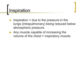
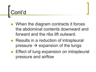
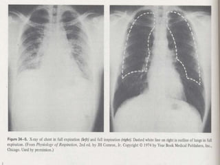
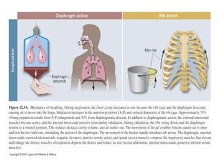
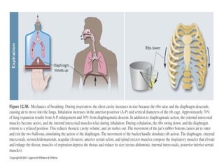
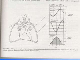
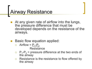
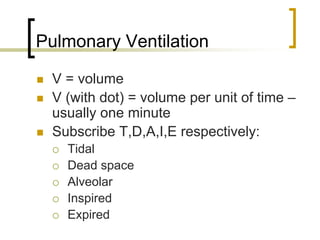
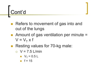
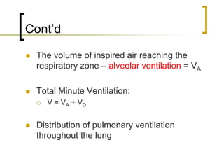
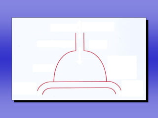
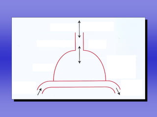
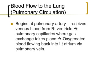
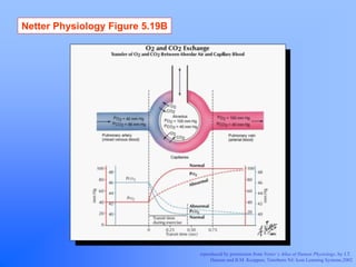
RESPIRATION II
Summary of pulmonary
ventilation

 Pulmonary ventilation primarily functions to
 maintain a fairly constant and favorable
 concentration of oxygen and carbon dioxide
 in the alveolar chambers during rest and
 exercise.

 Ensures complete gaseous exchange
 before the blood leaves the lungs for
 transport throughout the body.
Mechanics of Breathing

 Pulmonary Ventilation = movement of
 air from environment   lungs

 Process = bulk flow
   The movement of molecules along a
   passageway due to a pressure difference
   between the two ends of the
   passageway.
Inspiration

  Inspiration = due to the pressure in the
  lungs (intrapulmonary) being reduced below
  atmospheric pressure.
  Any muscle capable of increasing the
  volume of the chest = inspiratory muscle
Cont’d

 When the diagram contracts it forces
 the abdominal contents downward and
 forward and the ribs lift outward.
 Results in a reduction of intrapleural
 pressure     expansion of the lungs
 Effect of lung expansion on intrapleural
 pressure and airflow
Expiration

  Occurs when pressure within the lungs
  exceeds atmospheric pressure
  A passive process at rest
    Mechanism
  Expiration during exercise
    Ms involved – rectus abdominus, internal
    obliques
    Contraction    ↑ intrapulmonary pressure
    and expiration
Airway Resistance
 At any given rate of airflow into the lungs,
 the pressure difference that must be
 developed depends on the resistance of the
 airways.

 Basic flow equation applied:
   Airflow = P1-P2
           Resistance
   P1-P2 = pressure difference at the two ends of
   the airway
   Resistance is the resistance to flow offered by
   the airway
Cont’d

 Airflow is increased at any time there
 is an increase in the pressure gradient
 across the pulmonary system.
 Factors contributing to airway
 resistance:
   Diameter of airway
     COPD, asthma
3000


                        2000

                                               COMPLIANCE = ∆V / ∆P
                        1000
Volume above FRC (ml)


                                                           =1000/5

                                          ∆V               = 200 ml/cmH2O

                        FRC
                               ∆P


                           0        -10              -20              -30


                               Intrapleural pressure (cmH2O)
Pulmonary Ventilation

 V = volume
 V (with dot) = volume per unit of time –
 usually one minute
 Subscribe T,D,A,I,E respectively:
   Tidal
   Dead space
   Alveolar
   Inspired
   Expired
Cont’d

 Refers to movement of gas into and
 out of the lungs
 Amount of gas ventilation per minute =
 V = VT x f
 Resting values for 70-kg male:
   V = 7.5 L/min
     VT = 0.5 L
     f = 15
Cont’d

 Maximal Exercise
   V = 120-175 L/min
     VT = 3-3.5 L
     f = 40-50
Ventilation

  Not all the air breathed reaches the
  alveolar gas compartment.
  Part of each breath remains in the
  conducting pathways = VD = ventilation
  dead space
  The space that VD occupies =
  anatomical dead space
Cont’d

 The volume of inspired air reaching the
 respiratory zone – alveolar ventilation = VA

 Total Minute Ventilation:
   V = VA + VD


 Distribution of pulmonary ventilation
 throughout the lung
Pulmonary Volumes and
Capacities

 Spirometry
   Measures inspired and expired gas
   volumes
Definitions
    Tidal Volume (VT) = vol of gas
    inspired/expired during a normal
    respiration cycle
    Vital Capacity (VC) = max amt of gas
    expired after a max inspiration
    Residual Volume (RV) = vol of gas
    in the lungs after a max expiration
    Total Lung Capacity (TLC) = amt of
    gas in lungs after max inspiration
      VC + RV
Netter Physiology Figure 5.06


                                reproduced by permission from Netter’s Atlas of Human Physiology, by J.T.
                                     Hansen and B.M. Koeppen, Teterboro NJ: Icon Learning Systems,2002
VA vs VE
            TV     Breathing       Total VE Dead        VA
            (ml)   Rate                     Space       (ml/min)
                   (breaths/min)   (ml/min) Minute
                                            Ventilation
Shallow     150    40              6000     (150x40)    0
Breathing

Normal      500    12              6000     (150x12)     4200
Breathing

Deep        1000   6               6000     (150x6)      5100
Breathing
Dead Space vs Tidal Volume

 Effect of tidal volume on dead space

 Mechanism

 Bottom line
   Deeper breathing provides more effective
   alveolar ventilation than a similar minute
   ventilation achieved through an increased
   breathing rate.
Blood Flow to the Lung
(Pulmonary Circulation)

  Begins at pulmonary artery – receives
  venous blood from Rt ventricle
  pulmonary capillaries where gas
  exchange takes place      Oxygenated
  blood flowing back into Lt atrium via
  pulmonary vein.
Cont’d

 C.O. of Rt vs Lt heart

 Blood flow through Rt vs Lt heart

 Pressure in Rt vs Lt heart
   Mechanism
   Effect of increased blood flow
     Mechanism
Cont’d

 Effect of position on blood flow within
 lung
   Standing

   Exercise

   Supine

   Upside down
Ventilation

                           Concentration depends
                           on ventilation/blood
                           flow


Blood flow
Ventilation-Perfusion
Relationships

  Normal gas exchange requires a
  matching of ventilation to blood flow
  (perfusion, Q)
  The alveolus can be adequately
  ventilated, but if blood flow to the
  alveolus does not adequately match
  ventilation, normal gas exchange does
  not occur.
Cont’d

 Ideal ventilation to perfusion ratio
 (V/Q) = 1 or greater
 i.e. a one to one matching of
 ventilation to blood flow    optimal gas
 exchange

 V/Q differences throughout the lung
Fig 10-13 text
Cont’d
   A large V/Q represents a
   disproportionately high ventilation
   relative to blood flow   poor gas
   exchange
   A V/Q lower then 1 represents a
   greater blood flow vs ventilation in
   the region of the lung being
   considered
   A V/Q > 0.5 = adequate to meet gas
   exchange demands at rest
Physiologic Dead Space

 Definition
 Malfunctioning of alveoli:
   1. underperfusion of blood
   2. inadequate ventilation relative to
   alveolar surface


 Factors     its increase
Netter Physiology Figure 5.19B


                                 reproduced by permission from Netter’s Atlas of Human Physiology, by J.T.
                                      Hansen and B.M. Koeppen, Teterboro NJ: Icon Learning Systems,2002
Perfect Lung
Gas Exchange and Transport
Factors dictating supply of O2 to body

 1. Ambient air gas concentration
    Concentration of atmospheric gases
      O2
      CO2
      N2
 2. Ambient air gas pressure
    Barometric pressure
      Value at sea level
      Effect of weather and altitude
Diffusion of Gases

 Partial Pressure
   Dalton’s Law = the total pressure of a
   gas mixture = the sum of the
   pressures that each gas would exert
   independently

   Partial Pressure =
   Percentage concentration x Total pressure of gas
   mixture
Movement of Gas in Air and Fluids

 Henry’s Law

 Rate of gas diffusion:
   1. pressure differential between the
   gas above the fluid and the gas
   dissolved in the fluid
   2. solubility of the gas in the fluid
.
                                    Vo2
FICK PRINCIPLE


                   -
                 C vo
                        2                 Ca o
                                                 2


          .     .
                             -
          Vo2 = Q ( Ca o - C vo )
                         2     2
                       .
           .          Vo2
          Q =
                           -
                  Ca o - C vo
                            2   2
Blood and Circulation


O2 and CO2 in the Blood
Acid-base Balance
O2 and CO2 Transport in the Blood

 Some O2 and CO2 are transported
 as dissolved gases in the blood

 Majority of O2 - Hb

 Majority of CO2 – HCO-3
O2 in Solution

 Effect of solubility on PO2 in solution
 PAO2
 Quantity dissolved
 Typical blood volume = 5 L
   Total volume of O2 dissolved
   Ability to sustain life
Hemoglobin and O2 Transport

 99% O2 transported in blood =
 bound to Hb

 O2 carrying capacity of Hb
   Hb4 + 4 O2      Hb4O8


 Oxyhemoglobin
 Deoxyhemoglobin
 Effect of PO2 in solution on Hb state
Cont’d
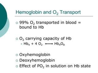
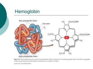
 Amt of O2 that can be transported
 per unit volume of blood is
 dependent upon [Hb]
 Normal [Hb]
   Gender differences (m=150g; f=130g)
 When completely saturated with O2,
 each g of Hb can transport 1.34 ml
 O2
Hemoglobin
PO2 and Hb Saturation

 Cooperative binding = the binding
 of one molecule to another
 progressively facilitates the binding
 of progressive molecules.
Oxyhemoglobin Dissociation Curve

 Illustrates the saturation of Hb with
 O2 at various PO2 values

 % saturation = O2 combined with Hb x 100
                 O2 capacity of Hb


 100 % saturation
Oxyhemoglobin Dissociation Curve
 Combination of O2 + Hb in lungs =
 loading
 Release of O2 from Hb at tissues =
 unloading
 Reversible Reaction:
   DeoxyHb + O2               OxyHb
 Factors affecting reaction direction
   1. PO2 in blood
   2. Affinity of Hb for O2
Cont’d
 Effect of high PO2

 Effect of low PO2

 Effect of ↓ affinity of Hb for O2

 High PO2 in lungs    high arterial PO2   ↑
 oxyHb formation
 Low PO2 in tissues    ↓ PO2 in systemic
 capillaries   unloading of O2 to be used by the
 tissues and increasing deoxyHb
O2 Transport Cascade
PO2 in the Lungs
 Actual Hb-O2 saturation at sea level
   Note sea-level alveolar PO2 of 100
   mmHg
   Effect of an ↑ alveolar PO2

 Effect of PO2 < 60 mmHg on O2-Hb
 saturation
   Importance
   Saturation at PO2 = 60 mmHg
PO2 in Tissues
 Resting PO2 in cells

 Dissolved O2 from the plasma diffuses
 across the capillary membrane into cell
   Effect on plasma PO2 and cellular PO2
   Effect on Hb saturation

 Hb saturation at cellular PO2

 Resting (a-v)O2 difference
(a-v)O2 Difference

 Definition
 Resting values
 Amount of O2 bound to Hb
   Importance


 Effect of exercise
 PO2 = 2-3 mmHg – effect on O2-Hb
O2 Transport in Muscle

 Myoglobin

 Location

 Composition

 Affinity for O2
Myoglobin

 Mb + O2      MbO2

 Primary function
CO2 Transport in the Blood

 CO2 transported in the blood in 3
 forms:

 1. dissolved CO2 (10%)
 2. bound to Hb =
 carbaminohemoglobin (20%)
 3. HCO-3 = bicarbonate (70%)
CO2 Transport as Bicarbonate
CO2 in solution combines with H2O to
form carbonic acid

CO2+H2O           H2CO3   (enzyme = carbonic anhydrase)


In tissues: CO2+H2O H2CO3 H++HCO-3

  As tissue PCO2 ↑, CO2 binds with H2O to form
  H2CO3 (carbonic acid). H2CO3 dissociates into
  HCO-3 + H+. H+ combines with Hb and HCO-3
  diffuses out of the RBC and into the plasma.
Control of blood acid-base balance


pH=6.1 + log (HCO-3 / CO2)
Respiration powerpoint

Respiration powerpoint

  • 1.
  • 2.
    Summary of pulmonary ventilation Pulmonary ventilation primarily functions to maintain a fairly constant and favorable concentration of oxygen and carbon dioxide in the alveolar chambers during rest and exercise. Ensures complete gaseous exchange before the blood leaves the lungs for transport throughout the body.
  • 3.
    Mechanics of Breathing Pulmonary Ventilation = movement of air from environment lungs Process = bulk flow The movement of molecules along a passageway due to a pressure difference between the two ends of the passageway.
  • 4.
    Inspiration Inspiration= due to the pressure in the lungs (intrapulmonary) being reduced below atmospheric pressure. Any muscle capable of increasing the volume of the chest = inspiratory muscle
  • 5.
    Cont’d When thediagram contracts it forces the abdominal contents downward and forward and the ribs lift outward. Results in a reduction of intrapleural pressure expansion of the lungs Effect of lung expansion on intrapleural pressure and airflow
  • 8.
    Expiration Occurswhen pressure within the lungs exceeds atmospheric pressure A passive process at rest Mechanism Expiration during exercise Ms involved – rectus abdominus, internal obliques Contraction ↑ intrapulmonary pressure and expiration
  • 12.
    Airway Resistance Atany given rate of airflow into the lungs, the pressure difference that must be developed depends on the resistance of the airways. Basic flow equation applied: Airflow = P1-P2 Resistance P1-P2 = pressure difference at the two ends of the airway Resistance is the resistance to flow offered by the airway
  • 13.
    Cont’d Airflow isincreased at any time there is an increase in the pressure gradient across the pulmonary system. Factors contributing to airway resistance: Diameter of airway COPD, asthma
  • 15.
    3000 2000 COMPLIANCE = ∆V / ∆P 1000 Volume above FRC (ml) =1000/5 ∆V = 200 ml/cmH2O FRC ∆P 0 -10 -20 -30 Intrapleural pressure (cmH2O)
  • 16.
    Pulmonary Ventilation V= volume V (with dot) = volume per unit of time – usually one minute Subscribe T,D,A,I,E respectively: Tidal Dead space Alveolar Inspired Expired
  • 17.
    Cont’d Refers tomovement of gas into and out of the lungs Amount of gas ventilation per minute = V = VT x f Resting values for 70-kg male: V = 7.5 L/min VT = 0.5 L f = 15
  • 18.
    Cont’d Maximal Exercise V = 120-175 L/min VT = 3-3.5 L f = 40-50
  • 19.
    Ventilation Notall the air breathed reaches the alveolar gas compartment. Part of each breath remains in the conducting pathways = VD = ventilation dead space The space that VD occupies = anatomical dead space
  • 20.
    Cont’d The volumeof inspired air reaching the respiratory zone – alveolar ventilation = VA Total Minute Ventilation: V = VA + VD Distribution of pulmonary ventilation throughout the lung
  • 21.
    Pulmonary Volumes and Capacities Spirometry Measures inspired and expired gas volumes
  • 22.
    Definitions Tidal Volume (VT) = vol of gas inspired/expired during a normal respiration cycle Vital Capacity (VC) = max amt of gas expired after a max inspiration Residual Volume (RV) = vol of gas in the lungs after a max expiration Total Lung Capacity (TLC) = amt of gas in lungs after max inspiration VC + RV
  • 24.
    Netter Physiology Figure5.06 reproduced by permission from Netter’s Atlas of Human Physiology, by J.T. Hansen and B.M. Koeppen, Teterboro NJ: Icon Learning Systems,2002
  • 30.
    VA vs VE TV Breathing Total VE Dead VA (ml) Rate Space (ml/min) (breaths/min) (ml/min) Minute Ventilation Shallow 150 40 6000 (150x40) 0 Breathing Normal 500 12 6000 (150x12) 4200 Breathing Deep 1000 6 6000 (150x6) 5100 Breathing
  • 31.
    Dead Space vsTidal Volume Effect of tidal volume on dead space Mechanism Bottom line Deeper breathing provides more effective alveolar ventilation than a similar minute ventilation achieved through an increased breathing rate.
  • 33.
    Blood Flow tothe Lung (Pulmonary Circulation) Begins at pulmonary artery – receives venous blood from Rt ventricle pulmonary capillaries where gas exchange takes place Oxygenated blood flowing back into Lt atrium via pulmonary vein.
  • 35.
    Cont’d C.O. ofRt vs Lt heart Blood flow through Rt vs Lt heart Pressure in Rt vs Lt heart Mechanism Effect of increased blood flow Mechanism
  • 39.
    Cont’d Effect ofposition on blood flow within lung Standing Exercise Supine Upside down
  • 41.
    Ventilation Concentration depends on ventilation/blood flow Blood flow
  • 42.
    Ventilation-Perfusion Relationships Normalgas exchange requires a matching of ventilation to blood flow (perfusion, Q) The alveolus can be adequately ventilated, but if blood flow to the alveolus does not adequately match ventilation, normal gas exchange does not occur.
  • 43.
    Cont’d Ideal ventilationto perfusion ratio (V/Q) = 1 or greater i.e. a one to one matching of ventilation to blood flow optimal gas exchange V/Q differences throughout the lung
  • 44.
  • 46.
    Cont’d A large V/Q represents a disproportionately high ventilation relative to blood flow poor gas exchange A V/Q lower then 1 represents a greater blood flow vs ventilation in the region of the lung being considered A V/Q > 0.5 = adequate to meet gas exchange demands at rest
  • 47.
    Physiologic Dead Space Definition Malfunctioning of alveoli: 1. underperfusion of blood 2. inadequate ventilation relative to alveolar surface Factors its increase
  • 48.
    Netter Physiology Figure5.19B reproduced by permission from Netter’s Atlas of Human Physiology, by J.T. Hansen and B.M. Koeppen, Teterboro NJ: Icon Learning Systems,2002
  • 49.
  • 54.
  • 55.
    Factors dictating supplyof O2 to body 1. Ambient air gas concentration Concentration of atmospheric gases O2 CO2 N2 2. Ambient air gas pressure Barometric pressure Value at sea level Effect of weather and altitude
  • 56.
    Diffusion of Gases Partial Pressure Dalton’s Law = the total pressure of a gas mixture = the sum of the pressures that each gas would exert independently Partial Pressure = Percentage concentration x Total pressure of gas mixture
  • 57.
    Movement of Gasin Air and Fluids Henry’s Law Rate of gas diffusion: 1. pressure differential between the gas above the fluid and the gas dissolved in the fluid 2. solubility of the gas in the fluid
  • 60.
    . Vo2 FICK PRINCIPLE - C vo 2 Ca o 2 . . - Vo2 = Q ( Ca o - C vo ) 2 2 . . Vo2 Q = - Ca o - C vo 2 2
  • 61.
    Blood and Circulation O2and CO2 in the Blood Acid-base Balance
  • 62.
    O2 and CO2Transport in the Blood Some O2 and CO2 are transported as dissolved gases in the blood Majority of O2 - Hb Majority of CO2 – HCO-3
  • 63.
    O2 in Solution Effect of solubility on PO2 in solution PAO2 Quantity dissolved Typical blood volume = 5 L Total volume of O2 dissolved Ability to sustain life
  • 64.
    Hemoglobin and O2Transport 99% O2 transported in blood = bound to Hb O2 carrying capacity of Hb Hb4 + 4 O2 Hb4O8 Oxyhemoglobin Deoxyhemoglobin Effect of PO2 in solution on Hb state
  • 65.
    Cont’d Amt ofO2 that can be transported per unit volume of blood is dependent upon [Hb] Normal [Hb] Gender differences (m=150g; f=130g) When completely saturated with O2, each g of Hb can transport 1.34 ml O2
  • 66.
  • 67.
    PO2 and HbSaturation Cooperative binding = the binding of one molecule to another progressively facilitates the binding of progressive molecules.
  • 68.
    Oxyhemoglobin Dissociation Curve Illustrates the saturation of Hb with O2 at various PO2 values % saturation = O2 combined with Hb x 100 O2 capacity of Hb 100 % saturation
  • 69.
    Oxyhemoglobin Dissociation Curve Combination of O2 + Hb in lungs = loading Release of O2 from Hb at tissues = unloading Reversible Reaction: DeoxyHb + O2 OxyHb Factors affecting reaction direction 1. PO2 in blood 2. Affinity of Hb for O2
  • 70.
    Cont’d Effect ofhigh PO2 Effect of low PO2 Effect of ↓ affinity of Hb for O2 High PO2 in lungs high arterial PO2 ↑ oxyHb formation Low PO2 in tissues ↓ PO2 in systemic capillaries unloading of O2 to be used by the tissues and increasing deoxyHb
  • 73.
  • 74.
    PO2 in theLungs Actual Hb-O2 saturation at sea level Note sea-level alveolar PO2 of 100 mmHg Effect of an ↑ alveolar PO2 Effect of PO2 < 60 mmHg on O2-Hb saturation Importance Saturation at PO2 = 60 mmHg
  • 76.
    PO2 in Tissues Resting PO2 in cells Dissolved O2 from the plasma diffuses across the capillary membrane into cell Effect on plasma PO2 and cellular PO2 Effect on Hb saturation Hb saturation at cellular PO2 Resting (a-v)O2 difference
  • 77.
    (a-v)O2 Difference Definition Resting values Amount of O2 bound to Hb Importance Effect of exercise PO2 = 2-3 mmHg – effect on O2-Hb
  • 78.
    O2 Transport inMuscle Myoglobin Location Composition Affinity for O2
  • 79.
    Myoglobin Mb +O2 MbO2 Primary function
  • 81.
    CO2 Transport inthe Blood CO2 transported in the blood in 3 forms: 1. dissolved CO2 (10%) 2. bound to Hb = carbaminohemoglobin (20%) 3. HCO-3 = bicarbonate (70%)
  • 83.
    CO2 Transport asBicarbonate CO2 in solution combines with H2O to form carbonic acid CO2+H2O H2CO3 (enzyme = carbonic anhydrase) In tissues: CO2+H2O H2CO3 H++HCO-3 As tissue PCO2 ↑, CO2 binds with H2O to form H2CO3 (carbonic acid). H2CO3 dissociates into HCO-3 + H+. H+ combines with Hb and HCO-3 diffuses out of the RBC and into the plasma.
  • 84.
    Control of bloodacid-base balance pH=6.1 + log (HCO-3 / CO2)