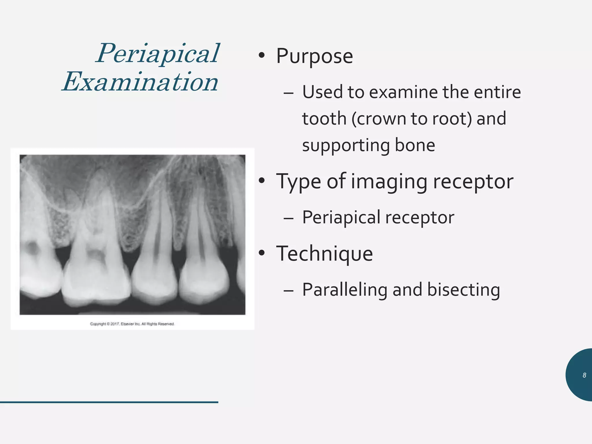
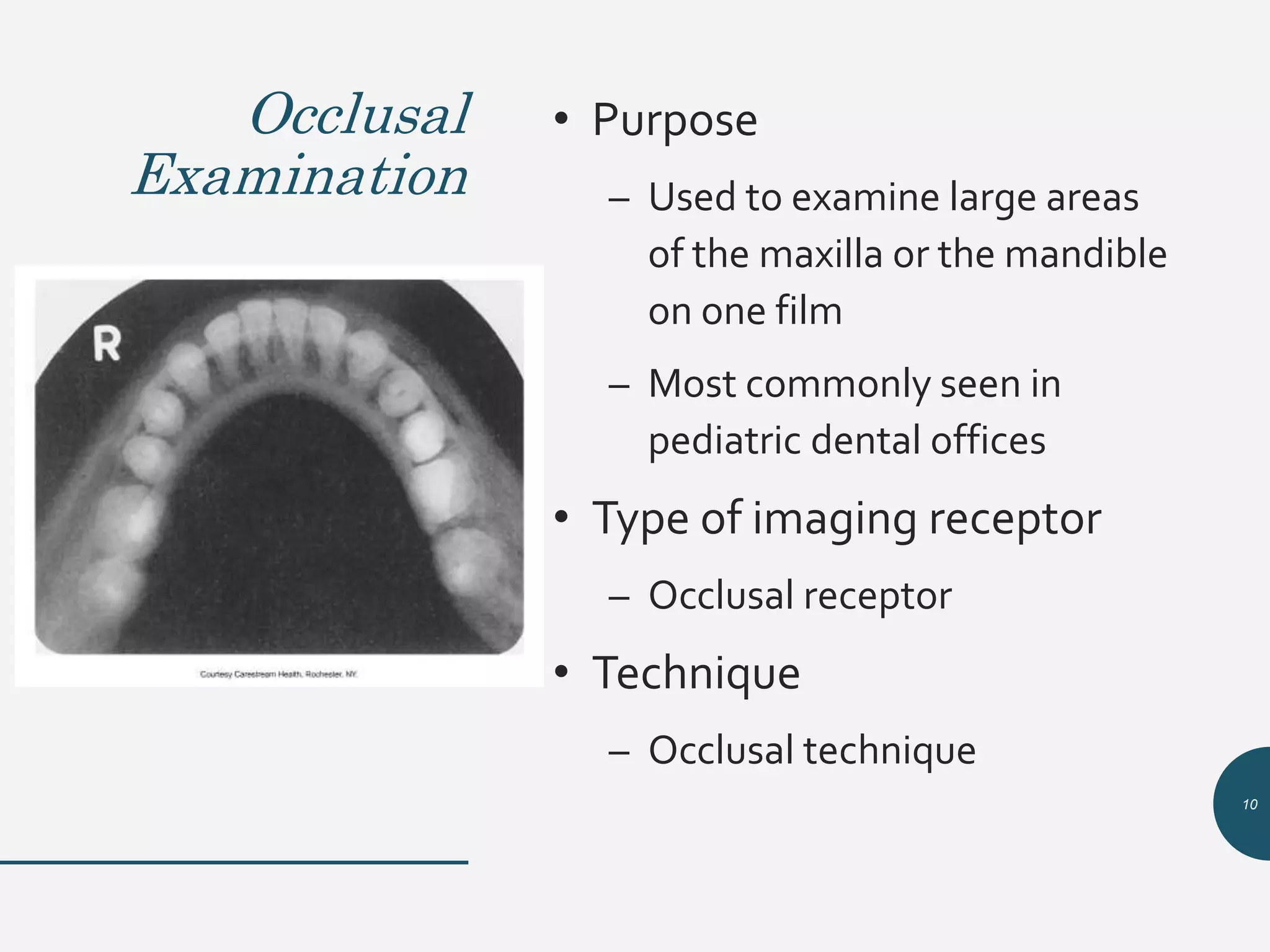
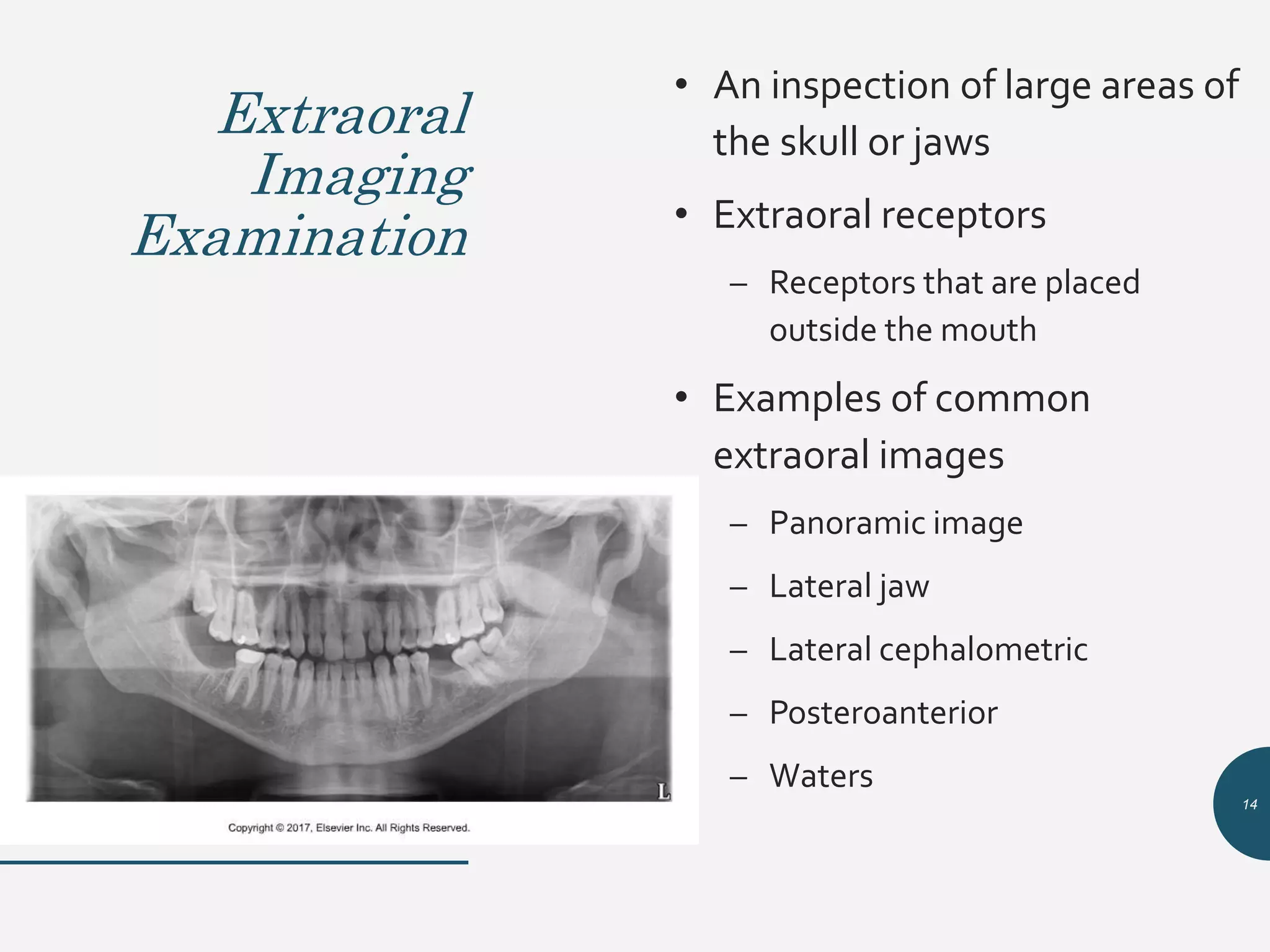
Introduction to Dental Imaging
Examinations
Chapter 16
1
Learning Objectives
Lesson 16.1: Introduction to Dental
Imaging Examinations
1. Define the key terms associated with dental imaging
examinations.
2. List the three types of intraoral imaging examinations.
3. Describe the purpose, the type of receptor, and the
technique used for each of the three types of intraoral
imaging examinations.
4. List the various projections that constitute a complete
mouth series (CMS).
2
Learning Objectives
Lesson 16.1: Introduction to Dental
Imaging Examinations (Cont.)
5. List the general diagnostic criteria for intraoral images.
6. List examples of extraoral imaging examinations.
7. Discuss the prescribing of dental images.
8. Describe when prescribing a CMS for a new patient is
warranted.
3
Introduction • Purpose
– To introduce the dental
radiographer to the different
intraoral radiographic
examinations used in
dentistry
– To define the complete mouth
series
– To describe in detail the
diagnostic criteria of intraoral
images
4
Intraoral
Imaging
Examination
• Types of intraoral imaging
examinations
• Complete mouth series
• Diagnostic criteria for intraoral
images
• Intraoral examinations are the
foundation of dental imaging.
5
Intraoral Imaging Examination (Cont.)
• An inspection of teeth and intraoral adjacent
structures
• Requires the use of intraoral image receptors
– Intraoral receptor
• Receptor that is placed inside the mouth and is used to examine
the teeth and supporting structures
• Intraoral film is the most commonly used x-ray film.
6
Types of Intraoral
Imaging Examinations
• Periapical examination
• Interproximal examination
• Occlusal examination
• Each of these examinations has a certain
purpose and requires the use of a specific type
of imaging receptor and technique.
7
Periapical
Examination
• Purpose
– Used to examine the entire
tooth (crown to root) and
supporting bone
• Type of imaging receptor
– Periapical receptor
• Technique
– Paralleling and bisecting
8
Interproximal
Examination
• Purpose
– To examine the crowns of both
the maxillary and mandibular
teeth on a single film
– This type of film is most often
taken to check for cavities
between teeth.
– Should show crestal bone
• Type of imaging receptor
– Bite-wing receptor
• Technique
– Bite-wing technique
9
Occlusal
Examination
• Purpose
– Used to examine large areas
of the maxilla or the mandible
on one film
– Most commonly seen in
pediatric dental offices
• Type of imaging receptor
– Occlusal receptor
• Technique
– Occlusal technique
10
Complete Mouth Series/Full Mouth
Series
• CMS, also known as FMS or FMX
• A full-mouth series or complete series
– Tooth-bearing areas
• Dentulous areas
• Edentulous areas
• A total of 14 to 20 films may be taken.
• Film size selection is important.
• Used to detect disease, foreign objects, retained roots.
• Patients usually have a full-mouth series of x-rays taken every 3
to 5 years.
11
12
Diagnostic Criteria
for Intraoral Images
• Images must have optimum density, contrast, definition, and
detail.
• Images must have the least amount of distortion possible.
• The CMS must include images that show all tooth-bearing
areas.
• Periapical images must show the entire crowns and roots of
teeth being examined, as well as 2 to 3 mm beyond the root
apices.
• Bite-wing images must show open contacts.
13
Extraoral
Imaging
Examination
• An inspection of large areas of
the skull or jaws
• Extraoral receptors
– Receptors that are placed
outside the mouth
• Examples of common
extraoral images
– Panoramic image
– Lateral jaw
– Lateral cephalometric
– Posteroanterior
– Waters
14
Prescribing
Dental
Images
• Prescribing is based on the
individual needs of the
patient.
• Dentist uses professional
judgment to make decisions
about the number, type, and
frequency of dental images.
15
Prescribing
Dental
Images
(Cont.)
• Not all patients need an FMX.
– FMX is appropriate when a new
adult patient presents with
clinical evidence of generalized
dental disease or a history of
extensive dental treatment.
– Otherwise, a combination of
bite-wings, selected
periapicals, and/or a panoramic
image should be prescribed on
the basis of a patient’s
individual needs. 16

Chapter 16: Intro to Dental Imaging Examinations

  • 1.
    Introduction to DentalImaging Examinations Chapter 16 1
  • 2.
    Learning Objectives Lesson 16.1:Introduction to Dental Imaging Examinations 1. Define the key terms associated with dental imaging examinations. 2. List the three types of intraoral imaging examinations. 3. Describe the purpose, the type of receptor, and the technique used for each of the three types of intraoral imaging examinations. 4. List the various projections that constitute a complete mouth series (CMS). 2
  • 3.
    Learning Objectives Lesson 16.1:Introduction to Dental Imaging Examinations (Cont.) 5. List the general diagnostic criteria for intraoral images. 6. List examples of extraoral imaging examinations. 7. Discuss the prescribing of dental images. 8. Describe when prescribing a CMS for a new patient is warranted. 3
  • 4.
    Introduction • Purpose –To introduce the dental radiographer to the different intraoral radiographic examinations used in dentistry – To define the complete mouth series – To describe in detail the diagnostic criteria of intraoral images 4
  • 5.
    Intraoral Imaging Examination • Types ofintraoral imaging examinations • Complete mouth series • Diagnostic criteria for intraoral images • Intraoral examinations are the foundation of dental imaging. 5
  • 6.
    Intraoral Imaging Examination(Cont.) • An inspection of teeth and intraoral adjacent structures • Requires the use of intraoral image receptors – Intraoral receptor • Receptor that is placed inside the mouth and is used to examine the teeth and supporting structures • Intraoral film is the most commonly used x-ray film. 6
  • 7.
    Types of Intraoral ImagingExaminations • Periapical examination • Interproximal examination • Occlusal examination • Each of these examinations has a certain purpose and requires the use of a specific type of imaging receptor and technique. 7
  • 8.
    Periapical Examination • Purpose – Usedto examine the entire tooth (crown to root) and supporting bone • Type of imaging receptor – Periapical receptor • Technique – Paralleling and bisecting 8
  • 9.
    Interproximal Examination • Purpose – Toexamine the crowns of both the maxillary and mandibular teeth on a single film – This type of film is most often taken to check for cavities between teeth. – Should show crestal bone • Type of imaging receptor – Bite-wing receptor • Technique – Bite-wing technique 9
  • 10.
    Occlusal Examination • Purpose – Usedto examine large areas of the maxilla or the mandible on one film – Most commonly seen in pediatric dental offices • Type of imaging receptor – Occlusal receptor • Technique – Occlusal technique 10
  • 11.
    Complete Mouth Series/FullMouth Series • CMS, also known as FMS or FMX • A full-mouth series or complete series – Tooth-bearing areas • Dentulous areas • Edentulous areas • A total of 14 to 20 films may be taken. • Film size selection is important. • Used to detect disease, foreign objects, retained roots. • Patients usually have a full-mouth series of x-rays taken every 3 to 5 years. 11
  • 12.
  • 13.
    Diagnostic Criteria for IntraoralImages • Images must have optimum density, contrast, definition, and detail. • Images must have the least amount of distortion possible. • The CMS must include images that show all tooth-bearing areas. • Periapical images must show the entire crowns and roots of teeth being examined, as well as 2 to 3 mm beyond the root apices. • Bite-wing images must show open contacts. 13
  • 14.
    Extraoral Imaging Examination • An inspectionof large areas of the skull or jaws • Extraoral receptors – Receptors that are placed outside the mouth • Examples of common extraoral images – Panoramic image – Lateral jaw – Lateral cephalometric – Posteroanterior – Waters 14
  • 15.
    Prescribing Dental Images • Prescribing isbased on the individual needs of the patient. • Dentist uses professional judgment to make decisions about the number, type, and frequency of dental images. 15
  • 16.
    Prescribing Dental Images (Cont.) • Not allpatients need an FMX. – FMX is appropriate when a new adult patient presents with clinical evidence of generalized dental disease or a history of extensive dental treatment. – Otherwise, a combination of bite-wings, selected periapicals, and/or a panoramic image should be prescribed on the basis of a patient’s individual needs. 16

Editor's Notes

  • #5 In addition, extraoral imaging examinations used in dentistry are introduced.
  • #6 Intraoral examinations are the foundation of dental imaging.
  • #7 Intraoral film is the most commonly used x-ray film.
  • #8 Each of these examinations has a certain purpose and requires the use of a specific type of imaging receptor and technique.
  • #9 Used to examine the tooth from crown to root. The word part peri means “around.” The word part apex refers to the end of the tooth root. Refer students to Figure 16-1.
  • #10 This type of film is most often taken to check for cavities between teeth. Should show crestal bone. Refer students to Figure 16-2.
  • #11 This type of film is most commonly seen in pediatric dental offices. Refer students to Figure 16-3.
  • #12 Also known as a full-mouth series or complete series. This series can include only periapicals or be a combination of periapicals and bite-wings. A total of 14 to 20 films may be taken. Film size selection is important. Used to detect disease, foreign objects, retained roots. Patients usually have a full-mouth series of x-rays taken every 3 to 5 years. Refer students to Figure 16-4.
  • #14 Refer students to Box 16-1.
  • #15 What does extraoral mean? Examples of common extraoral films include panoramic and cephalometric films. Refer students to Figure 16-5.
  • #16 All images are prescribed based on the individual needs of the patient.
  • #17 Refer students back to Table 5-1 as necessary.