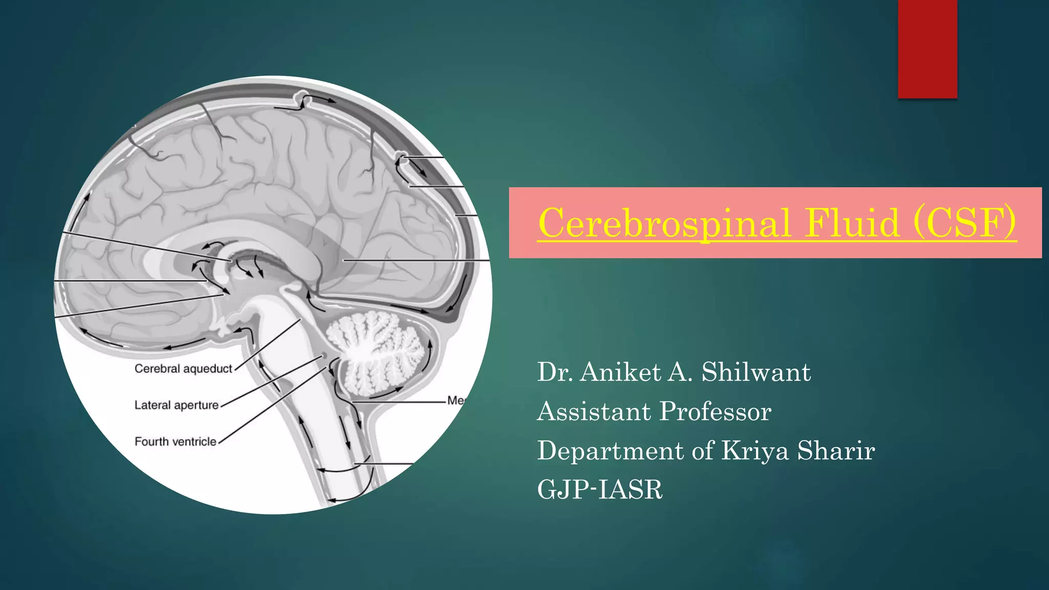
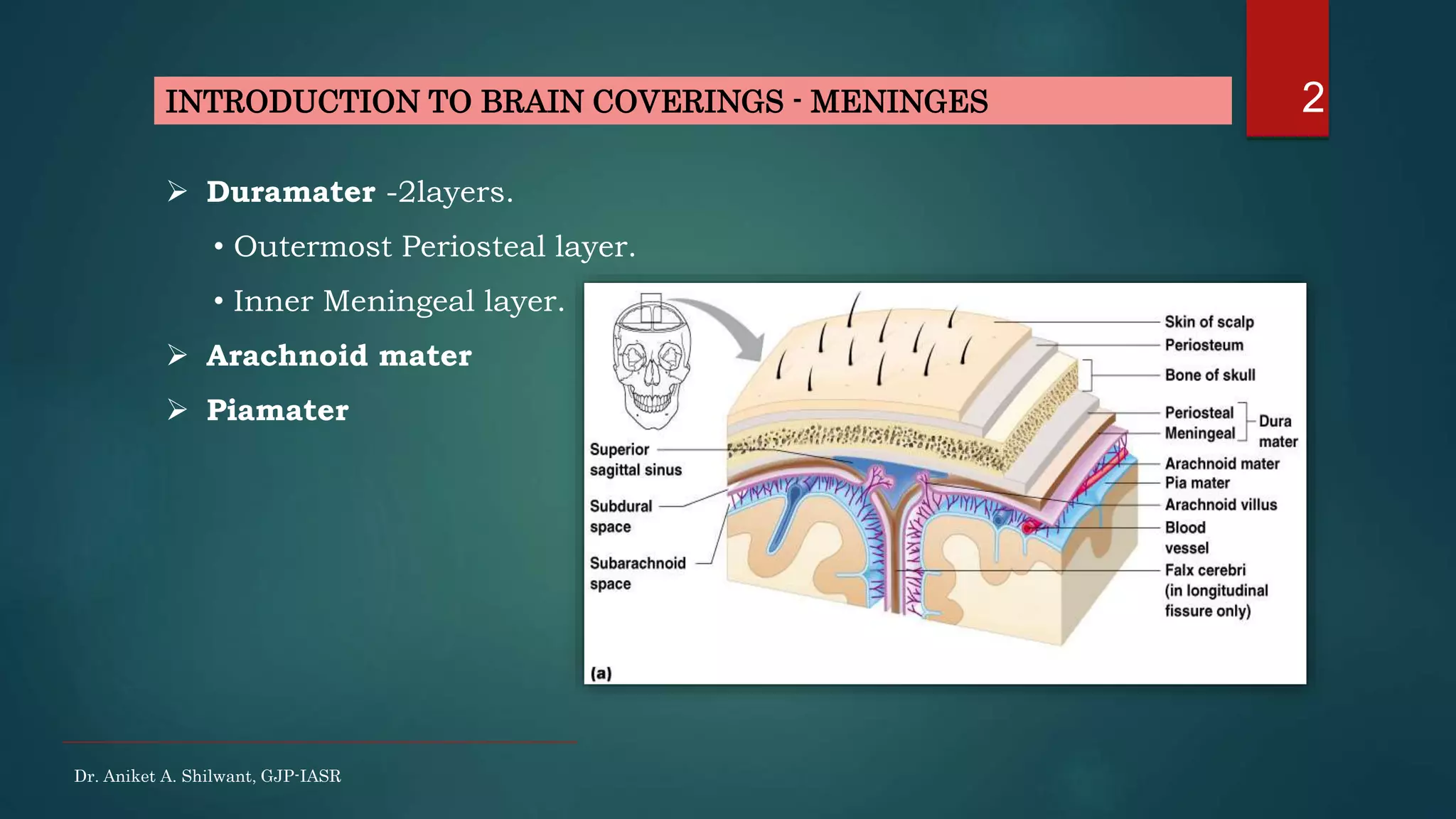
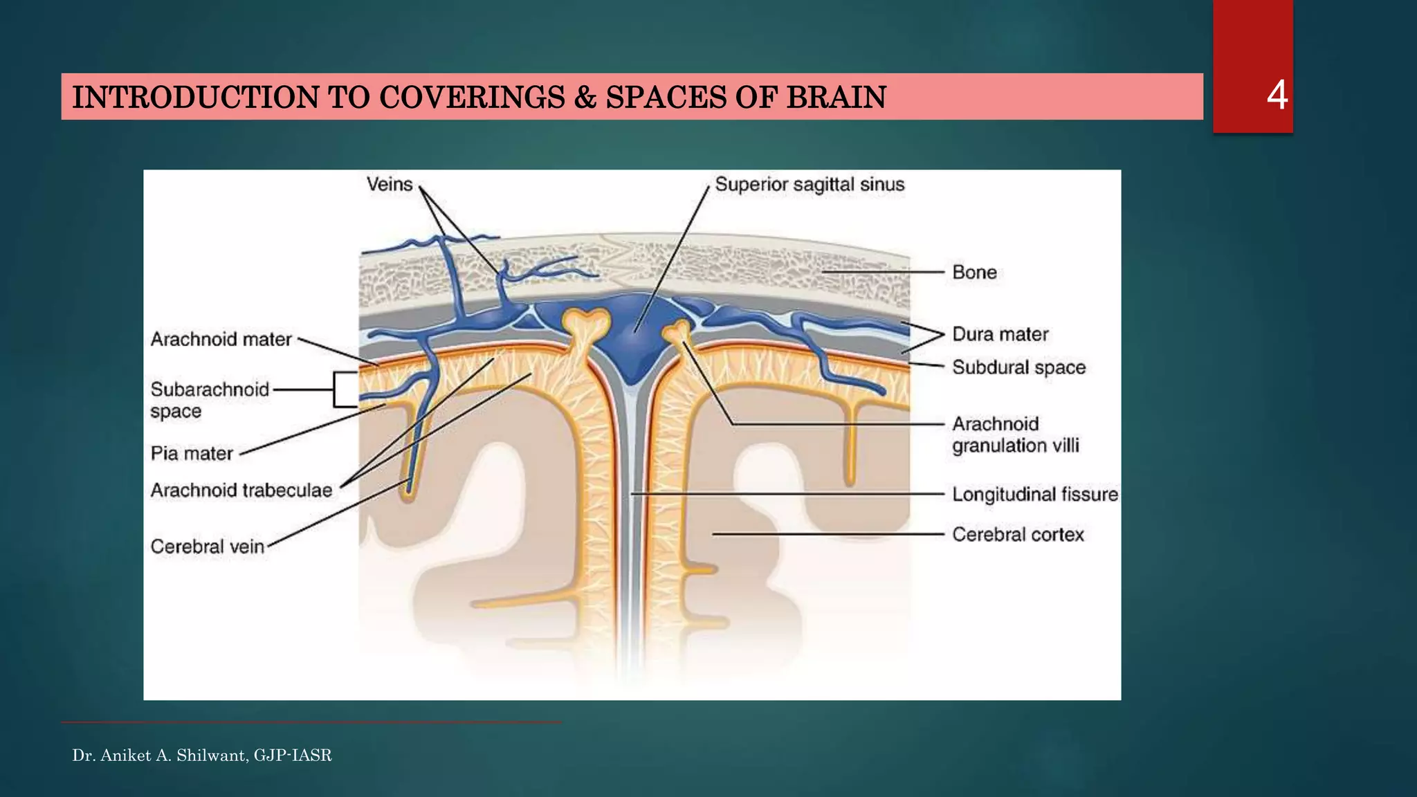
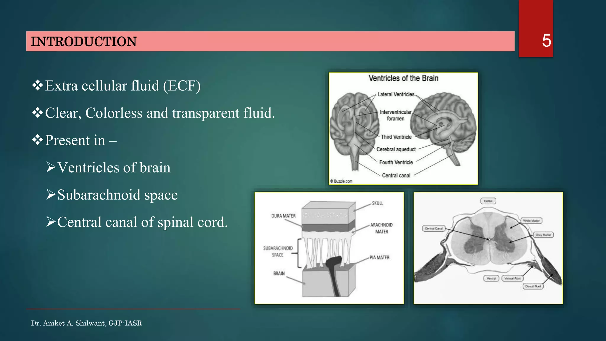
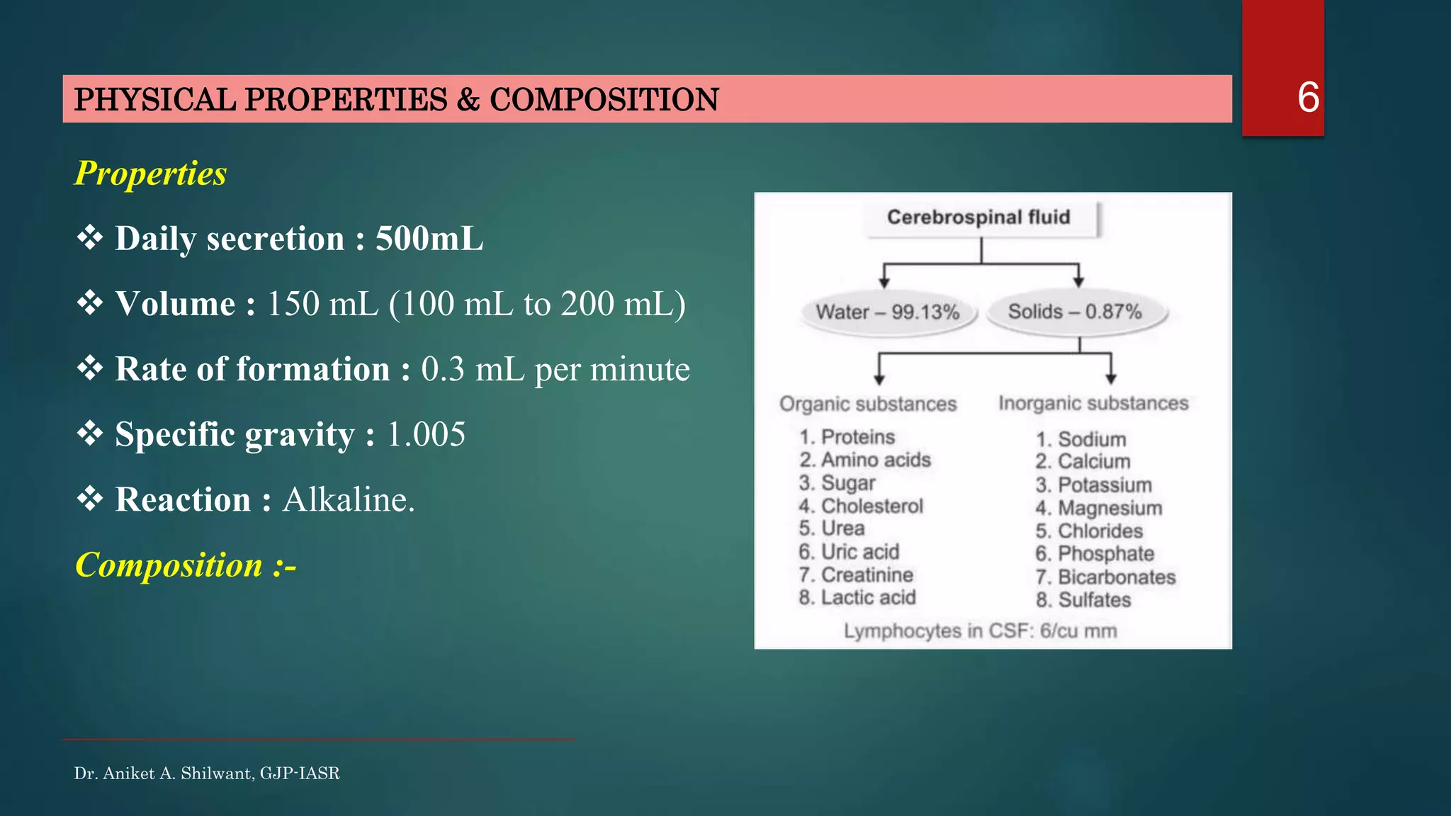
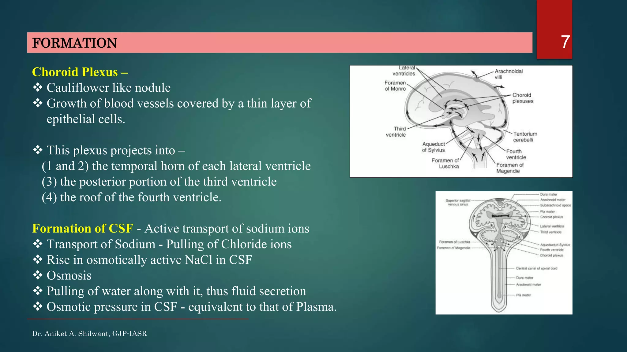
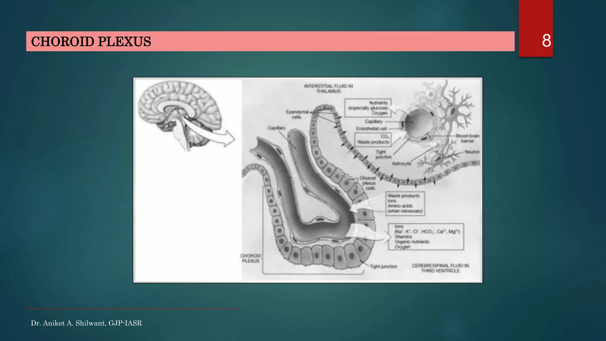
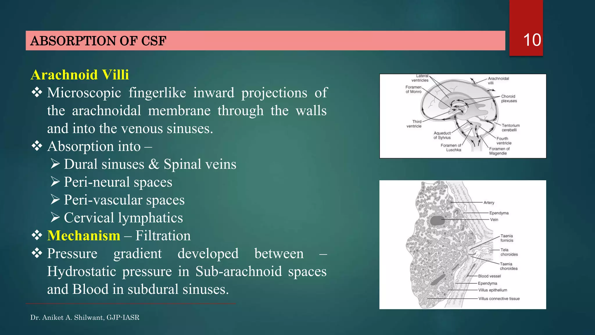
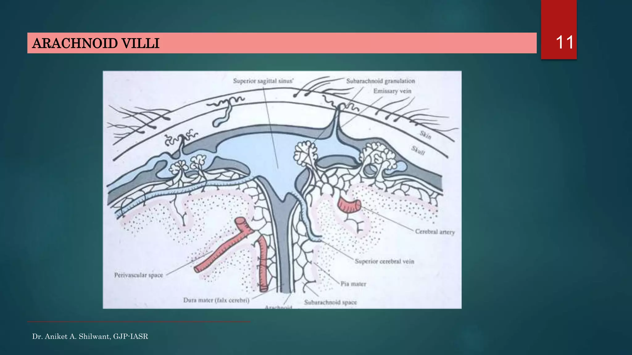
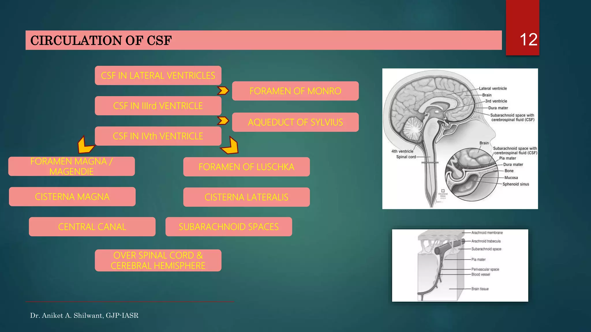
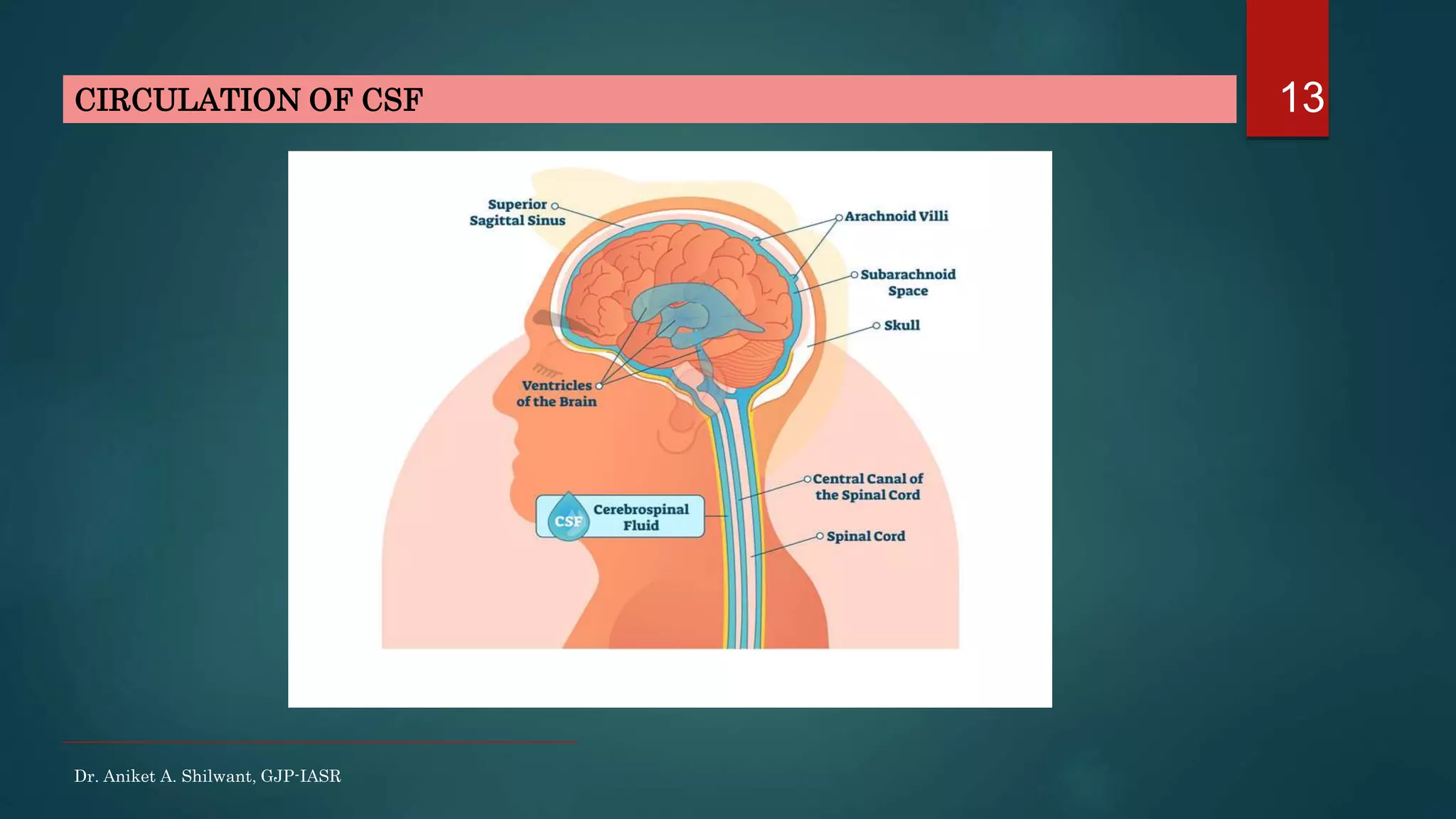
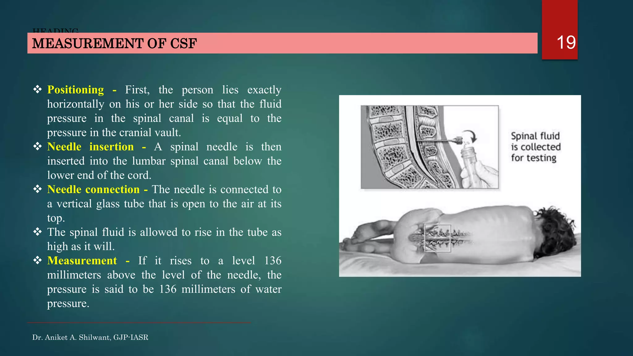
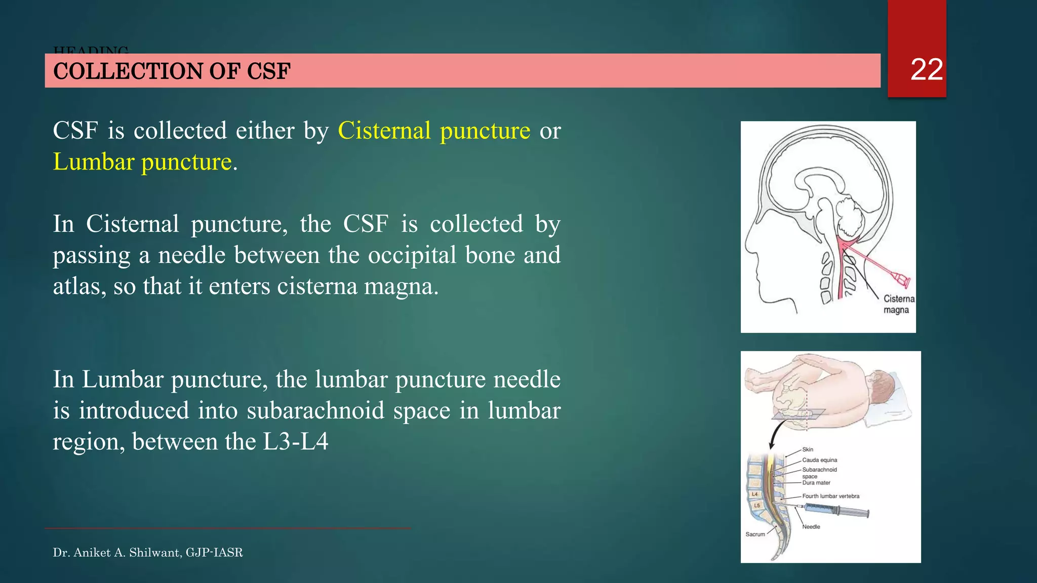
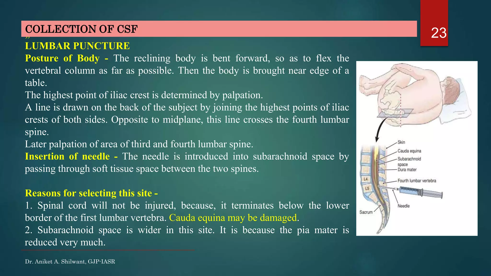
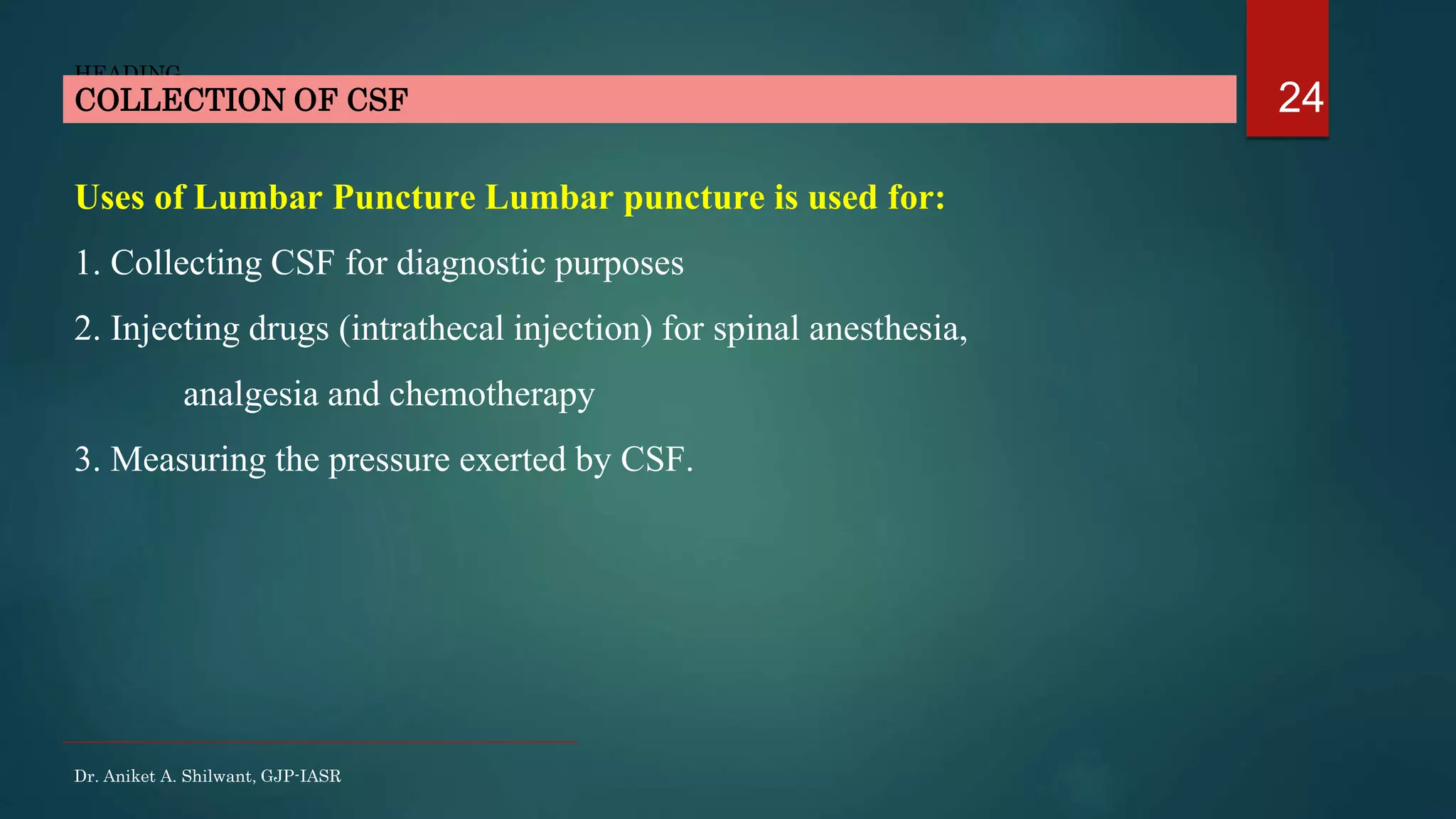
This document discusses cerebrospinal fluid (CSF), including its formation, circulation, absorption, and functions. It notes that CSF is formed by the choroid plexus in the ventricles of the brain at a rate of 0.3 mL per minute. CSF circulates through the ventricles and subarachnoid spaces before being absorbed into venous sinuses by arachnoid villi. The key functions of CSF are to protect the brain, serve as a medium of exchange, and help regulate cranial pressure. The document also describes how lumbar puncture is performed to measure CSF pressure or collect samples for diagnostic purposes.
2. Duramater -2layers.
• Outermost Periosteal layer.
• Inner Meningeal layer.
Arachnoid mater
Piamater
INTRODUCTION TO BRAIN COVERINGS - MENINGES
Dr. Aniket A. Shilwant, GJP-IASR
2
3. 3
Dr. Aniket A. Shilwant, GJP-IASR
INTRODUCTION TO COVERINGS & SPACES OF BRAIN
SUBDURAL SPACE
BONE LAYER OF SKULL
DURA MATER
DURA MATER
ARACHNOID MATER
ARACHNOID MATER
PIA MATER
SUBARACHNOID SPACE
EPIDURAL SPACE
4. 4
Dr. Aniket A. Shilwant, GJP-IASR
INTRODUCTION TO COVERINGS & SPACES OF BRAIN
5. 5INTRODUCTION
Extra cellular fluid (ECF)
Clear, Colorless and transparent fluid.
Present in –
Ventricles of brain
Subarachnoid space
Central canal of spinal cord.
Dr. Aniket A. Shilwant, GJP-IASR
6. 6
Properties
Daily secretion : 500mL
Volume : 150 mL (100 mL to 200 mL)
Rate of formation : 0.3 mL per minute
Specific gravity : 1.005
Reaction : Alkaline.
Composition :-
PHYSICAL PROPERTIES & COMPOSITION
Dr. Aniket A. Shilwant, GJP-IASR
7. 7FORMATION
Choroid Plexus –
Cauliflower like nodule
Growth of blood vessels covered by a thin layer of
epithelial cells.
This plexus projects into –
(1 and 2) the temporal horn of each lateral ventricle
(3) the posterior portion of the third ventricle
(4) the roof of the fourth ventricle.
Formation of CSF - Active transport of sodium ions
Transport of Sodium - Pulling of Chloride ions
Rise in osmotically active NaCl in CSF
Osmosis
Pulling of water along with it, thus fluid secretion
Osmotic pressure in CSF - equivalent to that of Plasma.
Dr. Aniket A. Shilwant, GJP-IASR
9. 9FORMATION & SECRETION BALANCE
When infused –
Hypertonic saline
CSF formation decreases, thus CSF pressure also decreased.
Hypotonic saline
CSF formation increases with increase in Intracranial pressure also.
Dr. Aniket A. Shilwant, GJP-IASR
10. Dr. Aniket A. Shilwant, GJP-IASR
10ABSORPTION OF CSF
Arachnoid Villi
Microscopic fingerlike inward projections of
the arachnoidal membrane through the walls
and into the venous sinuses.
Absorption into –
Dural sinuses & Spinal veins
Peri-neural spaces
Peri-vascular spaces
Cervical lymphatics
Mechanism – Filtration
Pressure gradient developed between –
Hydrostatic pressure in Sub-arachnoid spaces
and Blood in subdural sinuses.
12. Dr. Aniket A. Shilwant, GJP-IASR
12CIRCULATION OF CSF
CSF IN LATERAL VENTRICLES
CSF IN IIIrd VENTRICLE
CSF IN IVth VENTRICLE
FORAMEN MAGNA /
MAGENDIE
CISTERNA MAGNA
FORAMEN OF LUSCHKA
CISTERNA LATERALIS
CENTRAL CANAL SUBARACHNOID SPACES
OVER SPINAL CORD &
CEREBRAL HEMISPHERE
FORAMEN OF MONRO
AQUEDUCT OF SYLVIUS
14. Dr. Aniket A. Shilwant, GJP-IASR
14PRESSURE EXERTED BY CSF
Varies as per position.
Lateral recumbent position : 10 cm of H2O (7.3mm of Hg)
Lying position : 13 cm of H2O (9.5mm of Hg)
Sitting position : 30 cm of H2O (22.5mm of Hg)
CSF pressure increases :-
Coughing, Crying, Forceful Expiration
Compression of Internal Jugular vein
15. 15FUNCTIONS OF CSF
Dr. Aniket A. Shilwant, GJP-IASR
Protective Function
Contains – Proteins, WBC – provides Immunity
Serves as Buffer – Binds free H+ ions – Maintain Acid-Base balance
Act as like cushion preventing brain movements against skull bones and
prevents damage. Thus, Shock Absorber
Brains floats over it.
Countercoup injury.
Regulation of Cranial Content Volume
Increased intracranial pressure.
Cerebral hemorrhage, brain tumour.
Medium of Exchange
Serves as Lymphatics – Perivascular spaces
16. 16
Dr. Aniket A. Shilwant, GJP-IASR
High Cerebrospinal Fluid Pressure
Brain tumors – Decreased reabsorption of the CSF back into the blood.
Brain Hemorrhage or Bacterial or Viral Infections – Infiltration of RBC,
WBC, Proteins in CSF from blood which causes blockage of small absorption
channels through Arcahnoid villi.
Hydrocephalus – Congenitally increased ICP due to abnormally high resistance to
absorption mechanics through Arachnoid villi.
As Duramater extends around sheath of Optic nerve and further to the sclera so
chances of - Edema of the Optic Disc - Papilledema
Raised Intraocular pressure - Glaucoma
SIGNIFICANCE OF CSF
19. 19
HEADING
Positioning - First, the person lies exactly
horizontally on his or her side so that the fluid
pressure in the spinal canal is equal to the
pressure in the cranial vault.
Needle insertion - A spinal needle is then
inserted into the lumbar spinal canal below the
lower end of the cord.
Needle connection - The needle is connected to
a vertical glass tube that is open to the air at its
top.
The spinal fluid is allowed to rise in the tube as
high as it will.
Measurement - If it rises to a level 136
millimeters above the level of the needle, the
pressure is said to be 136 millimeters of water
pressure.
MEASUREMENT OF CSF
Dr. Aniket A. Shilwant, GJP-IASR
22. 22
HEADING
CSF is collected either by Cisternal puncture or
Lumbar puncture.
In Cisternal puncture, the CSF is collected by
passing a needle between the occipital bone and
atlas, so that it enters cisterna magna.
In Lumbar puncture, the lumbar puncture needle
is introduced into subarachnoid space in lumbar
region, between the L3-L4
COLLECTION OF CSF
Dr. Aniket A. Shilwant, GJP-IASR
23. 23
HEADING
„
LUMBAR PUNCTURE
Posture of Body - The reclining body is bent forward, so as to flex the
vertebral column as far as possible. Then the body is brought near edge of a
table.
The highest point of iliac crest is determined by palpation.
A line is drawn on the back of the subject by joining the highest points of iliac
crests of both sides. Opposite to midplane, this line crosses the fourth lumbar
spine.
Later palpation of area of third and fourth lumbar spine.
Insertion of needle - The needle is introduced into subarachnoid space by
passing through soft tissue space between the two spines.
Reasons for selecting this site -
1. Spinal cord will not be injured, because, it terminates below the lower
border of the first lumbar vertebra. Cauda equina may be damaged.
2. Subarachnoid space is wider in this site. It is because the pia mater is
reduced very much.
COLLECTION OF CSF
Dr. Aniket A. Shilwant, GJP-IASR
24. 24
HEADING
Uses of Lumbar Puncture Lumbar puncture is used for:
1. Collecting CSF for diagnostic purposes
2. Injecting drugs (intrathecal injection) for spinal anesthesia,
analgesia and chemotherapy
3. Measuring the pressure exerted by CSF.
COLLECTION OF CSF
Dr. Aniket A. Shilwant, GJP-IASR
25. Dr. Aniket A. Shilwant
Assistant Professor
Dept. of Kriya Sharir
GJP-IASR
Email – ayuraniket18@gmail.com
http://ayugjac.edu.in/Staff_CV.aspx?dl=dn3Mja19480dn3Mja19
http://scholar.google.co.in/citations?user=636K2sMAAAAJ&hl=en
https://www.researchgate.net/profile/Aniket_Shilwant
Thank You All !!!
26
Editor's Notes
Other facts –
Sodium pulls glucose in CSF
Exchanger mechanism – pulls K+ and HCO3- outside CSF into blood capillaries