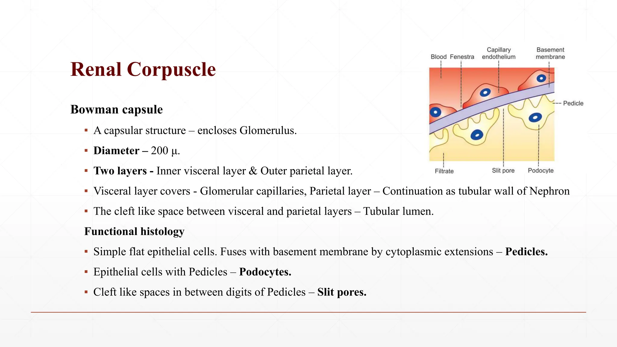
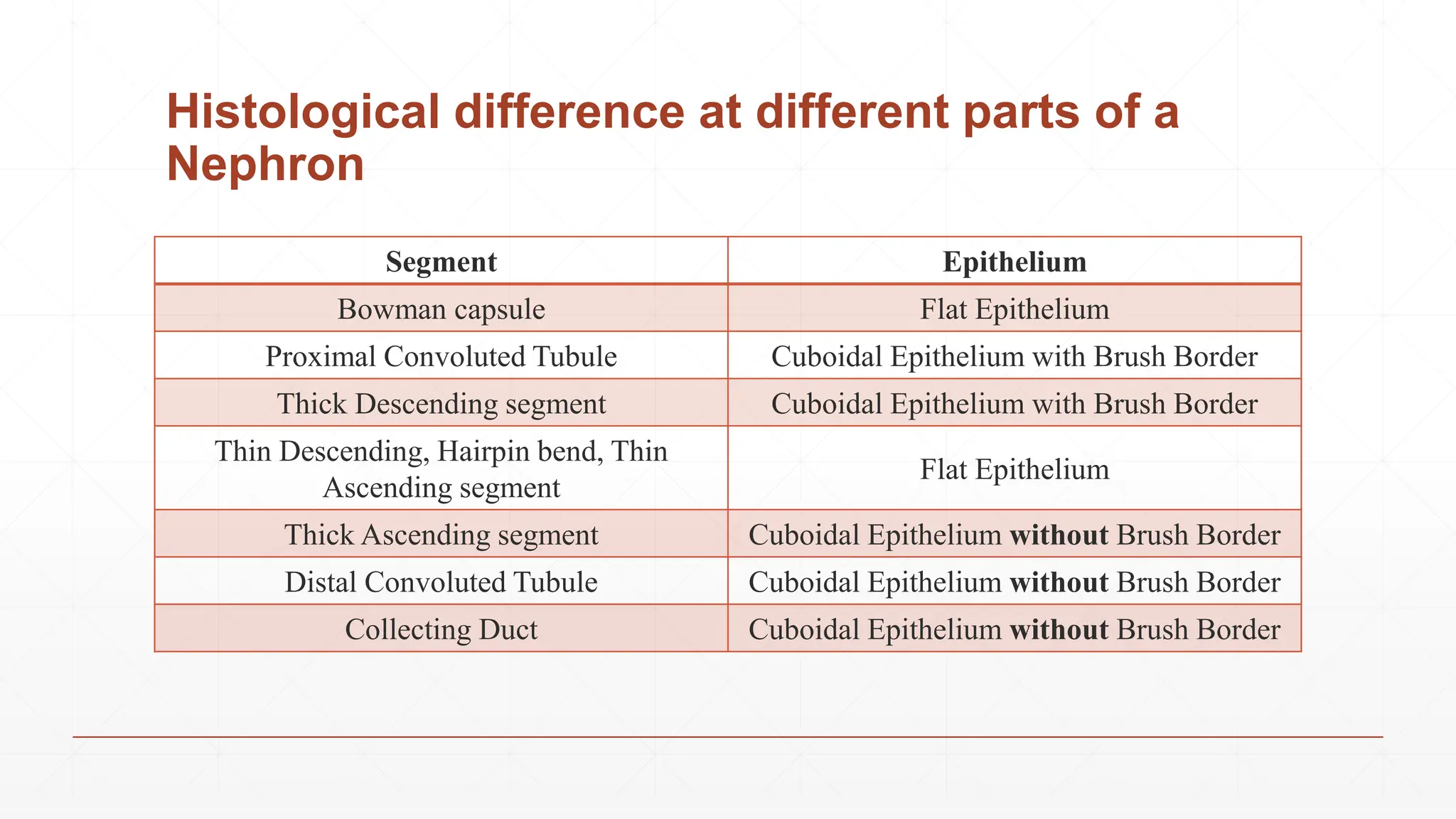
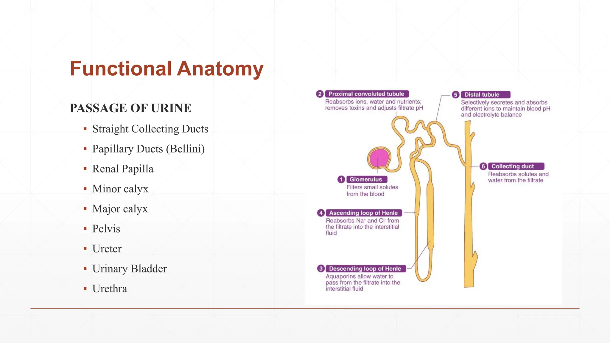
Nephron
Dr. Aniket Shilwant
BAMS. MD (Ayurveda Physiology)
Functional Anatomy
▪ Basic structural and functional unit of
kidney.
▪ Each Kidney - 1 to 1.3 millions
(Decreases in senile age)
▪ Two major parts:
▪ Malpighian corpuscle
▪ Renal tubule.
Functional Anatomy
RENAL CORPUSCLE
▪ Spheroidal and slightly flattened structure
▪ Diameter - 200 μ.
▪ Major function - Filtration of blood
▪ Location – Cortex of the kidney either near the
periphery or near the medulla.
▪ Types – Based on situation of Renal Corpuscle.
▪ Cortical nephrons or Superficial Nephrons - 85%
▪ Juxtamedullary nephrons - 15%
Characteristics of two types of Nephrons
Features Cortical Nephrons Juxtamedullary Nephrons
Quantity 85% 15%
Location Outer Cortex of Kidneys Inner Cortex near Medulla
Loop of
Henle
Short, Hairpin bend reaches
up to outer zone of Medulla
Long, Hairpin bend penetrates
till the tip of Papilla
Blood
supply
Peritubular Capillaries Vasa Recta
Function Urine Formation
Regulates Urine Concentration
Additional
Renal Corpuscle
Glomerulus
▪ A tuft of capillaries enclosed by Bowman capsule.
▪ Interposed between afferent and efferent arteriole
▪ Vascular system in the glomerulus is purely arterial
▪ Afferent Arterioles – Capillaries (Large & Small) – Efferent Arterioles
▪ Arrangement in loops forming Anastomosis.
▪ Dimensions – Afferent Arteriole > Efferent Arteriole
▪ Single layer of endothelial cells having multiple pores - Fenestrae or filtration pores.
Bowman capsule
▪ A capsular structure – encloses Glomerulus.
▪ Diameter – 200 μ.
▪ Two layers - Inner visceral layer & Outer parietal layer.
▪ Visceral layer covers - Glomerular capillaries, Parietal layer – Continuation as tubular wall of Nephron
▪ The cleft like space between visceral and parietal layers – Tubular lumen.
Functional histology
▪ Simple flat epithelial cells. Fuses with basement membrane by cytoplasmic extensions – Pedicles.
▪ Epithelial cells with Pedicles – Podocytes.
▪ Cleft like spaces in between digits of Pedicles – Slit pores.
Renal Corpuscle
▪ Proximal convoluted tubule
▪ Loop of Henle
▪ Distal convoluted tubule.
PROXIMAL CONVOLUTED TUBULE
▪ Coiled portion arising from Bowman capsule located in the cortex.
▪ It is continued as descending limb of loop of Henle.
▪ Dimensions – Length 14 mm and Diameter 55 μ.
Functional histology
▪ Single layer of cuboidal epithelial cells.
▪ Hair like projections pointing in to lumen - Brush-bordered cells.
Renal Tubules
LOOP OF HENLE
▪ Loop of Henle consists of - Descending limb - Hairpin bend - Ascending limb.
A. Descending Limb
▪ Two segments – a. Thick descending segment b. Thin descending segment.
Thick descending segment
▪ Brush-bordered Cuboidal Epithelial Cells.
Thin descending segment
▪ Flattened epithelial cells without brush border and continued as hairpin bend of the loop.
Renal Tubules
B. Hairpin Bend
▪ Flattened epithelial cells without brush border
C. Ascending Limb
Thin ascending segment
▪ Flattened epithelial cells without brush border.
Thick ascending segment
▪ Cuboidal epithelial cells without brush border.
▪ Its terminal portion runs between the afferent and efferent A. of same nephrons – Macula densa.
Length and Extent of Loop of Henle
▪ Cortical N – Short, hairpin bend penetrates only up to outer medulla
▪ Juxtamedullary N – Long, hairpin bend penetrates deep into inner medulla or even till Papilla too.
Renal Tubules
Histological difference at different parts of a
Nephron
Segment Epithelium
Bowman capsule Flat Epithelium
Proximal Convoluted Tubule Cuboidal Epithelium with Brush Border
Thick Descending segment Cuboidal Epithelium with Brush Border
Thin Descending, Hairpin bend, Thin
Ascending segment
Flat Epithelium
Thick Ascending segment Cuboidal Epithelium without Brush Border
Distal Convoluted Tubule Cuboidal Epithelium without Brush Border
Collecting Duct Cuboidal Epithelium without Brush Border
DISTAL CONVOLUTED TUBULE
▪ Occupies the cortex of kidney
▪ Further continued as collecting duct.
▪ Lined by single layer of cuboidal epithelial cells without brush border.
▪ Epithelial cells in DCT – Intercalated cells (I cells).
COLLECTING DUCT
▪ Continuation of DCT – Lies in Cortex.
▪ 8-10 collecting ducts – Single straight Collecting Duct
▪ Cuboidal or Columnar epithelial cells.
▪ Two types of epithelial cells: 1. Principal or P cells 2. Intercalated or I cells.
Renal Tubules
Functional Anatomy
PASSAGE OF URINE
▪ Straight Collecting Ducts
▪ Papillary Ducts (Bellini)
▪ Renal Papilla
▪ Minor calyx
▪ Major calyx
▪ Pelvis
▪ Ureter
▪ Urinary Bladder
▪ Urethra
Juxta-Glomerular Apparatus
▪ Near to Glomerulus of each Nephron
Structure -
1. Macula Densa
2. Extraglomerular mesangial cells
3. Juxtaglomerular cells
1. MACULA DENSA
▪ End portion of thick ascending segment before it opens into DCT.
▪ Location - Between afferent and efferent arterioles of the same nephron.
▪ Tightly packed cuboidal epithelial cells
Juxta-Glomerular Apparatus
2. EXTRAGLOMERULAR MESANGIAL CELLS
▪ Location - Triangular region bound by afferent arteriole, efferent
▪ arteriole and macula densa.
▪ Agranular cells, Lacis cells or Goormaghtigh cells
Glomerular Mesangial Cells
▪ Location – Between Glomerular capillaries
▪ Glomerular mesangial or Intraglomerular mesangial cells.
▪ Role – Supports by forming a cellular network. Regulates Filtration by their contractile property
▪ Phagocytic in nature. Secretes – Prostaglandin.
Juxta-Glomerular Apparatus
3. JUXTAGLOMERULAR CELLS
▪ Specialized smooth muscle cells
▪ Location – Wall of afferent arteriole before it enters Bowman capsule.
▪ Mostly present in Tunica Adventitia and Tunica Media of Afferent arteriole.
▪ Have secretory granules in cytoplasm thus - Granular cells
▪ JG cells forms a thick cuff called polar cushion or polkissen around the afferent arteriole.
Functions of JG Apparatus
A. Secretion of hormones.
B. Regulates the glomerular blood flow and thus glomerular filtration rate.
A. SECRETION OF HORMONES
1. Renin
2. Prostaglandin.
Functions of JG Apparatus
Renin
▪ Peptide with 340 AA
▪ Participates in Renin-Angiotensin System
▪ Regulates Arterial Blood Pressure.
Stimulants for renin secretion
▪ Fall in arterial blood pressure
▪ Reduction in the ECF volume
▪ Increased sympathetic activity
▪ Decreased load of sodium and chloride in Macula Densa.
Functions of JG Apparatus
Prostaglandin
▪ Stimulate JG apparatus
▪ Enhance secretion of renin.
▪ Causes Diuresis and Natriuresis.
Secretion of other substances
▪ Secretion of Cytokines – IL-2, TNF.
▪ Macula densa secretes Thromboxane A2.
Functions of JG Apparatus
REGULATION OF GLOMERULAR BLOOD FLOW AND
GLOMERULAR FILTRATION RATE
▪ Macula densa of JG apparatus - Feedback mechanism - Tubuloglomerular feedback
Resources
▪ Textbook of Medical Physiology – Guyton & Hall
▪ Ganong’s review of Medical Physiology
▪ Essentials of Medical Physiology – Sembulingam
▪ Images – Internet
Dr. Aniket A. Shilwant
Associate Professor, Sharir Kriya Dept.
GJPIASR, CVM University, New Vidyanagar, Anand
ayuraniket18@gmail.com
Thank You All !!!

STUDY OF BASIC FUNCTIONAL ANATOMY OF NEPHRON

  • 1.
    Nephron Dr. Aniket Shilwant BAMS.MD (Ayurveda Physiology)
  • 2.
    Functional Anatomy ▪ Basicstructural and functional unit of kidney. ▪ Each Kidney - 1 to 1.3 millions (Decreases in senile age) ▪ Two major parts: ▪ Malpighian corpuscle ▪ Renal tubule.
  • 3.
    Functional Anatomy RENAL CORPUSCLE ▪Spheroidal and slightly flattened structure ▪ Diameter - 200 μ. ▪ Major function - Filtration of blood ▪ Location – Cortex of the kidney either near the periphery or near the medulla. ▪ Types – Based on situation of Renal Corpuscle. ▪ Cortical nephrons or Superficial Nephrons - 85% ▪ Juxtamedullary nephrons - 15%
  • 4.
    Characteristics of twotypes of Nephrons Features Cortical Nephrons Juxtamedullary Nephrons Quantity 85% 15% Location Outer Cortex of Kidneys Inner Cortex near Medulla Loop of Henle Short, Hairpin bend reaches up to outer zone of Medulla Long, Hairpin bend penetrates till the tip of Papilla Blood supply Peritubular Capillaries Vasa Recta Function Urine Formation Regulates Urine Concentration Additional
  • 5.
    Renal Corpuscle Glomerulus ▪ Atuft of capillaries enclosed by Bowman capsule. ▪ Interposed between afferent and efferent arteriole ▪ Vascular system in the glomerulus is purely arterial ▪ Afferent Arterioles – Capillaries (Large & Small) – Efferent Arterioles ▪ Arrangement in loops forming Anastomosis. ▪ Dimensions – Afferent Arteriole > Efferent Arteriole ▪ Single layer of endothelial cells having multiple pores - Fenestrae or filtration pores.
  • 6.
    Bowman capsule ▪ Acapsular structure – encloses Glomerulus. ▪ Diameter – 200 μ. ▪ Two layers - Inner visceral layer & Outer parietal layer. ▪ Visceral layer covers - Glomerular capillaries, Parietal layer – Continuation as tubular wall of Nephron ▪ The cleft like space between visceral and parietal layers – Tubular lumen. Functional histology ▪ Simple flat epithelial cells. Fuses with basement membrane by cytoplasmic extensions – Pedicles. ▪ Epithelial cells with Pedicles – Podocytes. ▪ Cleft like spaces in between digits of Pedicles – Slit pores. Renal Corpuscle
  • 7.
    ▪ Proximal convolutedtubule ▪ Loop of Henle ▪ Distal convoluted tubule. PROXIMAL CONVOLUTED TUBULE ▪ Coiled portion arising from Bowman capsule located in the cortex. ▪ It is continued as descending limb of loop of Henle. ▪ Dimensions – Length 14 mm and Diameter 55 μ. Functional histology ▪ Single layer of cuboidal epithelial cells. ▪ Hair like projections pointing in to lumen - Brush-bordered cells. Renal Tubules
  • 8.
    LOOP OF HENLE ▪Loop of Henle consists of - Descending limb - Hairpin bend - Ascending limb. A. Descending Limb ▪ Two segments – a. Thick descending segment b. Thin descending segment. Thick descending segment ▪ Brush-bordered Cuboidal Epithelial Cells. Thin descending segment ▪ Flattened epithelial cells without brush border and continued as hairpin bend of the loop. Renal Tubules
  • 9.
    B. Hairpin Bend ▪Flattened epithelial cells without brush border C. Ascending Limb Thin ascending segment ▪ Flattened epithelial cells without brush border. Thick ascending segment ▪ Cuboidal epithelial cells without brush border. ▪ Its terminal portion runs between the afferent and efferent A. of same nephrons – Macula densa. Length and Extent of Loop of Henle ▪ Cortical N – Short, hairpin bend penetrates only up to outer medulla ▪ Juxtamedullary N – Long, hairpin bend penetrates deep into inner medulla or even till Papilla too. Renal Tubules
  • 10.
    Histological difference atdifferent parts of a Nephron Segment Epithelium Bowman capsule Flat Epithelium Proximal Convoluted Tubule Cuboidal Epithelium with Brush Border Thick Descending segment Cuboidal Epithelium with Brush Border Thin Descending, Hairpin bend, Thin Ascending segment Flat Epithelium Thick Ascending segment Cuboidal Epithelium without Brush Border Distal Convoluted Tubule Cuboidal Epithelium without Brush Border Collecting Duct Cuboidal Epithelium without Brush Border
  • 11.
    DISTAL CONVOLUTED TUBULE ▪Occupies the cortex of kidney ▪ Further continued as collecting duct. ▪ Lined by single layer of cuboidal epithelial cells without brush border. ▪ Epithelial cells in DCT – Intercalated cells (I cells). COLLECTING DUCT ▪ Continuation of DCT – Lies in Cortex. ▪ 8-10 collecting ducts – Single straight Collecting Duct ▪ Cuboidal or Columnar epithelial cells. ▪ Two types of epithelial cells: 1. Principal or P cells 2. Intercalated or I cells. Renal Tubules
  • 12.
    Functional Anatomy PASSAGE OFURINE ▪ Straight Collecting Ducts ▪ Papillary Ducts (Bellini) ▪ Renal Papilla ▪ Minor calyx ▪ Major calyx ▪ Pelvis ▪ Ureter ▪ Urinary Bladder ▪ Urethra
  • 14.
    Juxta-Glomerular Apparatus ▪ Nearto Glomerulus of each Nephron Structure - 1. Macula Densa 2. Extraglomerular mesangial cells 3. Juxtaglomerular cells 1. MACULA DENSA ▪ End portion of thick ascending segment before it opens into DCT. ▪ Location - Between afferent and efferent arterioles of the same nephron. ▪ Tightly packed cuboidal epithelial cells
  • 15.
    Juxta-Glomerular Apparatus 2. EXTRAGLOMERULARMESANGIAL CELLS ▪ Location - Triangular region bound by afferent arteriole, efferent ▪ arteriole and macula densa. ▪ Agranular cells, Lacis cells or Goormaghtigh cells Glomerular Mesangial Cells ▪ Location – Between Glomerular capillaries ▪ Glomerular mesangial or Intraglomerular mesangial cells. ▪ Role – Supports by forming a cellular network. Regulates Filtration by their contractile property ▪ Phagocytic in nature. Secretes – Prostaglandin.
  • 16.
    Juxta-Glomerular Apparatus 3. JUXTAGLOMERULARCELLS ▪ Specialized smooth muscle cells ▪ Location – Wall of afferent arteriole before it enters Bowman capsule. ▪ Mostly present in Tunica Adventitia and Tunica Media of Afferent arteriole. ▪ Have secretory granules in cytoplasm thus - Granular cells ▪ JG cells forms a thick cuff called polar cushion or polkissen around the afferent arteriole.
  • 17.
    Functions of JGApparatus A. Secretion of hormones. B. Regulates the glomerular blood flow and thus glomerular filtration rate. A. SECRETION OF HORMONES 1. Renin 2. Prostaglandin.
  • 18.
    Functions of JGApparatus Renin ▪ Peptide with 340 AA ▪ Participates in Renin-Angiotensin System ▪ Regulates Arterial Blood Pressure. Stimulants for renin secretion ▪ Fall in arterial blood pressure ▪ Reduction in the ECF volume ▪ Increased sympathetic activity ▪ Decreased load of sodium and chloride in Macula Densa.
  • 19.
    Functions of JGApparatus Prostaglandin ▪ Stimulate JG apparatus ▪ Enhance secretion of renin. ▪ Causes Diuresis and Natriuresis. Secretion of other substances ▪ Secretion of Cytokines – IL-2, TNF. ▪ Macula densa secretes Thromboxane A2.
  • 20.
    Functions of JGApparatus REGULATION OF GLOMERULAR BLOOD FLOW AND GLOMERULAR FILTRATION RATE ▪ Macula densa of JG apparatus - Feedback mechanism - Tubuloglomerular feedback
  • 22.
    Resources ▪ Textbook ofMedical Physiology – Guyton & Hall ▪ Ganong’s review of Medical Physiology ▪ Essentials of Medical Physiology – Sembulingam ▪ Images – Internet
  • 23.
    Dr. Aniket A.Shilwant Associate Professor, Sharir Kriya Dept. GJPIASR, CVM University, New Vidyanagar, Anand ayuraniket18@gmail.com Thank You All !!!