INDEX
• Introduction
• Anatomyof cerebral venous system
• Conditions associated with cerebral venous thrombosis
• Etiopathogenesis
• Pathophysiology
• Clinical features
• Clinical manifestations of cerebral venous sinus thrombosis according to occlusion site
• Investigations
• Treatment
• Complications
• CVT and Pregnancy
• Prognosis
3.
INTRODUCTION
• Most strokesare caused by occlusion or rupture of brain supplying arteries.
• Brain infarcts, edema and hemorrhages are sometimes caused by cerebral venous thrombosis
(CVT), defined by thrombosis of dural sinuses and cerebral and cerebellar veins.
• CVT is a rare variety of venous thromboembolic disease.
• Its incidence is estimated to range from 0.2 to 1.32 per 100 000 person-years.
• CVT accounts for 0.5% of all strokes.
Dural venous sinuses
•Endothelium lined channels contained between outer periosteal and
inner meningeal layer of dura.
• Often contain arachnoid granulations that are CSF containing
projections that extend from subarachnoid space into dural venous
sinuses.
• Falx cerebriis dural fold that separates
the two cerebral hemispheres.
• Superior sagittal sinus is present in the
superior border of falx and inferior
sagittal sinus in the bottom.
• Straight sinus is formed by junction of
inferior sagittal sinus and vein of galen.
• Straight sinus terminates by joining the
transverse sinus and superior sagittal
sinus to form sinus confluence (torcula
herophili)
8.
• Transverse sinusa/k/a lateral sinuses are contained
between attachments of tentorium cerebelli to inner
table of skull.
• Anatomic variation in transverse sinus is very
common.
• The two transverse sinus are asymmetric and
hypoplastic/stenotic segments are seen in one third
population.
• Transverse sinus curve and turn inferiorly to become
sigmoid sinus.
• Sigmoid sinus follow s shape curve.
• Terminate by becoming the internal jugular veins.
9.
• Cavernous sinus
•Lie along sides of sella turcica.
• Formed by prominent lateral and
much thinner medial dural wall.
• Contains the two internal carotid
arteries and abducens nerve(CN VI).
• CN III, CN IV, CN V1 and CN V2 lie
within lateral dural wall.
• Both the cavernous sinus
communicate with each other.
10.
• Superior andinferior petrosal sinus
course along top of petrous temporal
bone and extend from cavernous sinus
to sigmoid sinus.
• Sphenoparietal sinus courses along the
lesser wing of sphenoid at rim of
middle cranial fossa and drains into
cavernous sinus or inferior petrosal
sinus.
11.
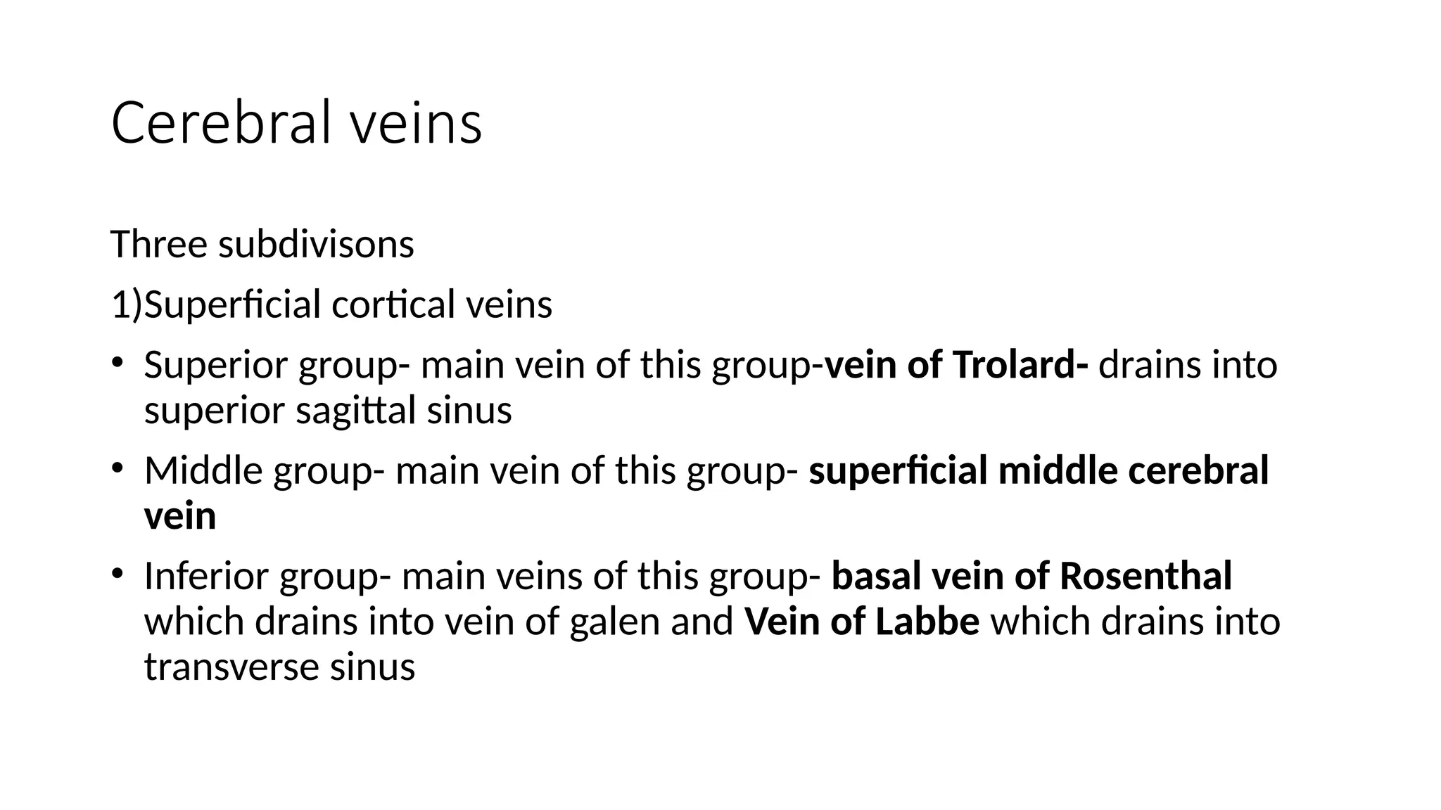
Cerebral veins
Three subdivisons
1)Superficialcortical veins
• Superior group- main vein of this group-vein of Trolard- drains into
superior sagittal sinus
• Middle group- main vein of this group- superficial middle cerebral
vein
• Inferior group- main veins of this group- basal vein of Rosenthal
which drains into vein of galen and Vein of Labbe which drains into
transverse sinus
12.
2)Deep cerebral veins
•Medullary veins- originate 1-2 cm below cortex, course through white
matter towards ventricles and drain into subependymal vein
• Subependymal veins- courses under ventricular ependyma
• Internal cerebral vein-
• Vein of Galen
13.
3) Brainstem/posterior fossavein
• Precentral cerebellar vein and anterior pontomesencephalic vein
(drain into vein of galen)
• Petrosal vein
• Inferior vermian vein
17.
Conditions Associated WithCerebral Venous
Thrombosis
Predisposing
• Alcohol consumption
• Hypercholesterolemia
• Hyperhomocysteinemia
• Antiphospholipid antibodies present and antiphospholipid syndrome
• Autoimmune disease
• Anemia
• Malignancy (particularly within the first year of cancer diagnosis as well as among
patients with hematologic malignancies)19
18.
• Pregnancy andthe puerperium
• Factor V G1691A polymorphism
• Methylenetetrahydrofolate reductase (MTHFR) C677T polymorphism
• Prothrombin G20210A polymorphism
• Protein C deficiency
• Protein S deficiency
• Antithrombin III deficiency
• Obesity (in women who used oral contraceptives, being overweight or obese was associated with an
increased risk of CVT in a dose-dependent manner)
• Elevated factor VIII serum levels
• Behçet disease
19.
Precipitating
• Glucocorticosteroids
• Trauma
•Infection (particularly central nervous system or ear and face)
• Surgical procedures
• Combined oral contraception pill treatment
• Vaccine-induced immune thrombotic thrombocytopenia
• L-asparaginase therapy
• All-transretinoic acid in acute promyelocytic leukemia
• Lumbar puncture
20.
ETIOPATHOGENESIS
• Risk factorsfor venous thrombosis in general are linked classically to
VIRCHOWS TRIAD
• Among all risk factors imp.. Prothrombotic conditions
21.
PATHOPHYSIOLOGY
Two main mechanisms
1.Obstruction of veins causes increased venous pressure, reduced capillary perfusion, and
locally increased cerebral blood volume. Initially, this is compensated by dilatation of
cerebral veins and the recruitment of collateral vessels; however, continued elevation of
venous pressure causes decreased cerebral perfusion pressure and cerebral blood flow
resulting in ischemic injury and cytotoxic edema. Disruption of the blood- brain barrier
leads to vasogenic edema, and venous and capillary rupture culminates in parenchymal
hemorrhage. Thus, both cytotoxic and vasogenic edema can occur.
2. CVT also hampers cerebrospinal fluid absorption through the arachnoid villi, which then
leads to raised intracranial pressure (ICP) typically seen in association with superior sagittal
sinus obstruction.
23.
CLINICAL FEATURES
CVT hasa highly variable clinical presentation
It can present as… Acute, Subacute, Chronic
Presentation as TIA also reported
Signs and Symptoms can be grouped into 4 main syndromes
1) ISOLATED INTRACRANIAL HYPERTENSION SYNDROME( Headache +/- vomiting, papilledema, visual disturbances)
2) FOCAL SYNDROME( focal deficits, seizures, both)
3) ENCEPHALOPATHY( multifocal signs, mental status changes, stupor or coma)
4) Cavernous Sinus Syndrome with Multiple Cranial Neuropathies
Variables affecting presentation and course
1) Age 2) Gender 3)Presence of parenchymal lesions 4)Site and number of occluded sinuses and veins
5)Interval from CVT onset and presentation
24.
Headache
• Head discomfortor pain is an extremely common symptom in patients with CVT and is most often the initial one.
• CD had headaches before and at the onset of his neurological symptoms.
• Headache is much more common in patients with venous thrombosis than in patients with arterial thromboembolic
disease.
• The presence of headache is best explained by two major factors:
(1) the local process within the veins and dural sinuses;
(2) the development of increased intracranial pressure.
• Headache in CVT has no specific diagnostic characteristics.
• Most often diffuse, it can be unilateral, localized to any region of the head, or even limited to the neck as in jugular
vein thrombosis.
• The severity is also highly variable, ranging from a mild sensation of heaviness to a severe excruciating abrupt
headache (“thunderclap” headache).
25.
Focal neurological signsand symptoms
• Venous occlusive disease leads to focal parenchymal abnormalities, including edema, hemorrhage,
ischemia, and infarction, which cause focal neurological signs and symptoms.
• Edema, whether localized or generalized, is the most common brain abnormality in CVT.
• Focal neurological symptoms and signs are present in approximately one-half of patients with CVT.
26.
Seizure
• Seizures arequite common in patients with CVT.
• Seizures are the presenting symptom in about 7–15% of patients, and often occur during the early course
of the illness.
• Seizures are approximately equally divided between focal and generalized.
27.
Encephalopathy
• May developa subacute encephalopathy with confusion and lethargy
• Or experience a rapid neurologic deterioration progressing to coma due to edema of
bilateral thalami, basal ganglia, or other deep structures typically drained by these veins.
28.
Clinical manifestations ofcerebral venous
sinus thrombosis according to occlusion site
• Occclusion of transverse sinus
• If isolated without infarction: asymptomatic or headache
• Seizures
• Contralateral pyramidal symptoms and signs
• If left transverse sinus with venous infarction and vein of Labbe occlusion:
aphasia
• If extending into the contiguous sinuses: intracranial hypertension,
consciousness disturbance, focal cerebral signs and cranial nerve palsies (IX –
XXI)
• If extending into cerebellar veins: headache, vomiting, and limb or gait ataxia.
• Occclusion ofsigmoid sinus
• Pain in mastoid region.
• Combination of VI-VII-VIII cranial nerve palsies.
31.
• Occclusion ofdeep venous system
• Mental status disturbances- reduced arousal
• Diffuse encephalopathy or coma
• Motor deficits (bilateral or fluctuating alternating paresis)
32.
• Occclusion ofcortical veins
• Focal neurological symptoms and signs according to location.
• Seizures
• Occclusion of cavernous sinus
• Headache, ocular pain, chemosis, proptosis, ocular nerve palsy (III, IV,
Vi and the ophthalmic division of V)
• Fever( when there is an infective cause)
33.
INVESTIGATIONS
1) NEURO IMAGING
a)NON INVASIVE- CT,CTV, MRI ,MRV , ULTASOUND
b) INVASIVE - CEREBRAL ANGIOGRAPHY, DIRECT CEREBRAL VENOGRAPHY
NON INVASIVE
CT,CTV
• CT is initial modality in new onset neurological symptoms, often normal
• Only around 30% cases abnormal
• Signs in CT associated with CVT
o Dense triangle sign/Empty delta sign/ Cord sign
o Other findings--Hemorrhagic leisons/ non-hemorrhagic leisons like edema, venous infarction
34.
MRI, MRV
MRI usinggradient echo T2* susceptibility-weighted sequences in combination with MR venography is the
most sensitive imaging
In the first five days, the thrombosed sinuses appear isointense on T1-weighted images and hypointense on
T2-weighted images
Beyond five days, venous thrombus becomes more apparent because signal is increased on both T1 and
T2- weighted images
After the first month, thrombosed sinuses exhibit a variable pattern of signal, which may appear isointense
MRV, usually performed using the time-of-flight (TOF) technique, is useful for demonstrating absence of flow
in cerebral venous sinuses, though interpretation can be confounded by normal anatomic variants such as
sinus hypoplasia and asymmetric flow
35.
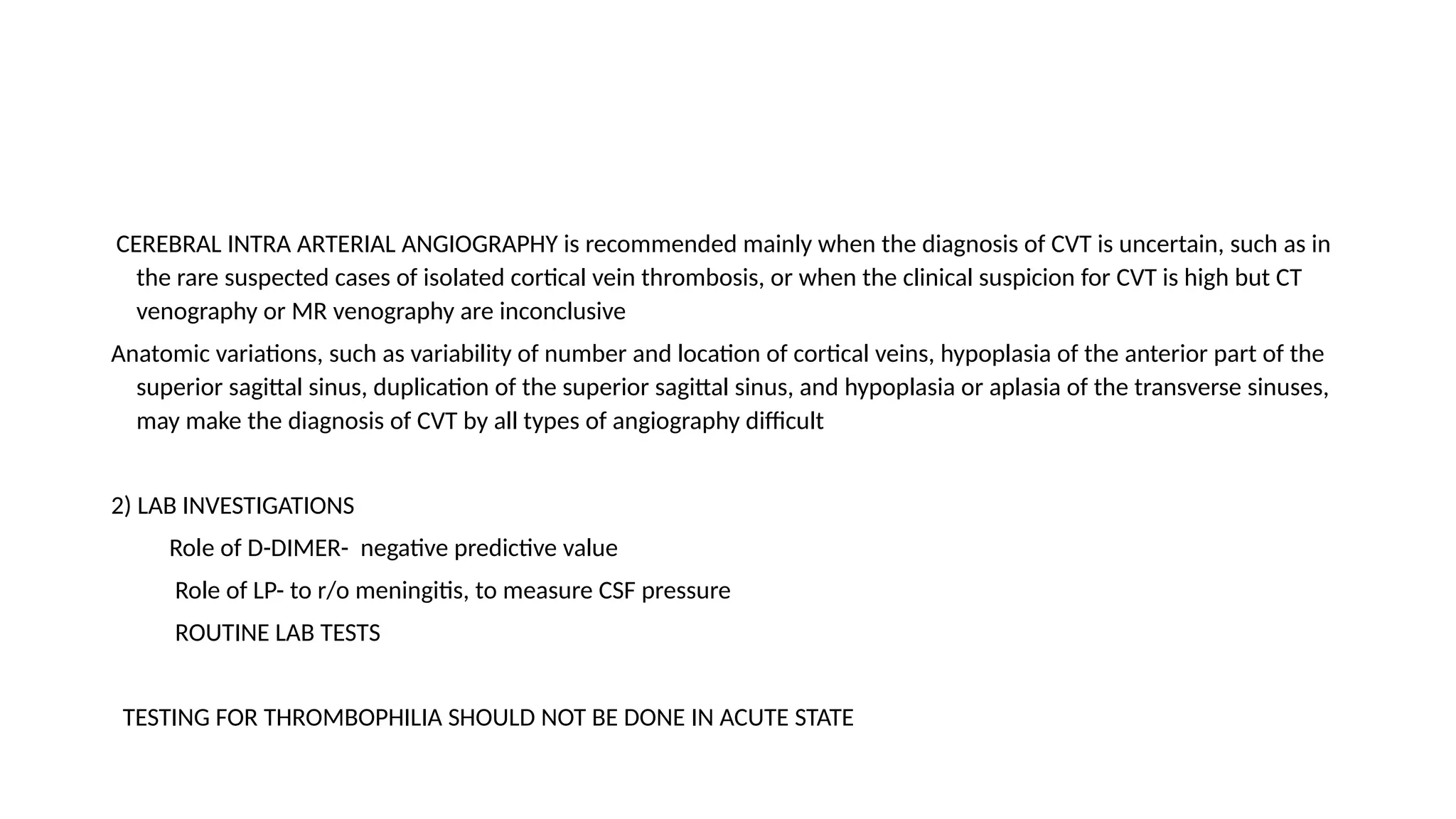
CEREBRAL INTRA ARTERIALANGIOGRAPHY is recommended mainly when the diagnosis of CVT is uncertain, such as in
the rare suspected cases of isolated cortical vein thrombosis, or when the clinical suspicion for CVT is high but CT
venography or MR venography are inconclusive
Anatomic variations, such as variability of number and location of cortical veins, hypoplasia of the anterior part of the
superior sagittal sinus, duplication of the superior sagittal sinus, and hypoplasia or aplasia of the transverse sinuses,
may make the diagnosis of CVT by all types of angiography difficult
2) LAB INVESTIGATIONS
Role of D-DIMER- negative predictive value
Role of LP- to r/o meningitis, to measure CSF pressure
ROUTINE LAB TESTS
TESTING FOR THROMBOPHILIA SHOULD NOT BE DONE IN ACUTE STATE
46.
TREATMENT
1) ANTICOAGULATION
2) OTHERTREATMENTS
• FIBRINOLYTIC THERAPY
• DIRECT CATHETER ABLATION
• MECHANICAL THROMBECTOMY/ THROMBOLYSIS
• SURGERY
• STEROIDS.. Useful in vasogenic edema but not recommended routinely. Steroid medications are
not recommended, even in the presence of parenchymal brain lesions on CT/MRI, unless needed
for another underlying disease.
• ANTIBIOTICS.. Local infection can cause CVT so appropriate antibiotics should be adminsitered if
needed surgical intervention should be done
47.
ANTICOAGULATION
Initial anticoagulation by..LMWH/ UFH( dose adjusted with a goal of 2-3 times control APTT)
Later.. oral Vit K Antagonists
Target INR- 2.0-3.0
DURATION..
1) Provoked CVT- 3-6 mon
(associated with transient risk factor)
2) Unprovoked CVT- 6-12 mon
3) Recurrent/ CVT with severe THROMBOPHILIA / VTE after CVT- indefinite anticoagulation
Severe thrombophilia- deficeincy of protein c/ protein S/ antithrombin, APLA, homozygous factor V
leiden, homozygous prothrombin G20210A
48.
Testing for proteinC, protein S, and antithrombin deficiency is generally indicated 2 to 4 weeks
after completion of anticoagulation. There is a very limited value of testing in the acute setting or
if patient is taking warfarin.
Special situation of CVT with cerebral haemorrhage on presentation.
Anticoagulants appear to be safe to use in adult patients with CVT who have intracranial
haemorrhages, either intracerebral or subarachnoid
51.
ENDOVASCULAR THROMBOLYSIS
Some patientswith CVT worsen despite anticoagulant therapy.
Direct endovascular thrombolysis has been used as an alternative treatment in such cases.
Direct thrombolysis aims to dissolve the venous clot by delivering a thrombolytic substance
(urokinase or rt-PA) within the occluded sinus through an intravenous catheter.
In some cases, mechanical endovascular disruption of the thrombus has also been used
DECOMPRESSIVE HEMICRANIECTOMY
In patients with neurological deterioration due to severe mass effect or intracranial hemorrhage
causing intractable intracranial hypertension, decompressive hemicraniectomy may be considered
52.
COMPLICATIONS
1)EARLY..
a)SEIZURES-
37% cases
Rx.. Controversial..To initiate or await initial seizures before treatment
RECOMMENDATIONS
Early initiation of AED in pts with CVT and single seizure with parenchymal lesions for definite period is recommended to
prevent further seizure
CVT with seizures without parenchymal lesion AED initiation is probably recommended
Pts without seizures routine use of AED not recommended
b) HYDROCEPHALUS
Communicating/ Obstructive
If obstructive- ventriculostomy/ VP shunt
53.
c) INTRACRANIAL HYPERTENSION-40%
Rx.. Anticoagulation, LP, Acetazolamide, Decompressive craniotomy
2)LATE..
a) HEADACHE
50%
Common complaint in follow up
Persistent/severe headache- r/o recurrence or intracranial HTN
In patients with a history of CVT who complain of new, persisting, or severe headache, evaluation for CVT recurrence and
intracranial hypertension should be considered
b) VISUAL LOSS
c) SEIZURES
d) DURAL ARTERIOVENOUS FISTULA
54.
CVT and PREGNANCY
Oneof risk factor because of hypercoagulable state.
The greatest risk periods for CVT include the third trimester and the first 4 postpartum weeks
Caesarean delivery appears to be associated with a higher risk of CVT
Women with a history of VTE appear to have an increased risk of thrombotic events
CVT is not a contraindication for future pregnancies
55.
RECOMMENDATIONS
For women withCVT during pregnancy, LMWH in full anticoagulant doses should be continued throughout pregnancy, and
LMWH or vitamin K antagonist with a target INR of 2.0 to 3.0 should be continued for at least 6 weeks postpartum (for a
total minimum duration of therapy of 6 months)
It is reasonable to advise women with a history of CVT that future pregnancy is not contraindicated. Further investigations
regarding the underlying cause and a formal consultation with a hematologist and/or maternal fetal medicine specialist are
reasonable.
It is reasonable to treat acute CVT during pregnancy with full-dose LMWH rather than UFH
For women with a history of CVT, prophylaxis with LMWH during future pregnancies and the postpartum period is probably
recommended
56.
PROGNOSIS
CVT is associatedwith a good outcome (complete recovery or minor residual symptoms or signs) in close to 80 % of patients.
Nevertheless, approximately 5% of patients die in the acute phase of the disorder, and longer-term mortality is nearly 10%.
The main cause of acute death with CVT is neurologic, most often from brain herniation.
After the acute phase, most deaths are related to underlying disorders such as cancer
Causes of death in acute phase… Transtentorial herniation, Diffuse brain edema, Status epilepticus, Medical complications,
Pulmonary embolism
Cause of death in later phase… is generally due to underlying cause like cancer
Neurological worsening may occur in 23% of patients, even several days after diagnosis. Approximately one third of patients
with neurological deterioration will have new parenchymal lesions when neuroimaging is repeated.