Downloaded 766 times


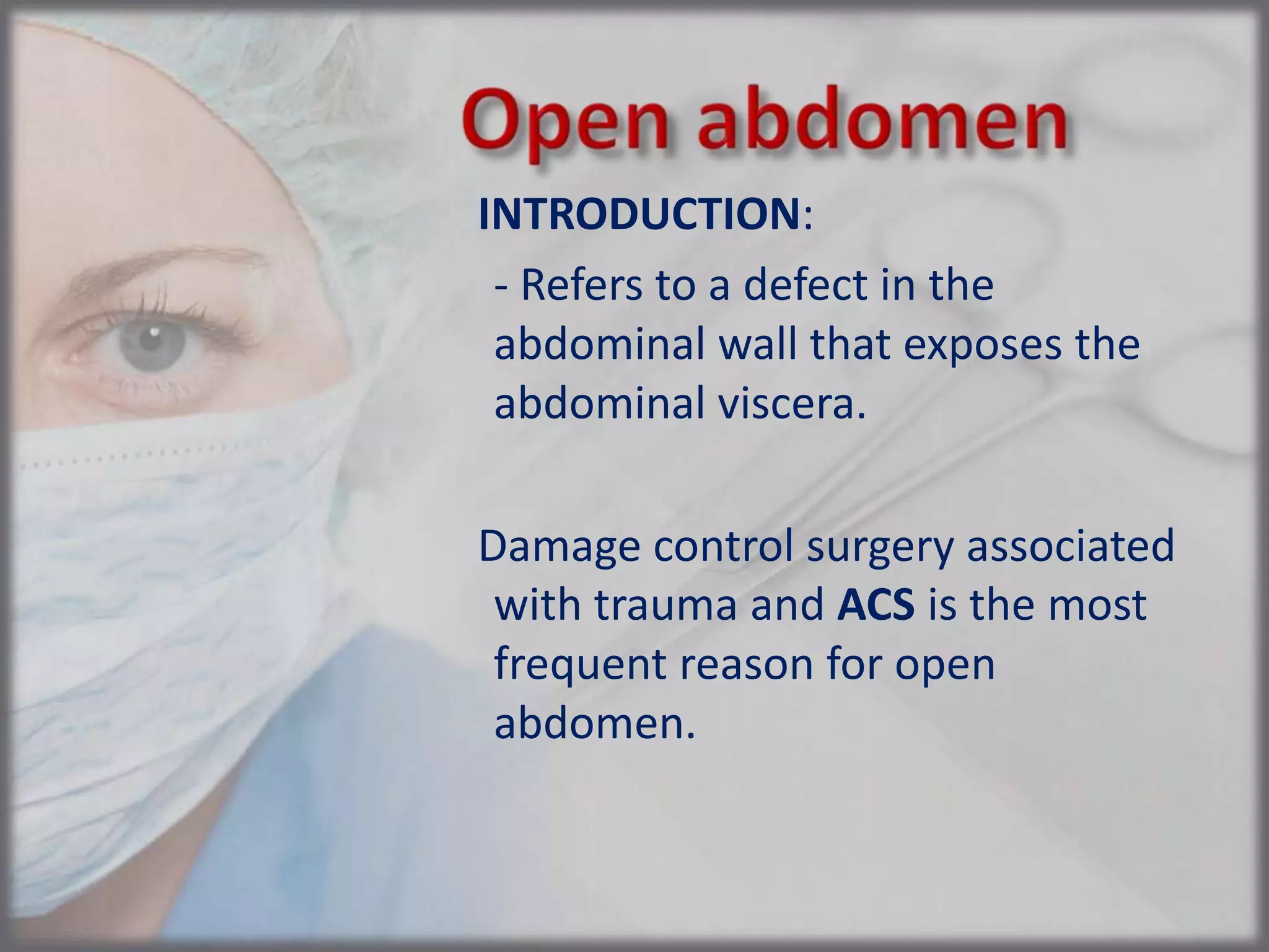


This document discusses abdominal compartment syndrome (ACS), which refers to organ dysfunction caused by increased intra-abdominal pressure known as intra-abdominal hypertension (IAH). The key points are: 1. ACS can impair nearly every organ system but is often underdiagnosed. Diagnosis requires measuring intra-abdominal pressure via bladder catheter. 2. Management consists of supportive care initially but may require surgical decompression of the abdomen in severe cases. 3. Following decompression, temporary abdominal closure techniques are used until definitive closure can be achieved or a planned hernia results.


![Abdominal compartment syndrome[1]](https://cdn.slidesharecdn.com/ss_thumbnails/abdominalcompartmentsyndrome1-121012075429-phpapp02-thumbnail.jpg?width=640&height=640&fit=bounds)


