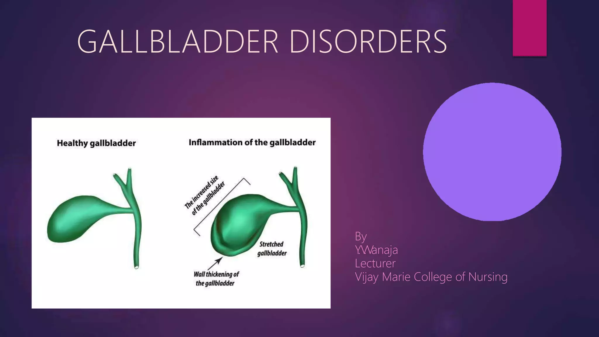
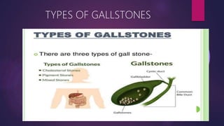
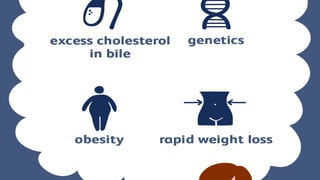
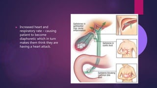
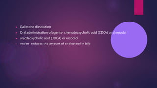
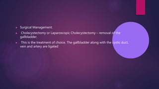
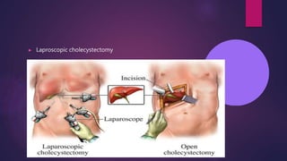
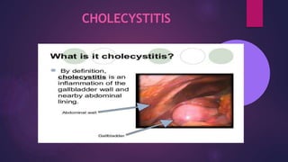
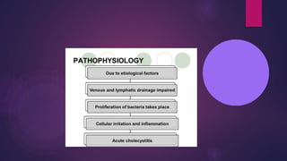
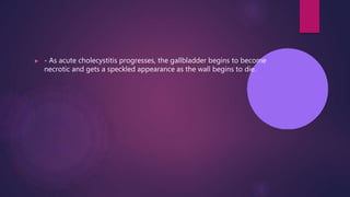
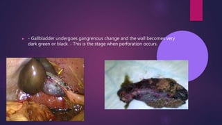
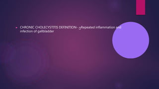
GALLBLADDER DISORDERS
By
Y
.V
.V
anaja
Lecturer
Vijay Marie College of Nursing
Biliary tract disorders includes
▶ Cholelithiasis
▶ Acute cholecystitis
▶ Chronic cholecystitis
CHOLELITHIASIS/ GALLSTONES
DEFINITION
▶ Gallstones are collections of cholesterol, bile pigment or a combination of
the two, which can form in the gallbladder or within the bile ducts of the
liver.
▶ calculi or gallstones, usually form in the gallbladder from the solid
constituents of bile; they vary greatly in size, shape and composition.
“ Brunner’
’
INCIDENCE
▶ The incidence increases with age
▶ The incidence of cholelithiasis is higher in women, multiparous women
▶ Post menopausal women on estrogen therapy
▶ Oral contraceptive pills
▶ Familial tendency
▶ Obesity
TYPES OF GALLSTONES
Pigment stones: pigment stones probably form when when unconjugated pigments in the bile
precipitate to form stones
May be black colour
Cholesterol stones:
▶ In gall stone prone patients there is decreased bile acid synthesis and increased cholesterol synthesis
the liver resulting in bile supersaturated with cholesterol , which precipitate out of the bile to form bile
stones.
▶ Smooth & whitish yellow to tan colour
Mixed stones
▶ Combination of cholesterol and pigment stones or other substance
▶ Calcium carbonate, phosphate, bile salts, and palmitate
ETIOLOGY
▶ Change in bile composition-
▶ Gallbladder stasis
▶ supersaturation of bile with cholesterol
▶ Infection and tissue injury
▶ Genetics
▶ Cirrhosis
▶ Hemolysis and infections of the biliary tract
Pathophysiology
▶ In gallstone prone patients there is decreased bile acid synthesis and
increased cholesterol synthesis in the liver
▶ Super saturation of the bile with cholesterol or calcium
▶ Precipitation of solute from solvent to form solid crystal
▶ Crystals come together and fuse to form stones
▶ Venous and lymphatic drainage impaired
▶ Proliferation of bacteria occur
▶ Localized cellular irritation & infiltration
▶ Ischemia and necrosis
▶ Complaints of indigestion after
eating high fat foods.
▶ Localized pain in the right-
upper quadrant epigastric
region.
▶
fl
Anorexia, nausea, vomiting and
atulence
▶ Increased heart and
respiratory rate – causing
patient to become
diaphoretic which in turn
makes them think they are
having a heart attack.
▶ Abdominal distention
▶ Changes in the urine and stool color
▶ Vitamin deficiency
▶ Low grade fever.
▶ Elevated leukocyte count.
▶ Mild jaundice.
▶ Stools that contain fat – steatorrhea.
▶ Clay colored stools caused by a lack of bile in the intestinal tract.
▶ Urine may be dark amber- to tea-colored.
DIAGNOSTICS TEST
▶ History of patient
▶ Physical examination
▶ Laboratory test for- Elevated conjugated bilirubin.
▶ Elevated alkaline phosphate
▶ Serum amylase and lipase
▶ Elevated WBC count
▶ Fecal studies.
▶ Ultrasound of the gallbladder
▶ CT Scan
▶ MRI
▶ HIDA (hepato- iminodiacetic
acid)
▶ Cholangiography
▶ ERCP (endoscopic retrograde
cholangiopancreatography)
▶
MEDICAL MANAGEMENT
▶ MEDICAL MANAGEMENT GOALS-
▶ T
o resolve symptoms
▶ T
o remove stones
▶ T
o prevent complications
▶ PAIN MANAGEMENT
▶ Give analgesics
▶ Antacids,
▶ H2 blockers or proton pump inhibitors- to neutralize gastric acid
▶ For nausea and vomiting, Antiemetics given
▶ antibiotics
▶ Gall stone dissolution
▶ Oral administration of agents- chenodeoxycholic acid (CDCA) or chenodal
▶ ursodeoxycholic acid (UDCA) or ursodiol
▶ Action- reduces the amount of cholesterol in bile
▶ Lithotripsy
▶ Extracorporeal shock wave
lithotripsy (ESWL)
▶ 1500 shock waves directed
at stones
▶ Used for fewer than 4
stones,
▶ each smaller than 3cm.
▶ If stones are present in the common bile duct, an endoscopic
sphincterotomy must be performed to remove them BEFORE a
cholecystectomy is done.
▶ A number of various instruments are inserted through the endoscope in
order to "cut" or stretch the sphincter.
▶ Once this is done, additional instruments are passed that enable the
removal of stones and the stretching of narrowed regions of the ducts.
▶ Drains (stents) can also be used to prevent a narrowed area from rapidly
returning to its previously narrowed state.
▶ Surgical Management.
▶ Cholecystectomy or Laparoscopic Cholecystectomy – removal of the
gallbladder.
▶ This is the treatment of choice. The gallbladder along with the cystic duct,
vein and artery are ligated
▶ Laproscopic cholecystectomy
CHOLECYSTITIS
TYPES OF CHOLECYTITIS
▶ Acute cholecystitis
▶ Chronic cholecystitis
ACUTE CHOLECYSTITIS
▶ DEFINITION;
▶ Acute cholecystitis refers to acute inflammation of the gallbladder wall.
TYPES OF ACUTE CHOLECYSTITIS
▶ Two types of acute cholecystitis can occur
▶ Calculous cholecystitis
▶ Acalculous cholecystitis
▶ 1) Calculous: -It is the obstructive cholecystitis due to gall stones having
the most common variety in which around 90% of people having gall
stones suffers.
▶ 2) Acalculous: -It is the non-obstructive type which is common in person
suffering from major illness like sever sepsis, burns, DM, dehydration,
multiple injury etc.
ETIOLOGY
▶ Gall stone in cystic duct
▶ Obstruction in cystic duct
▶ Bacterial infection (gram positive and gram negative aerobes and
anaerobes:- E. Coli, klebsiella, Clostredium and streptococcus)
Pathophysiology
Gall stones
irritation and inflammmartion
Obstruct the cystic duct , gallbladder neck, or a common bile duct
When the gallbladder is inflamed trapped bile is reabsorbed and acts as a chemical irritant to the
gallbladder wall
reabsorbed bile, in combination with impaired circulation, edema, and distention of gallbladder, causes
ischemia and infection
The result is tissue sloughing with necrosis and gangrene
the gallbladder wall may eventually perforate peritonitis
PROGRESSION OF ACUTE
CHOLECYSTITIS.
▶ - Gallbladder has a grayish appearance &
is edematous.
▶ -There is an obstruction of the cystic duct
and the gallbladder begins to swell.
▶ - It no longer has the "robin egg blue"
appearance of a normal gallbladder.
▶ - As acute cholecystitis progresses, the gallbladder begins to become
necrotic and gets a speckled appearance as the wall begins to die.
▶ - Gallbladder undergoes gangrenous change and the wall becomes very
dark green or black. - This is the stage when perforation occurs.
▶ SIGNS AND SYMPTOMS Complain of pain In right upper quadrant In
epigastric region In right subscapular Onset sudden Peak in 30min
▶ Nausea and vomiting Low grade fever Mild jaundice
▶ CHRONIC CHOLECYSTITIS DEFINITION- Repeated inflammation and
infection of gallbladder
▶ SIGNS AND SYMPTOMS Epigastric pain Indigestion Fat intolerance
Heart burn Fibrosis of gall tissues Inability to concentrate bile
▶ MEDICAL MANAGEMENT GOAL- to treat symptomatic causes to
prevent complication
▶ Antibiotic therapy- Ampicillin Ureidopenicillins – piperacillin or
mezlocillin Third generation cephalosporins- Ceftriaxone, cefixime,
Cefotaxime Aminoglycosides – Gentamicin, Amikacin, Neomycin
▶ SURGICAL MANAGEMENT cholecystectomy
▶ Nursing Interventions Post Op - Cholesystectomy 1. Administer oral
analgesics to facilitate movement and deep breathing – and to stay ahead
of pts pain. 2. Observe dressings frequently for exudate and hemorrhage.
3. Vitals are routinely checked. 4. Patient teaching: -Must understand how
to splint the abd. before coughing. -Report any abnormalities such as,
severe pain, tenderness in RUQ, increase in pulse, etc . . -Instructed that
they usually can return to work in 3 days & can resume full activity in 1
week. 5. Fluid balance is maintained IV – potassium added to compensate
for loss from surgery.
▶ Nursing Interventions 1. Urine and stool should be observed for alterations
in the presence of bilirubin. 2. NG tube must be monitored for amount,
color & consistency of output. Also, tube must be on LOW suction and
nasal area should be monitored for irritation and necrosis. 3. Anti-emetics
may be administered if nausea persists. 4. I & O are measured and
described carefully. 5. Pt. must understand how to splint the abdomen for
post op coughing, turning and deep breathing. Interventions center on
keeping patient comfortable by carefully administering meds and watching
for reactions.

cholecystitis-200522171937.pptx

  • 1.
  • 2.
    Biliary tract disordersincludes ▶ Cholelithiasis ▶ Acute cholecystitis ▶ Chronic cholecystitis
  • 3.
  • 4.
    DEFINITION ▶ Gallstones arecollections of cholesterol, bile pigment or a combination of the two, which can form in the gallbladder or within the bile ducts of the liver. ▶ calculi or gallstones, usually form in the gallbladder from the solid constituents of bile; they vary greatly in size, shape and composition. “ Brunner’ ’
  • 5.
    INCIDENCE ▶ The incidenceincreases with age ▶ The incidence of cholelithiasis is higher in women, multiparous women ▶ Post menopausal women on estrogen therapy ▶ Oral contraceptive pills ▶ Familial tendency ▶ Obesity
  • 6.
  • 7.
    Pigment stones: pigmentstones probably form when when unconjugated pigments in the bile precipitate to form stones May be black colour Cholesterol stones: ▶ In gall stone prone patients there is decreased bile acid synthesis and increased cholesterol synthesis the liver resulting in bile supersaturated with cholesterol , which precipitate out of the bile to form bile stones. ▶ Smooth & whitish yellow to tan colour Mixed stones ▶ Combination of cholesterol and pigment stones or other substance ▶ Calcium carbonate, phosphate, bile salts, and palmitate
  • 9.
    ETIOLOGY ▶ Change inbile composition- ▶ Gallbladder stasis ▶ supersaturation of bile with cholesterol ▶ Infection and tissue injury ▶ Genetics ▶ Cirrhosis ▶ Hemolysis and infections of the biliary tract
  • 11.
    Pathophysiology ▶ In gallstoneprone patients there is decreased bile acid synthesis and increased cholesterol synthesis in the liver ▶ Super saturation of the bile with cholesterol or calcium ▶ Precipitation of solute from solvent to form solid crystal ▶ Crystals come together and fuse to form stones ▶ Venous and lymphatic drainage impaired ▶ Proliferation of bacteria occur
  • 12.
    ▶ Localized cellularirritation & infiltration ▶ Ischemia and necrosis
  • 14.
    ▶ Complaints ofindigestion after eating high fat foods. ▶ Localized pain in the right- upper quadrant epigastric region. ▶ fl Anorexia, nausea, vomiting and atulence
  • 15.
    ▶ Increased heartand respiratory rate – causing patient to become diaphoretic which in turn makes them think they are having a heart attack.
  • 16.
    ▶ Abdominal distention ▶Changes in the urine and stool color ▶ Vitamin deficiency ▶ Low grade fever. ▶ Elevated leukocyte count. ▶ Mild jaundice. ▶ Stools that contain fat – steatorrhea. ▶ Clay colored stools caused by a lack of bile in the intestinal tract. ▶ Urine may be dark amber- to tea-colored.
  • 17.
    DIAGNOSTICS TEST ▶ Historyof patient ▶ Physical examination ▶ Laboratory test for- Elevated conjugated bilirubin. ▶ Elevated alkaline phosphate ▶ Serum amylase and lipase ▶ Elevated WBC count ▶ Fecal studies.
  • 18.
    ▶ Ultrasound ofthe gallbladder ▶ CT Scan ▶ MRI ▶ HIDA (hepato- iminodiacetic acid) ▶ Cholangiography ▶ ERCP (endoscopic retrograde cholangiopancreatography) ▶
  • 20.
    MEDICAL MANAGEMENT ▶ MEDICALMANAGEMENT GOALS- ▶ T o resolve symptoms ▶ T o remove stones ▶ T o prevent complications ▶ PAIN MANAGEMENT ▶ Give analgesics ▶ Antacids, ▶ H2 blockers or proton pump inhibitors- to neutralize gastric acid ▶ For nausea and vomiting, Antiemetics given ▶ antibiotics
  • 21.
    ▶ Gall stonedissolution ▶ Oral administration of agents- chenodeoxycholic acid (CDCA) or chenodal ▶ ursodeoxycholic acid (UDCA) or ursodiol ▶ Action- reduces the amount of cholesterol in bile
  • 22.
    ▶ Lithotripsy ▶ Extracorporealshock wave lithotripsy (ESWL) ▶ 1500 shock waves directed at stones ▶ Used for fewer than 4 stones, ▶ each smaller than 3cm.
  • 23.
    ▶ If stonesare present in the common bile duct, an endoscopic sphincterotomy must be performed to remove them BEFORE a cholecystectomy is done. ▶ A number of various instruments are inserted through the endoscope in order to "cut" or stretch the sphincter. ▶ Once this is done, additional instruments are passed that enable the removal of stones and the stretching of narrowed regions of the ducts. ▶ Drains (stents) can also be used to prevent a narrowed area from rapidly returning to its previously narrowed state.
  • 24.
    ▶ Surgical Management. ▶Cholecystectomy or Laparoscopic Cholecystectomy – removal of the gallbladder. ▶ This is the treatment of choice. The gallbladder along with the cystic duct, vein and artery are ligated
  • 25.
  • 26.
  • 27.
    TYPES OF CHOLECYTITIS ▶Acute cholecystitis ▶ Chronic cholecystitis
  • 28.
    ACUTE CHOLECYSTITIS ▶ DEFINITION; ▶Acute cholecystitis refers to acute inflammation of the gallbladder wall.
  • 29.
    TYPES OF ACUTECHOLECYSTITIS ▶ Two types of acute cholecystitis can occur ▶ Calculous cholecystitis ▶ Acalculous cholecystitis ▶ 1) Calculous: -It is the obstructive cholecystitis due to gall stones having the most common variety in which around 90% of people having gall stones suffers. ▶ 2) Acalculous: -It is the non-obstructive type which is common in person suffering from major illness like sever sepsis, burns, DM, dehydration, multiple injury etc.
  • 30.
    ETIOLOGY ▶ Gall stonein cystic duct ▶ Obstruction in cystic duct ▶ Bacterial infection (gram positive and gram negative aerobes and anaerobes:- E. Coli, klebsiella, Clostredium and streptococcus)
  • 32.
    Pathophysiology Gall stones irritation andinflammmartion Obstruct the cystic duct , gallbladder neck, or a common bile duct When the gallbladder is inflamed trapped bile is reabsorbed and acts as a chemical irritant to the gallbladder wall reabsorbed bile, in combination with impaired circulation, edema, and distention of gallbladder, causes ischemia and infection The result is tissue sloughing with necrosis and gangrene the gallbladder wall may eventually perforate peritonitis
  • 33.
    PROGRESSION OF ACUTE CHOLECYSTITIS. ▶- Gallbladder has a grayish appearance & is edematous. ▶ -There is an obstruction of the cystic duct and the gallbladder begins to swell. ▶ - It no longer has the "robin egg blue" appearance of a normal gallbladder.
  • 34.
    ▶ - Asacute cholecystitis progresses, the gallbladder begins to become necrotic and gets a speckled appearance as the wall begins to die.
  • 35.
    ▶ - Gallbladderundergoes gangrenous change and the wall becomes very dark green or black. - This is the stage when perforation occurs.
  • 36.
    ▶ SIGNS ANDSYMPTOMS Complain of pain In right upper quadrant In epigastric region In right subscapular Onset sudden Peak in 30min ▶ Nausea and vomiting Low grade fever Mild jaundice
  • 37.
    ▶ CHRONIC CHOLECYSTITISDEFINITION- Repeated inflammation and infection of gallbladder
  • 38.
    ▶ SIGNS ANDSYMPTOMS Epigastric pain Indigestion Fat intolerance Heart burn Fibrosis of gall tissues Inability to concentrate bile
  • 39.
    ▶ MEDICAL MANAGEMENTGOAL- to treat symptomatic causes to prevent complication
  • 40.
    ▶ Antibiotic therapy-Ampicillin Ureidopenicillins – piperacillin or mezlocillin Third generation cephalosporins- Ceftriaxone, cefixime, Cefotaxime Aminoglycosides – Gentamicin, Amikacin, Neomycin
  • 41.
    ▶ SURGICAL MANAGEMENTcholecystectomy
  • 42.
    ▶ Nursing InterventionsPost Op - Cholesystectomy 1. Administer oral analgesics to facilitate movement and deep breathing – and to stay ahead of pts pain. 2. Observe dressings frequently for exudate and hemorrhage. 3. Vitals are routinely checked. 4. Patient teaching: -Must understand how to splint the abd. before coughing. -Report any abnormalities such as, severe pain, tenderness in RUQ, increase in pulse, etc . . -Instructed that they usually can return to work in 3 days & can resume full activity in 1 week. 5. Fluid balance is maintained IV – potassium added to compensate for loss from surgery.
  • 43.
    ▶ Nursing Interventions1. Urine and stool should be observed for alterations in the presence of bilirubin. 2. NG tube must be monitored for amount, color & consistency of output. Also, tube must be on LOW suction and nasal area should be monitored for irritation and necrosis. 3. Anti-emetics may be administered if nausea persists. 4. I & O are measured and described carefully. 5. Pt. must understand how to splint the abdomen for post op coughing, turning and deep breathing. Interventions center on keeping patient comfortable by carefully administering meds and watching for reactions.