1. Monocytes and macrophages are part of the immune system's mononuclear cells. Monocytes circulate in the blood before migrating into tissues and differentiating into macrophages.
2. Macrophages are larger than monocytes and have more organelles that allow them to carry out phagocytosis and antigen presentation more effectively. They are found in various tissues throughout the body where they play important roles in innate immunity.
3. Macrophages recognize and phagocytose pathogens via pattern recognition receptors like opsonic receptors, toll-like receptors, and scavenger receptors. Opsonic receptors bind pathogens that have been opsonized with antibodies or complement proteins to enhance phagocytosis.
This presentation gives you the detailed description of various cells & organs of immune systems that participates (particularly, in combination), make communication between themselves to regulate the whole immune system very precisely.
Lymphoid organs concerned with development and differentiation of lymphocyte.gives specific and non specific immune response against pathogen enters into the body .Lymphoid organs are classified into three categories ;primary lymphoid organs e.g.thymus and bone marrow (involved in lymphocyte development ):secondary lymphoid organs e.g.Lymph nodes ,Spleen,MALT(involved in Ag and lymphocyte interaction and elimination of pathogen).Tertiary lymphoid organs e.g.CALT(import lymphocyte during inflammation ).
Overview of the Immune System: Innate vs. Adaptive Defenses
Innate-Nonspecific Defenses
First Line of defense: Physical barriers
Second Line of defense:
- Major cellular components
Phagocytes
Basophils
Eosinophils
NK cells
- Chemical signals
Interferons
Complement Proteins
Inflammation
Fever (pyrogens)
The main effector cells of innate immunity are macrophages, neutrophils, dendritic cells, and natural killer (NK) cells .
Phagocytosis, release of inflammatory mediators, activation of complement system proteins, as well as synthesis of acute phase proteins, cytokines and chemokines are the main mechanisms in innate immunity
Immune system and its functions
The main effector cells of innate immunity are macrophages, neutrophils, dendritic cells, and natural killer (NK) cells .
Histology of group of immune cells that mediate the cellular immune response by processing and presenting antigens for recognition by certain lymphocytes such as T cells.
Phagocytosis begins with adhesion of the phagocyte surface receptors to the pathogen, which then is internalized into vesicles called phagosomes.
Inside the phagocyte, the phagosome fuses to lysosomes, whose contents are released with consequent digestion and pathogen elimination.
Changes in the oxidase’s gene system components present in phagolysosome membrane lead to disability in respiratory burst and generation of reactive oxygen species (ROS).
Seminar of U.V. Spectroscopy by SAMIR PANDASAMIR PANDA
Spectroscopy is a branch of science dealing the study of interaction of electromagnetic radiation with matter.
Ultraviolet-visible spectroscopy refers to absorption spectroscopy or reflect spectroscopy in the UV-VIS spectral region.
Ultraviolet-visible spectroscopy is an analytical method that can measure the amount of light received by the analyte.
(May 29th, 2024) Advancements in Intravital Microscopy- Insights for Preclini...Scintica Instrumentation
Intravital microscopy (IVM) is a powerful tool utilized to study cellular behavior over time and space in vivo. Much of our understanding of cell biology has been accomplished using various in vitro and ex vivo methods; however, these studies do not necessarily reflect the natural dynamics of biological processes. Unlike traditional cell culture or fixed tissue imaging, IVM allows for the ultra-fast high-resolution imaging of cellular processes over time and space and were studied in its natural environment. Real-time visualization of biological processes in the context of an intact organism helps maintain physiological relevance and provide insights into the progression of disease, response to treatments or developmental processes.
In this webinar we give an overview of advanced applications of the IVM system in preclinical research. IVIM technology is a provider of all-in-one intravital microscopy systems and solutions optimized for in vivo imaging of live animal models at sub-micron resolution. The system’s unique features and user-friendly software enables researchers to probe fast dynamic biological processes such as immune cell tracking, cell-cell interaction as well as vascularization and tumor metastasis with exceptional detail. This webinar will also give an overview of IVM being utilized in drug development, offering a view into the intricate interaction between drugs/nanoparticles and tissues in vivo and allows for the evaluation of therapeutic intervention in a variety of tissues and organs. This interdisciplinary collaboration continues to drive the advancements of novel therapeutic strategies.
What is greenhouse gasses and how many gasses are there to affect the Earth.moosaasad1975
What are greenhouse gasses how they affect the earth and its environment what is the future of the environment and earth how the weather and the climate effects.
Deep Behavioral Phenotyping in Systems Neuroscience for Functional Atlasing a...Ana Luísa Pinho
Functional Magnetic Resonance Imaging (fMRI) provides means to characterize brain activations in response to behavior. However, cognitive neuroscience has been limited to group-level effects referring to the performance of specific tasks. To obtain the functional profile of elementary cognitive mechanisms, the combination of brain responses to many tasks is required. Yet, to date, both structural atlases and parcellation-based activations do not fully account for cognitive function and still present several limitations. Further, they do not adapt overall to individual characteristics. In this talk, I will give an account of deep-behavioral phenotyping strategies, namely data-driven methods in large task-fMRI datasets, to optimize functional brain-data collection and improve inference of effects-of-interest related to mental processes. Key to this approach is the employment of fast multi-functional paradigms rich on features that can be well parametrized and, consequently, facilitate the creation of psycho-physiological constructs to be modelled with imaging data. Particular emphasis will be given to music stimuli when studying high-order cognitive mechanisms, due to their ecological nature and quality to enable complex behavior compounded by discrete entities. I will also discuss how deep-behavioral phenotyping and individualized models applied to neuroimaging data can better account for the subject-specific organization of domain-general cognitive systems in the human brain. Finally, the accumulation of functional brain signatures brings the possibility to clarify relationships among tasks and create a univocal link between brain systems and mental functions through: (1) the development of ontologies proposing an organization of cognitive processes; and (2) brain-network taxonomies describing functional specialization. To this end, tools to improve commensurability in cognitive science are necessary, such as public repositories, ontology-based platforms and automated meta-analysis tools. I will thus discuss some brain-atlasing resources currently under development, and their applicability in cognitive as well as clinical neuroscience.
Comparing Evolved Extractive Text Summary Scores of Bidirectional Encoder Rep...University of Maribor
Slides from:
11th International Conference on Electrical, Electronics and Computer Engineering (IcETRAN), Niš, 3-6 June 2024
Track: Artificial Intelligence
https://www.etran.rs/2024/en/home-english/
Slide 1: Title Slide
Extrachromosomal Inheritance
Slide 2: Introduction to Extrachromosomal Inheritance
Definition: Extrachromosomal inheritance refers to the transmission of genetic material that is not found within the nucleus.
Key Components: Involves genes located in mitochondria, chloroplasts, and plasmids.
Slide 3: Mitochondrial Inheritance
Mitochondria: Organelles responsible for energy production.
Mitochondrial DNA (mtDNA): Circular DNA molecule found in mitochondria.
Inheritance Pattern: Maternally inherited, meaning it is passed from mothers to all their offspring.
Diseases: Examples include Leber’s hereditary optic neuropathy (LHON) and mitochondrial myopathy.
Slide 4: Chloroplast Inheritance
Chloroplasts: Organelles responsible for photosynthesis in plants.
Chloroplast DNA (cpDNA): Circular DNA molecule found in chloroplasts.
Inheritance Pattern: Often maternally inherited in most plants, but can vary in some species.
Examples: Variegation in plants, where leaf color patterns are determined by chloroplast DNA.
Slide 5: Plasmid Inheritance
Plasmids: Small, circular DNA molecules found in bacteria and some eukaryotes.
Features: Can carry antibiotic resistance genes and can be transferred between cells through processes like conjugation.
Significance: Important in biotechnology for gene cloning and genetic engineering.
Slide 6: Mechanisms of Extrachromosomal Inheritance
Non-Mendelian Patterns: Do not follow Mendel’s laws of inheritance.
Cytoplasmic Segregation: During cell division, organelles like mitochondria and chloroplasts are randomly distributed to daughter cells.
Heteroplasmy: Presence of more than one type of organellar genome within a cell, leading to variation in expression.
Slide 7: Examples of Extrachromosomal Inheritance
Four O’clock Plant (Mirabilis jalapa): Shows variegated leaves due to different cpDNA in leaf cells.
Petite Mutants in Yeast: Result from mutations in mitochondrial DNA affecting respiration.
Slide 8: Importance of Extrachromosomal Inheritance
Evolution: Provides insight into the evolution of eukaryotic cells.
Medicine: Understanding mitochondrial inheritance helps in diagnosing and treating mitochondrial diseases.
Agriculture: Chloroplast inheritance can be used in plant breeding and genetic modification.
Slide 9: Recent Research and Advances
Gene Editing: Techniques like CRISPR-Cas9 are being used to edit mitochondrial and chloroplast DNA.
Therapies: Development of mitochondrial replacement therapy (MRT) for preventing mitochondrial diseases.
Slide 10: Conclusion
Summary: Extrachromosomal inheritance involves the transmission of genetic material outside the nucleus and plays a crucial role in genetics, medicine, and biotechnology.
Future Directions: Continued research and technological advancements hold promise for new treatments and applications.
Slide 11: Questions and Discussion
Invite Audience: Open the floor for any questions or further discussion on the topic.
Professional air quality monitoring systems provide immediate, on-site data for analysis, compliance, and decision-making.
Monitor common gases, weather parameters, particulates.
Cancer cell metabolism: special Reference to Lactate PathwayAADYARAJPANDEY1
Normal Cell Metabolism:
Cellular respiration describes the series of steps that cells use to break down sugar and other chemicals to get the energy we need to function.
Energy is stored in the bonds of glucose and when glucose is broken down, much of that energy is released.
Cell utilize energy in the form of ATP.
The first step of respiration is called glycolysis. In a series of steps, glycolysis breaks glucose into two smaller molecules - a chemical called pyruvate. A small amount of ATP is formed during this process.
Most healthy cells continue the breakdown in a second process, called the Kreb's cycle. The Kreb's cycle allows cells to “burn” the pyruvates made in glycolysis to get more ATP.
The last step in the breakdown of glucose is called oxidative phosphorylation (Ox-Phos).
It takes place in specialized cell structures called mitochondria. This process produces a large amount of ATP. Importantly, cells need oxygen to complete oxidative phosphorylation.
If a cell completes only glycolysis, only 2 molecules of ATP are made per glucose. However, if the cell completes the entire respiration process (glycolysis - Kreb's - oxidative phosphorylation), about 36 molecules of ATP are created, giving it much more energy to use.
IN CANCER CELL:
Unlike healthy cells that "burn" the entire molecule of sugar to capture a large amount of energy as ATP, cancer cells are wasteful.
Cancer cells only partially break down sugar molecules. They overuse the first step of respiration, glycolysis. They frequently do not complete the second step, oxidative phosphorylation.
This results in only 2 molecules of ATP per each glucose molecule instead of the 36 or so ATPs healthy cells gain. As a result, cancer cells need to use a lot more sugar molecules to get enough energy to survive.
introduction to WARBERG PHENOMENA:
WARBURG EFFECT Usually, cancer cells are highly glycolytic (glucose addiction) and take up more glucose than do normal cells from outside.
Otto Heinrich Warburg (; 8 October 1883 – 1 August 1970) In 1931 was awarded the Nobel Prize in Physiology for his "discovery of the nature and mode of action of the respiratory enzyme.
WARNBURG EFFECT : cancer cells under aerobic (well-oxygenated) conditions to metabolize glucose to lactate (aerobic glycolysis) is known as the Warburg effect. Warburg made the observation that tumor slices consume glucose and secrete lactate at a higher rate than normal tissues.
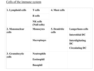
1. 1. Lymphoid cells T cells 4. Mast cells
B cells
NK cells
(Null cells)
2. Mononuclear
cells
Monocytes 5. Dendritic
cells
Langerhans cells
Interstitial DC
Macrophages Interdigitating
DC
Circulating DC
3. Granulocytic
cells
Neutrophils
Eosinophil
Basophil
Cells of the immune system
4. Bone marrow haematopoietic stem cell precursors
called as monoblasts
Differentiate into
promonocytes
Enter into blood
promonocytes
Blood
Differentiate into
Mature monocytes
(circulates in blood for app 8 hrs and enlarges
during that time)
6. Mechanism of pathogen clearance by monocytes
- By phagocytosis of Ag
2 subpopulations of monocytes
1. CD14++ monocyte (the classical monocyte, which is
characterized by high level expression of the CD14 cell
surface receptor)
2. CD14+CD16+ monocyte (the non-classical, pro-
inflammatory monocyte with low level expression of
CD14 and with additional co-expression of the CD16
receptor)
7. Monocytes in diagnosis
Monocytosis - is the state of excess monocytes in the
peripheral blood. It may be indicative of various disease states
A high count of CD14+CD16+ monocytes is found in severe
infection (sepsis)
a very low count of these cells is found after therapy with
immuno-suppressive glucocorticoids
9. Macrophages are
5-10 times larger than monocytes
has more organelles like lysosomes
Has increased phagocytic activity
Produce large no of hydrolytic enzymes
Secrete large variety of soluble factors
Macrophage
hydrolytic
enzymes
soluble factors
10. Macrophages
Some are fixed at
certain tissues (not
motile)
Some are free and motile
(travel by ameboid movement
in the tissues)
Nomenclature of Macrophages in tissues
Alveolar macrophages - lung
Histiocytes - connective tissues
Kupffer cells - liver
Mesangial cells - kidney
Microglial cells - brain (neural tissue)
Osteoclasts - bone
Epitheliod cells - granulomas
Sinusoidal lining cells - Spleen
11. Alveolar macrophages (in lungs)
- First line of defense against both living pathogens
and nonliving particulates, that enter from the
inspired air.
- Alveolus of lung contains both dendritic cells and
macrophages, as major immune cells
- Dendritic cells of alveolar lining of lung-
responsible for-adaptive immunity (antigen
specific response)
- Macrophages of alveolar lining of lung-
responsible for-innate immunity (not antigen
specific)
12. Resting macrophages
Activated macrophages
Stimulus for activation
-phagocytosis of a pathogen
-cytokines (INF-g) secreted by activated TH
-mediators of the inflammatory response
-components of bacterial cell walls
13. Resting Vs Activated Macrophages
Activated macrophages – more effective in eliminating pathogen
Activated
macrophage
greater
phagocytic
activity
increased ability
to kill ingested
microbes
increased
ability to
activate T
cells
increased
secretion of
inflammatory
mediators
secrete various
cytotoxic
proteins for
eliminating
broad range of
pathogens
14. The photomicrographs below show a human macrophage
engulfing and internalising the yeast Candida albicans.
15. Macrophages
-Their role is to phagocytose (engulf and then digest) cellular
debris and pathogens and
-to stimulate lymphocytes and other immune cells to respond
to the pathogen.
16. What is opsonisation?
Coating of Ag/pathogen with Ab or C3b
C3b
AntigenAb Ab
Ab
Ab Ab
Antigen
C3bC3b
C3b
C3b
Opsonin
1. C3b
2. Ab
What are opsonins?
17. How are the pathogens phagocytosed by macrophages?
Through Fc and Complement receptors
19. Receptors on macrophage Opsonic
Receptors
for
phagocytosis
1. Integrins
2. Fc receptors
Called as opsonic
receptors
They function in
phagocytosis and
endocytosis of
complement- or
antibody-
opsonised
particles,
respectively
AntigenAb Ab
Ab
Ab Ab
Antigen
C3bC3b
C3b
C3b
20. non-Toll-like receptors
(NTLR)
which include the family
of scavenger receptors
and the C-type lectins
Opsonic R and NTLRs
have effect on NF-kB
regulate production of
pro-inflammatory
mediators
21. Toll-Like receptors (TLR)
Are non-opsonic surface
receptors
do not mediate
phagocytosis/endocytosis
but are important sensors
of bacteria, fungi and
viruses
22. Some TLR are located
within vacuoles and
play a role in
recognition of
intracellular pathogens