1. Phagocytosis by macrophages
Step 1
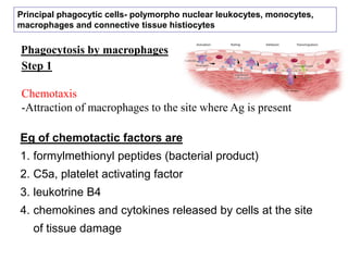
Chemotaxis
-Attraction of macrophages to the site where Ag is present
Eg of chemotactic factors are
1. formylmethionyl peptides (bacterial product)
2. C5a, platelet activating factor
3. leukotrine B4
4. chemokines and cytokines released by cells at the site
of tissue damage
Principal phagocytic cells- polymorpho nuclear leukocytes, monocytes,
macrophages and connective tissue histiocytes
2. Step 2
- adherence of the antigen to the macrophage cell membrane.
- Better adherance for- Complex antigens (such as whole bacterial
cells or viral particles) - readily phagocytosed
- Less adherence for-isolated proteins and encapsulated bacteria
- less readily phagocytosed.
3. Attachment of Ag and further internalisation
Depends on 2 receptors
1. Pattern receptors
2. Receptor for opsonins
5. OPSONISATION
Coating of antigen (e.g., a
bacterium) with the appropriate
antibody /C3b
Ag-Ab
Ab and C3b- called as opsonin
Ag-Ab/ Ag-C3b- bind to receptor for Ab on
Macrophage
Phagocytosis- enhanced by opsonisation of Ag
6. Coating of Ag with Ab or C3
Marks it as foreign
So, they bind to Fcg R or C3 R
Opsonin receptors
2 major classes of opsonins are
1. Ig A
2. Ig G
3rd class is- complement fragment
(C3b)
Receptor in
macrophage is
Fcg receptors
specific for
IgA,G and E
C3b receptors
7. Kinds of Fc receptors on phagocytes
1. FcgRI (CD64)
2. FcgRII (CD32)
3. FcgRIII (CD16)
4. FcaR (CD 89)
5. FceRI
6. FceRII (CD 23)
9. Different types of PRR recognizes different PAMP
PRR - Pattern Recognition Receptor
PAMP - Pathogen associated molecular pattern
MacrophagePathogen
Pam3CSK4 - Porphyromonas gingivalis lipopolysaccharide and lipoteichoic acid
PGN - Peptidoglycan
Pathogen PAMP PRR Macrophage
10. CpG sites
CpG" stands for cytosine and guanine separated by a
phosphate, which links the two nucleosides together in DNA
CpG site
The "CpG" notation is used to distinguish a cytosine followed
by guanine from a cytosine base paired to a guanine.
Base pairing
11. Binding of a Pathogen via Its PAMP (Pathogen Associated Molecular
Pattern) to a TLR's PRR (Pattern Recognition Receptor) Domain
Toll Like Receptors (TLR)
On October 3, 2011, Dr. Beutler and Dr. Hoffmann were
awarded the Nobel Prize in Medicine or Physiology for their
work on TLR
14. Surface lectins
pathogen
CHO molecules (Mannose and Fructose on microbial surface)
Macrophage
Surface lectins (Mannose Specific Macrophage Receptor)
pathogen
CHO molecules (galactose residues in senescent erythrocytes)
Macrophage
Surface lectins (Galactose specific lectin in liver macrophages (Kupffer
cells)
15. Scavenger receptors
-Mainly recognizes cells undergoing apoptosis
-Have broad binding specificity for oxidised lipoproteins,
polyribonucleotides, anionic polysaccharides and
bacterial LPS
Apoptotic cells
- Have high anionic polysaccharides on their surface
Bind to anionic polymers
16. Step 3
- Formation of membrane protrusions called as pseudopodia
- It extends around the attached material
17.
18. Step 4
Fusion of the pseudopodia
To encloses the material
within a membrane-
bounded structure called a
phagosome
then it enters the
endocytic processing
pathway
pH in phagosome and
lysosome- below 4, so
bacteria cannot multiply
19. Endocytic processing pathway
phagosome moves toward the
cell interior
Fuses with
with a lysosome
to form a phagolysosome.
Lysosomes contain
1. lysozyme and
2. a variety of other hydrolytic
enzymes
They
digest the ingested material.
20. 1. Lysozyme
also known as muramidase or N-acetylmuramide
glycanhydrolase
It hydrolyses the polysaccharides of bacterial cell wall
catalyzing hydrolysis of 1,4-beta-linkages between N-
acetylmuramic acid and N-acetyl-D-glucosamine
residues in a peptidoglycan
Components in lysosomes
21. 2. Lactoferrin
- Binds to Iron and sequesters it, so that Iron is not
available for the microrganism
Components in lysosomes
25. 1. OXYGEN-DEPENDENT KILLING MECHANISMS
Activated macrophages produces
a number of
reactive oxygen intermediates (ROIs) and
reactive nitrogen intermediates
that have
antimicrobial activity
26. Macrophage
Phagocytosis of Ag
Process called respiratory burst occurs
Activates
membrane-bound oxidase
reduction
oxygen superoxide anion
(ROI)
toxic to ingested
microorganisms.
SO- also generates other powerful
oxidizing
Agents like hydroxyl radicals and
hydrogen peroxide
27. When the lysosome fuses with the phagosome
the activity of myeloperoxidase – is increased
hydrogen peroxide + chloride ions hypochlorite
toxic to ingested microbes
28. Activated macrophages
Produce nitric oxide synthetase (NOS)
Has antimicrobial activity
Combines with superoxide
Produces
More toxic antimicrobial
substances
30. Each Defensin peptide has six invariant
cysteines
forms a circular molecule that is stabilized by
intramolecular disulfide bonds
These circularized defensin peptides
form ion-permeable channels in bacterial cell membranes
31. Most defensins (shown as large
ovals) are amphipathic molecules
They have
1. positively charged amino-acid side
chains (pink) and
2. hydrophobic amino-acid side
chains (green).
This allows them to interact with
microbial membranes, shown
schematically with their negatively
charged phospholipid headgroups
(blue) and hydrophobic fatty acid
chains (green).
Thus, forms pores in microbial
membrane
Hydrophobic side chain
+vely charged chain
-vely charged head gp
32. SOLUBLE FACORS SECRETED BY ACTIVATED
MACROPHAGES- eliminate the pathogen
Activated
macrophage
Soluble factors
33. Cytokines
1. interleukin 1 (IL-1)
2. TNF-a
3. interleukin 6 (IL-6)
promote inflammatory responses.
1. IL-1 activates lymphocytes
2. IL-1, IL-6, and TNF- promote fever by affecting
the thermoregulatory center in the hypothalamus.