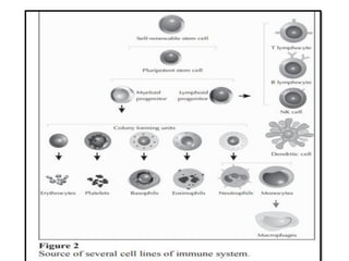
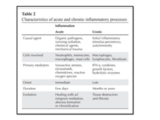
Immune system and its functions
Assistant lecture of Oral Medicine, Periodontology, Diagnosis
and Dental Radiology (Al-Azhar Univerisity)
Content
Soluble molecules and cells of the immune system
Immunity by equilibrium
The Mechanism of Stimulating and Mobilizing the Immune
System Enhancing the Anti-Tumor Immunity
Influence of adjuvant or delivery system on the induction of
humoral or cell-mediated immunity to mucosally delivered
vaccines
The main effector cells of innate immunity are
macrophages, neutrophils, dendritic cells, and natural
killer (NK) cells .
Phagocytosis, release of inflammatory mediators,
activation of complement system proteins, as well as
synthesis of acute phase proteins, cytokines and
chemokines are the main mechanisms in innate
immunity
These mechanisms are activated by specific stimuli,
represented by molecular structures of ubiquitous
occurrence in microorganisms, but not in human species.
Molecules commonly found on the surface of
microorganisms, such as lipopolysaccharides, mannose
and teichoic acids constitute pathogen-associated
molecular patterns (PAMPs) and activate the innate
immune response by interaction with different receptors
known as pattern recognition receptors (PRR), among
which is the family of Toll-like receptors (TLRs).
Phagocytosis begins with adhesion of the phagocyte
surface receptors to the pathogen, which then is
internalized into vesicles called phagosomes.
Inside the phagocyte, the phagosome fuses to
lysosomes, whose contents are released with
consequent digestion and pathogen elimination.
Changes in the oxidase’s gene system components present
in phagolysosome membrane lead to disability in respiratory
burst and generation of reactive oxygen species (ROS).
The absence of ROS determines serious deficiency in the
destructive capacity of phagocytes, being responsible for a
significant primary immunodeficiency called chronic
granulomatous disease
Unlike innate response, adaptive or acquired immune
response depends on activation of specialized cells (the
lymphocytes) and soluble molecules produced by
lymphocytes.
The main features of acquired response are:
specificity and diversity of recognition, memory,
specialized response, self-restraint, and tolerance to
components of the organism itself.
 Although the main cells involved in acquired
immune response are lymphocytes, antigen
presenting cells (APCs) play a key role in its activation,
presenting antigens associated with molecules of the
major histocompatibility complex (MHC) to T
lymphocyte (TL).6
Dendritic cells
Dendritic cells, specialized in capturing and presenting
antigens to lymphocytes, are considered a bridge between
innate and adaptive immunity because they are attracted
and activated by elements of innate response and permit
TL sensibilization of adaptive immune response
During their lifetime, the immature DCs migrate from bone
marrow into bloodstream, reaching peripheral tissues as
skin, where they become residents (Langerhans cells).
A curious aspect is that DCs are the first cells to
arrive at a site of infection, preceding even the
neutrophils.
After contact with antigen, DCs become activated
and migrate through lymphatic vessels to secondary
lymphoid organs.
There are two pathways of DCs differentiation from a
common progenitor.
The myeloid pathway generates myeloid DCs (mDCs),
among which there are the Langerhans cells, the main
DCs in skin, and the interstitial DCs found in other
tissues.
The other pathway of differentiation generates
plasmacytoid DCs (pDCs), which predominate in the
peripheral blood and secrete large amounts of type I
interferon (IFN-α/β) in the presence of viral infections.
DCs are crucial for determining the activation and type
of immunity mediated by TLs.
In general, immature DCs are tolerogenic, while
mature DCs are immunostimulatory.
 However, in some contexts, mature DCs can expand
the population of TLs regulators
Induction of tolerance or immune response depends
on the set of signals received by DCs, such as activation
of TLRs and cytokines present in the environment.
 DCs can coordinate LBs response via TL activation or
directly by soluble substances such as INF-α.
Figure 3 Dendritic cells and generation of TLs specific
antigens.
(A) Characteristics of immature dendritic cells (iDCs).
(B) Activation and uptake of pathogens through cytokine
microenvironment and interaction with pattern recognition
receptors, with consequent migration of DCs to lymph nodes.
(C) Maturation of dendritic cells.
(D) Migration of naive T cells to paracortical area of lymph
node. Entry through high endothelial venules (HEV) and
chemokine-driven migration of lymphoid tissue.
(E) Presentation of processed antigens to T lymphocytes,
generating activated effector cells
Neutrophils
Neutrophils are the most abundant leukocytes in
peripheral blood, with an important role in the early
stages of inflammatory reaction and sensitive to
chemotactic agents, such as cleavage products of
complement fractions (C3a and C5a) and substances
released by mast cells and basophils.
They are among the first cells to migrate from vessels
to tissues attracted by chemokines, such as IL-8, and
are activated by various stimuli, such as bacterial
products, complement proteins (C5a), immune
complex (IC), chemokines, and cytokines.
The neutrophil phagocytic capacity is stimulated by
binding of its receptors for opsonins, IgG-Fc, C3b, and
TLRs. These cells also undergo degranulation, releasing
three classes of granules in the extracellular
environment:
Primary or azurophilic granules that contain important
mediators, such as myeloperoxidase, defensins, neutrophil
elastase, permeability-increasing protein, and bacterial
cathepsin G.
Secondary granules with components specifically secreted
by neutrophils, with lactoferrin is a prime example.
Tertiary granules with cathepsins and gelatinases as main
proteins
Recent studies have shown that neutrophils can also
generate the so-called neutrophil extracellular traps
(NETs) formed by granule substances and nuclear
components capable of calling off the virulence factors
and destroying extracellular bacteria.
The NETs are present in large quantity in inflammatory
sites, acting directly on microorganisms and also serving
as a physical barrier that prevents spreading.
Macrophage
Monocytes constitute 3-8% of circulating leukocytes
and, in connective tissue or parenchyma of organs, give
rise to macrophages and myeloid dendritic cells.
Monocytes and macrophages are efficient phagocytes,
engulfing pathogens and cellular debris
 Unlike neutrophils, macrophages can remain in tissue
for months to years, acting as true sentinels. Besides
having a role in innate immunity, macrophages process
and present antigens via MHC molecules, thus
stimulating the response mediated by TL.
Recently, the existence of three subpopulations of
macrophages was proposed: activated, tissue repair, and
regulator macrophages..
The first would be the classic macrophages with
tumoricidal and microbicidal activity, which secrete
large amounts of proinflammatory mediators and
cytokines, present antigens to TLs, and are involved in
cellular immune response
The second type, activated by IL4, would be primarily
involved in tissue repair by stimulating fibroblasts and
promoting extracellular matrix deposition.
The third type would exert regulatory activity through
release of IL-10, an anti-inflammatory cytokine.
Some microorganisms, like Mycobacterium
tuberculosis, are resistant to the microbicidal action and
remain viable for a long time in macrophages’
phagosomes .
These macrophages become large and multinucleated
(giant cells) and, together with lymphocytes and
fibroblasts that accumulate around them, form
granulomas, which are the body’s attempt to prevent
the spread of the pathogen
Natural killer cells
Natural killer cells (NK) originate in bone marrow from a
common progenitor to TLs, constituting 5% to 20% of
blood mononuclear cells.
They are an important line of nonspecific defense,
recognizing and lysing cells infected by viruses, bacteria
and protozoa, as well as tumor cells. Furthermore, they
recruit neutrophils and macrophages, activate DCs and
T and B lymphocytes.
The expansion and activation of NKs are stimulated by
IL-15, produced by macrophages, and IL-12, potent
inducer of IFN-γ and cytolytic action.
Once activated, the NKs lyse infected and tumoral cells
and secrete proinflammatory cytokines (IL-1, IL-2, and
especially IFN-γ).
 The cytolysis mediated by NKs occurs through the
action of the enzymes perforins, which create pores in
membrane of target cells, and granzymes, which
penetrate into cells and trigger cell death by apoptosis
Another effector action of NK is the
destruction of cells coated with IgG, via Fc
receptors (FcγRIII or CD16), by a mechanism of
antibody-dependent cellular cytotoxicity (ADCC)
Figure 4 Function of receptor activation (ITAM) and inhibition
(ITIM) in the physiology of NK cells.
(A) Interaction of NK cells with a normal body cell expressing MHC
class I, with consequent inhibition of NK cytolysis dependent
induction.
(B) (B) Interaction of NK cells with virus-infected cell, with
consequent MHC class I loss of expression, which results in
activation of NK cells with concomitant release of lethal
products.
Mast cells
Mast cells are derived from CD34+ hematopoietic
progenitors in bone marrow and, in general, are not
found in the circulation.
From bone marrow, the progenitors migrate to
peripheral tissues as immature cells and differentiate
in situ according to the particular characteristics of the
microenvironment
Mast cells have surface receptors of high
affinity, FcεRI, bound to IgE molecules, and are
activated by multivalent antigen recognition by
IgE.
Stimuli such as products of complement activation,
basic substances, including some animals’ poisons,
certain neuropeptides, and several physical agents
(mechanical trauma, heat, and cold) can activate mast
cells independently of IgE binding.
The binding of bacterial components to TLRs 1, 2,
4, and 6, and other specific receptors such as CD48,
also activates mast cells, leading to mediators’
release
The mediators formed following activation include
platelet activating factor (PAF), arachidonic acid
derivatives, and a series of cytokines.
The release of these mediators induces inflammatory
cell migration (neutrophils and macrophages), increased
vascular permeability, mucus secretion, increased
gastrointestinal motility, and bronchoconstriction, which
are the signs and symptoms of allergy and anaphylaxis.
Effector functions of mast cells in synovia suggest their
involvement in leukocyte recruitment, fibroblast
activation and hyperplasia, angiogenesis, and destruction
of cartilage and bone.
 They also participate in joint destruction by inducing
fibroblasts and chondrocytes to secrete matrix
metalloproteinases and promoting osteoclast
differentiation.
basophils
The granules found in basophils have mediators
similar to those of mast cells.
 Basophils also express FcεRI, bind IgE, and are
activated by IgE-antigen complexes and may
contribute to immediate hypersensitivity reactions.
Eosinophil
Eosinophil cationic protein creates pores in target-cell
membrane, allowing the entry of other cytotoxic
molecules; inhibits TL proliferation; suppresses
antibody production by LB; induces degranulation of
mast cells; and stimulates secretion of
glucosaminoglycans by fibroblasts.
Eosinophil peroxidase forms ROS and NO, promoting
oxidative stress in target-cell and causing cell death by
apoptosis and necrosis.
Other effector mechanisms that contribute to the
inflammatory process include production of a
variety of cytokines, such as IL-1, IL-2, IL-4, IL-5, IL-
6, IL-8, IL-13, and TNF- α,25 and release of
proinflammatory lipid mediators, such as
leukotrienes (LTC4, LTD4, LTE4), and prostaglandins
(PGE2).
Elastase enzymes and the growth factor TGF-β,
growth factor derived from platelets (PDGF), and
endothelial vessels growth factor (VEGF)
contributes to tissue remodeling.
The complement system
Complement system (CS) consists of a family of
more than 20 plasma glycoproteins, synthesized in
the liver, but also by macrophages and fibroblasts.
There are three forms of CS activation: classical,
alternative, and via mannose-binding lectin (MBL).
 The activation of these pathways contributes to the
integration of effector mechanisms of innate and
adaptive immunity.
 In the innate immune response, pathogens that
invade the organism encounter soluble substances
of innate immune response, such as CS proteins,
C-reactive protein, and others.
In adaptive immunity, CS is activated by binding of
preformed antibodies to pathogen or antigen (immune
complex).
 Lectin pathway begins by recognizing mannose on
microorganism surface by MBL bound to MASP1 and
MASP2 serine proteases.
Activation of these proteases results in the
breakdown of CS components C2 and C4 into smaller
fragments (C4a and C2b) and larger fragments (C4b
and C2a).
C4bC2a complex is the C3 convertase of the classical
pathway, which cleaves C3 into soluble C3a and C3b, which
in turn binds to C4bC2a at the surface of microorganism.
The C4bC2aC3b complex, called C5 convertase, cleaves the
C5 component, following on this pathway and culminating in
the formation of MAC.
.The classical pathway resembles the lectin
pathway and is initiated by the binding of C1q
component to two molecules of IgG or to one
molecule of IgM, complexed with the target antigen
(immune complexes).
This binding activates the proteases R (C1r) and S
(C1s) associated with C1q, cleaving components C2 and
C4 and following the pathway, as described.
 Because the classical pathway depends on the prior
production of specific antibodies attached to the surface
of pathogens, it is associated with specific humoral
immune response.
The alternative pathway begins with the spontaneous
rupture of the C3 component into C3a and C3b
fragments .
A thioester binding in fragment C3b is exposed with
this cleavage, which allows their covalent binding to the
surface of invading microorganisms.
If there is no binding of C3b component, thioesters
binding site is rapidly hydrolyzed and the fragment is
inactivated.
The binding of C3b enables binding to Factor B, which
is then cleaved into fragments Ba and Bb by Factor D.
The C3bBb complex (alternative pathway C3
convertase) cleaves more C3 molecules and remains on
the surface.
This complex is stabilized by properdin (Factor P),
amplifying the breakdown of C3.
 C3bBb component cleaves C3, generating C3bBbC3b, a
protease able to cleave C5, the last step of the
alternative pathway.
The major histocompatibility complex
The human major histocompatibility complex (MHC) is
composed of a set of highly polymorphic genes called
human leukocyte antigen (HLA) and comprises more than
120 functional genes, of which about 20% are associated
with immunity.
The association between autoimmune diseases and
MHC genes reflects the important role of these
molecules in directing the immune response.
For its role in antigen presentation, MHC provides a link
between innate response and adaptive response.
In humans, these genes are located on chromosome 6
and are traditionally divided into classes I, II, and III.
Only the genes of classes I and II are involved in presenting
antigen protein to LT.
Class I molecules are present on the surface of all
nucleated cells, while class II are found primarily on APCs
(macrophages, DCs, and LB).
All MHC molecules found in the surface of a cell have an
associated peptide.
Although the molecules of classes I and II exhibit
different structural characteristics, both are expressed as
heterotrimers in which two chains are from MHC
molecule and the third is the peptide presented to TL
Innate immunity in the context of
inflammatory response
The acute inflammatory response evolves from a phase
started by vascular cells in the tissue immediately after
the injury.
At baseline, only a fraction of the capillaries comprising
the tissue is permeable, but after injury, local vasodilation
and increased capillary permeability occur mediated by
vasoactive amines, histamine, and serotonin released from
mast cells and monocytes minutes after the injury.
 Initially, electrolytes and small molecules leave the
capillary bed, forming the transudate. Subsequently, larger
molecules, such as albumin and fibrinogen, also leave the
capillary bed, forming the exudate.
Protein output to extravascular space is
accompanied by water loss and marginalization of
leukocytes, which start circulating by the
endothelium.
Figure 7 Mechanisms of leukocyte migration to inflammatory site.
Macrophages stimulated by inductors of inflammatory response produce
cytokines, such as TNF-α and IL-1, which induce the endothelial venules to
express selectins, integrins, and chemokines ligands .
Selectins mediate the weak adhesion of neutrophils; integrins promote strong
adhesion; and chemokines activate and stimulate the migration of neutrophils
to inflammatory focus.
 Monocytes and activated T lymphocytes use the same mechanisms to migrate
to infection sites.
Persistence of harmful agent leads to the chronicity of
the process, with concomitant destruction and tissue
repair.
In chronic inflammation, tissue characteristically
presents an infiltrate composed mainly of mononuclear
cells (monocytes, macrophages, and lymphocytes), signs
of angiogenesis, and fibrosis (Table 2)
Several stimuli can induce persistent chronic
inflammation, such as intracellular bacteria (e.g.,
Mycobacterium tuberculosis), chemical substances such
as silica, and even physical agents such as ultraviolet
radiation, and repetitive trauma.
The mediators of inflammatory response
Immunity by equilibrium
The Mechanism of Stimulating and Mobilizing the
Immune System Enhancing the Anti-Tumor Immunity
FIGURE 1 | The tumor microenvironment is composed of cellular and
non-cellular components that support tumor growth. Tumor and its
microenvironment interact and promote each other through
angiogenesis and immunosuppression.
Therefore, targeting tumor microenvironment in anti-tumor therapy
can make greater progress, such as inhibiting tumor angiogenesis and
tumor immunity
FIGURE 2 | Immune T cells were isolated from patients and genetically engineered in
vitro to be fitted with chimeric antigen receptors (CAR) that recognize cancer cell
surface antigens. The modified cells are amplified in large Numbers in vitro and
injected back into the patient to achieve the therapeutic effect of accurately
identifying and killing cancer cell
Influence of adjuvant or delivery system on the
induction of humoral or cell-mediated immunity to
mucosally delivered vaccines
immunology.pdf
immunology.pdf
immunology.pdf
immunology.pdf
immunology.pdf
immunology.pdf

immunology.pdf

  • 2.
    Immune system andits functions Assistant lecture of Oral Medicine, Periodontology, Diagnosis and Dental Radiology (Al-Azhar Univerisity)
  • 3.
    Content Soluble molecules andcells of the immune system Immunity by equilibrium The Mechanism of Stimulating and Mobilizing the Immune System Enhancing the Anti-Tumor Immunity Influence of adjuvant or delivery system on the induction of humoral or cell-mediated immunity to mucosally delivered vaccines
  • 5.
    The main effectorcells of innate immunity are macrophages, neutrophils, dendritic cells, and natural killer (NK) cells .
  • 6.
    Phagocytosis, release ofinflammatory mediators, activation of complement system proteins, as well as synthesis of acute phase proteins, cytokines and chemokines are the main mechanisms in innate immunity
  • 7.
    These mechanisms areactivated by specific stimuli, represented by molecular structures of ubiquitous occurrence in microorganisms, but not in human species. Molecules commonly found on the surface of microorganisms, such as lipopolysaccharides, mannose
  • 8.
    and teichoic acidsconstitute pathogen-associated molecular patterns (PAMPs) and activate the innate immune response by interaction with different receptors known as pattern recognition receptors (PRR), among which is the family of Toll-like receptors (TLRs).
  • 9.
    Phagocytosis begins withadhesion of the phagocyte surface receptors to the pathogen, which then is internalized into vesicles called phagosomes. Inside the phagocyte, the phagosome fuses to lysosomes, whose contents are released with consequent digestion and pathogen elimination.
  • 10.
    Changes in theoxidase’s gene system components present in phagolysosome membrane lead to disability in respiratory burst and generation of reactive oxygen species (ROS).
  • 11.
    The absence ofROS determines serious deficiency in the destructive capacity of phagocytes, being responsible for a significant primary immunodeficiency called chronic granulomatous disease
  • 12.
    Unlike innate response,adaptive or acquired immune response depends on activation of specialized cells (the lymphocytes) and soluble molecules produced by lymphocytes.
  • 13.
    The main featuresof acquired response are: specificity and diversity of recognition, memory, specialized response, self-restraint, and tolerance to components of the organism itself.
  • 14.
     Although themain cells involved in acquired immune response are lymphocytes, antigen presenting cells (APCs) play a key role in its activation, presenting antigens associated with molecules of the major histocompatibility complex (MHC) to T lymphocyte (TL).6
  • 17.
  • 18.
    Dendritic cells, specializedin capturing and presenting antigens to lymphocytes, are considered a bridge between innate and adaptive immunity because they are attracted and activated by elements of innate response and permit TL sensibilization of adaptive immune response
  • 19.
    During their lifetime,the immature DCs migrate from bone marrow into bloodstream, reaching peripheral tissues as skin, where they become residents (Langerhans cells).
  • 20.
    A curious aspectis that DCs are the first cells to arrive at a site of infection, preceding even the neutrophils. After contact with antigen, DCs become activated and migrate through lymphatic vessels to secondary lymphoid organs.
  • 21.
    There are twopathways of DCs differentiation from a common progenitor. The myeloid pathway generates myeloid DCs (mDCs), among which there are the Langerhans cells, the main DCs in skin, and the interstitial DCs found in other tissues.
  • 22.
    The other pathwayof differentiation generates plasmacytoid DCs (pDCs), which predominate in the peripheral blood and secrete large amounts of type I interferon (IFN-α/β) in the presence of viral infections.
  • 23.
    DCs are crucialfor determining the activation and type of immunity mediated by TLs. In general, immature DCs are tolerogenic, while mature DCs are immunostimulatory.  However, in some contexts, mature DCs can expand the population of TLs regulators
  • 24.
    Induction of toleranceor immune response depends on the set of signals received by DCs, such as activation of TLRs and cytokines present in the environment.  DCs can coordinate LBs response via TL activation or directly by soluble substances such as INF-α.
  • 27.
    Figure 3 Dendriticcells and generation of TLs specific antigens. (A) Characteristics of immature dendritic cells (iDCs). (B) Activation and uptake of pathogens through cytokine microenvironment and interaction with pattern recognition receptors, with consequent migration of DCs to lymph nodes. (C) Maturation of dendritic cells. (D) Migration of naive T cells to paracortical area of lymph node. Entry through high endothelial venules (HEV) and chemokine-driven migration of lymphoid tissue. (E) Presentation of processed antigens to T lymphocytes, generating activated effector cells
  • 28.
  • 29.
    Neutrophils are themost abundant leukocytes in peripheral blood, with an important role in the early stages of inflammatory reaction and sensitive to chemotactic agents, such as cleavage products of complement fractions (C3a and C5a) and substances released by mast cells and basophils.
  • 30.
    They are amongthe first cells to migrate from vessels to tissues attracted by chemokines, such as IL-8, and are activated by various stimuli, such as bacterial products, complement proteins (C5a), immune complex (IC), chemokines, and cytokines.
  • 31.
    The neutrophil phagocyticcapacity is stimulated by binding of its receptors for opsonins, IgG-Fc, C3b, and TLRs. These cells also undergo degranulation, releasing three classes of granules in the extracellular environment:
  • 32.
    Primary or azurophilicgranules that contain important mediators, such as myeloperoxidase, defensins, neutrophil elastase, permeability-increasing protein, and bacterial cathepsin G. Secondary granules with components specifically secreted by neutrophils, with lactoferrin is a prime example. Tertiary granules with cathepsins and gelatinases as main proteins
  • 33.
    Recent studies haveshown that neutrophils can also generate the so-called neutrophil extracellular traps (NETs) formed by granule substances and nuclear components capable of calling off the virulence factors and destroying extracellular bacteria.
  • 34.
    The NETs arepresent in large quantity in inflammatory sites, acting directly on microorganisms and also serving as a physical barrier that prevents spreading.
  • 35.
  • 36.
    Monocytes constitute 3-8%of circulating leukocytes and, in connective tissue or parenchyma of organs, give rise to macrophages and myeloid dendritic cells. Monocytes and macrophages are efficient phagocytes, engulfing pathogens and cellular debris
  • 37.
     Unlike neutrophils,macrophages can remain in tissue for months to years, acting as true sentinels. Besides having a role in innate immunity, macrophages process and present antigens via MHC molecules, thus stimulating the response mediated by TL.
  • 38.
    Recently, the existenceof three subpopulations of macrophages was proposed: activated, tissue repair, and regulator macrophages..
  • 39.
    The first wouldbe the classic macrophages with tumoricidal and microbicidal activity, which secrete large amounts of proinflammatory mediators and cytokines, present antigens to TLs, and are involved in cellular immune response
  • 40.
    The second type,activated by IL4, would be primarily involved in tissue repair by stimulating fibroblasts and promoting extracellular matrix deposition. The third type would exert regulatory activity through release of IL-10, an anti-inflammatory cytokine.
  • 41.
    Some microorganisms, likeMycobacterium tuberculosis, are resistant to the microbicidal action and remain viable for a long time in macrophages’ phagosomes .
  • 42.
    These macrophages becomelarge and multinucleated (giant cells) and, together with lymphocytes and fibroblasts that accumulate around them, form granulomas, which are the body’s attempt to prevent the spread of the pathogen
  • 43.
  • 44.
    Natural killer cells(NK) originate in bone marrow from a common progenitor to TLs, constituting 5% to 20% of blood mononuclear cells.
  • 45.
    They are animportant line of nonspecific defense, recognizing and lysing cells infected by viruses, bacteria and protozoa, as well as tumor cells. Furthermore, they recruit neutrophils and macrophages, activate DCs and T and B lymphocytes.
  • 46.
    The expansion andactivation of NKs are stimulated by IL-15, produced by macrophages, and IL-12, potent inducer of IFN-γ and cytolytic action. Once activated, the NKs lyse infected and tumoral cells and secrete proinflammatory cytokines (IL-1, IL-2, and especially IFN-γ).
  • 47.
     The cytolysismediated by NKs occurs through the action of the enzymes perforins, which create pores in membrane of target cells, and granzymes, which penetrate into cells and trigger cell death by apoptosis
  • 48.
    Another effector actionof NK is the destruction of cells coated with IgG, via Fc receptors (FcγRIII or CD16), by a mechanism of antibody-dependent cellular cytotoxicity (ADCC)
  • 50.
    Figure 4 Functionof receptor activation (ITAM) and inhibition (ITIM) in the physiology of NK cells. (A) Interaction of NK cells with a normal body cell expressing MHC class I, with consequent inhibition of NK cytolysis dependent induction. (B) (B) Interaction of NK cells with virus-infected cell, with consequent MHC class I loss of expression, which results in activation of NK cells with concomitant release of lethal products.
  • 51.
  • 52.
    Mast cells arederived from CD34+ hematopoietic progenitors in bone marrow and, in general, are not found in the circulation. From bone marrow, the progenitors migrate to peripheral tissues as immature cells and differentiate in situ according to the particular characteristics of the microenvironment
  • 53.
    Mast cells havesurface receptors of high affinity, FcεRI, bound to IgE molecules, and are activated by multivalent antigen recognition by IgE.
  • 54.
    Stimuli such asproducts of complement activation, basic substances, including some animals’ poisons, certain neuropeptides, and several physical agents (mechanical trauma, heat, and cold) can activate mast cells independently of IgE binding.
  • 55.
    The binding ofbacterial components to TLRs 1, 2, 4, and 6, and other specific receptors such as CD48, also activates mast cells, leading to mediators’ release
  • 56.
    The mediators formedfollowing activation include platelet activating factor (PAF), arachidonic acid derivatives, and a series of cytokines.
  • 57.
    The release ofthese mediators induces inflammatory cell migration (neutrophils and macrophages), increased vascular permeability, mucus secretion, increased gastrointestinal motility, and bronchoconstriction, which are the signs and symptoms of allergy and anaphylaxis.
  • 58.
    Effector functions ofmast cells in synovia suggest their involvement in leukocyte recruitment, fibroblast activation and hyperplasia, angiogenesis, and destruction of cartilage and bone.  They also participate in joint destruction by inducing fibroblasts and chondrocytes to secrete matrix metalloproteinases and promoting osteoclast differentiation.
  • 59.
  • 60.
    The granules foundin basophils have mediators similar to those of mast cells.  Basophils also express FcεRI, bind IgE, and are activated by IgE-antigen complexes and may contribute to immediate hypersensitivity reactions.
  • 61.
  • 62.
    Eosinophil cationic proteincreates pores in target-cell membrane, allowing the entry of other cytotoxic molecules; inhibits TL proliferation; suppresses antibody production by LB; induces degranulation of mast cells; and stimulates secretion of glucosaminoglycans by fibroblasts.
  • 63.
    Eosinophil peroxidase formsROS and NO, promoting oxidative stress in target-cell and causing cell death by apoptosis and necrosis.
  • 64.
    Other effector mechanismsthat contribute to the inflammatory process include production of a variety of cytokines, such as IL-1, IL-2, IL-4, IL-5, IL- 6, IL-8, IL-13, and TNF- α,25 and release of proinflammatory lipid mediators, such as leukotrienes (LTC4, LTD4, LTE4), and prostaglandins (PGE2).
  • 65.
    Elastase enzymes andthe growth factor TGF-β, growth factor derived from platelets (PDGF), and endothelial vessels growth factor (VEGF) contributes to tissue remodeling.
  • 66.
  • 67.
    Complement system (CS)consists of a family of more than 20 plasma glycoproteins, synthesized in the liver, but also by macrophages and fibroblasts.
  • 68.
    There are threeforms of CS activation: classical, alternative, and via mannose-binding lectin (MBL).  The activation of these pathways contributes to the integration of effector mechanisms of innate and adaptive immunity.
  • 71.
     In theinnate immune response, pathogens that invade the organism encounter soluble substances of innate immune response, such as CS proteins, C-reactive protein, and others.
  • 72.
    In adaptive immunity,CS is activated by binding of preformed antibodies to pathogen or antigen (immune complex).  Lectin pathway begins by recognizing mannose on microorganism surface by MBL bound to MASP1 and MASP2 serine proteases.
  • 73.
    Activation of theseproteases results in the breakdown of CS components C2 and C4 into smaller fragments (C4a and C2b) and larger fragments (C4b and C2a).
  • 74.
    C4bC2a complex isthe C3 convertase of the classical pathway, which cleaves C3 into soluble C3a and C3b, which in turn binds to C4bC2a at the surface of microorganism. The C4bC2aC3b complex, called C5 convertase, cleaves the C5 component, following on this pathway and culminating in the formation of MAC.
  • 75.
    .The classical pathwayresembles the lectin pathway and is initiated by the binding of C1q component to two molecules of IgG or to one molecule of IgM, complexed with the target antigen (immune complexes).
  • 76.
    This binding activatesthe proteases R (C1r) and S (C1s) associated with C1q, cleaving components C2 and C4 and following the pathway, as described.  Because the classical pathway depends on the prior production of specific antibodies attached to the surface of pathogens, it is associated with specific humoral immune response.
  • 77.
    The alternative pathwaybegins with the spontaneous rupture of the C3 component into C3a and C3b fragments . A thioester binding in fragment C3b is exposed with this cleavage, which allows their covalent binding to the surface of invading microorganisms.
  • 78.
    If there isno binding of C3b component, thioesters binding site is rapidly hydrolyzed and the fragment is inactivated.
  • 79.
    The binding ofC3b enables binding to Factor B, which is then cleaved into fragments Ba and Bb by Factor D. The C3bBb complex (alternative pathway C3 convertase) cleaves more C3 molecules and remains on the surface.
  • 80.
    This complex isstabilized by properdin (Factor P), amplifying the breakdown of C3.  C3bBb component cleaves C3, generating C3bBbC3b, a protease able to cleave C5, the last step of the alternative pathway.
  • 81.
  • 82.
    The human majorhistocompatibility complex (MHC) is composed of a set of highly polymorphic genes called human leukocyte antigen (HLA) and comprises more than 120 functional genes, of which about 20% are associated with immunity.
  • 83.
    The association betweenautoimmune diseases and MHC genes reflects the important role of these molecules in directing the immune response. For its role in antigen presentation, MHC provides a link between innate response and adaptive response. In humans, these genes are located on chromosome 6 and are traditionally divided into classes I, II, and III.
  • 84.
    Only the genesof classes I and II are involved in presenting antigen protein to LT. Class I molecules are present on the surface of all nucleated cells, while class II are found primarily on APCs (macrophages, DCs, and LB). All MHC molecules found in the surface of a cell have an associated peptide.
  • 85.
    Although the moleculesof classes I and II exhibit different structural characteristics, both are expressed as heterotrimers in which two chains are from MHC molecule and the third is the peptide presented to TL
  • 88.
    Innate immunity inthe context of inflammatory response
  • 89.
    The acute inflammatoryresponse evolves from a phase started by vascular cells in the tissue immediately after the injury.
  • 90.
    At baseline, onlya fraction of the capillaries comprising the tissue is permeable, but after injury, local vasodilation and increased capillary permeability occur mediated by vasoactive amines, histamine, and serotonin released from mast cells and monocytes minutes after the injury.
  • 91.
     Initially, electrolytesand small molecules leave the capillary bed, forming the transudate. Subsequently, larger molecules, such as albumin and fibrinogen, also leave the capillary bed, forming the exudate.
  • 92.
    Protein output toextravascular space is accompanied by water loss and marginalization of leukocytes, which start circulating by the endothelium.
  • 94.
    Figure 7 Mechanismsof leukocyte migration to inflammatory site. Macrophages stimulated by inductors of inflammatory response produce cytokines, such as TNF-α and IL-1, which induce the endothelial venules to express selectins, integrins, and chemokines ligands . Selectins mediate the weak adhesion of neutrophils; integrins promote strong adhesion; and chemokines activate and stimulate the migration of neutrophils to inflammatory focus.  Monocytes and activated T lymphocytes use the same mechanisms to migrate to infection sites.
  • 95.
    Persistence of harmfulagent leads to the chronicity of the process, with concomitant destruction and tissue repair. In chronic inflammation, tissue characteristically presents an infiltrate composed mainly of mononuclear cells (monocytes, macrophages, and lymphocytes), signs of angiogenesis, and fibrosis (Table 2)
  • 96.
    Several stimuli caninduce persistent chronic inflammation, such as intracellular bacteria (e.g., Mycobacterium tuberculosis), chemical substances such as silica, and even physical agents such as ultraviolet radiation, and repetitive trauma.
  • 98.
    The mediators ofinflammatory response
  • 102.
  • 109.
    The Mechanism ofStimulating and Mobilizing the Immune System Enhancing the Anti-Tumor Immunity
  • 111.
    FIGURE 1 |The tumor microenvironment is composed of cellular and non-cellular components that support tumor growth. Tumor and its microenvironment interact and promote each other through angiogenesis and immunosuppression. Therefore, targeting tumor microenvironment in anti-tumor therapy can make greater progress, such as inhibiting tumor angiogenesis and tumor immunity
  • 112.
    FIGURE 2 |Immune T cells were isolated from patients and genetically engineered in vitro to be fitted with chimeric antigen receptors (CAR) that recognize cancer cell surface antigens. The modified cells are amplified in large Numbers in vitro and injected back into the patient to achieve the therapeutic effect of accurately identifying and killing cancer cell
  • 113.
    Influence of adjuvantor delivery system on the induction of humoral or cell-mediated immunity to mucosally delivered vaccines