Download as PDF, PPTX

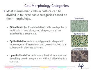
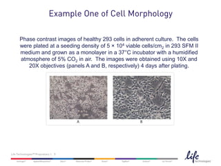


The document discusses the importance of regularly examining cell morphology in culture for successful experiments, highlighting its role in early contamination detection and identifying signs of cell deterioration. It categorizes mammalian cells into fibroblastic, epithelial-like, and lymphoblast-like based on their shape and growth patterns. Additionally, it includes examples and resources for further information on cell culture.