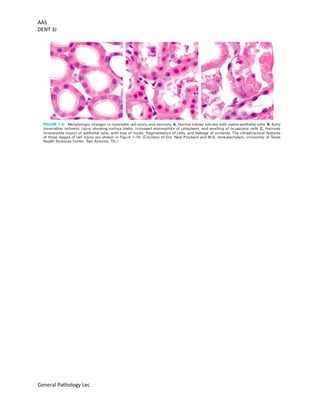
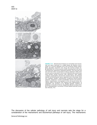
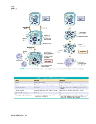
AAS
DENT 3J
General Pathology Lec
What causes cell injury?
Overview of Cell Injury and Cell Death
Cell injury results when cells are stressed so severely that they are no longer able to adapt
or when cells are exposed to inherently damaging agents or suffer from intrinsic abnormalities.
Injury may progress through a reversible stage and culminate in cell death.
Reversible cell injury. In early stages or mild forms of injury, the functional and morphologic
changes are reversible if the damaging stimulus is removed. The hall-marks of reversible
injury are reduced oxidative phosphorylation with resultant depletion of energy stores in
the form of adenosine triphosphate (ATP), and cellular swelling caused by changes in ion
concentrations and water influx. In addition, various intracellular organelles, such
as mitochondria and the cytoskeleton, may also show alterations.
Cell death. With continuing damage the injury becomes irreversible, at which time the cell
cannot recover and it dies. There are two principal types of cell death, necrosis and apoptosis,
which differ in their morphology, mechanisms, and roles in physiology and disease.
When damage to membranes is severe, lysosomal enzymes enter the cytoplasm and digest the
cell, and cellular contents leak out, resulting in necrosis.
In situations when the cell’s DNA or proteins are damaged beyond repair, the cell kills itself
by apoptosis, a form of cell death that is characterized by nuclear dissolution, fragmentation
of the cell without complete loss of membrane integrity, and rapid removal of the cellular
debris.
Whereas necrosis is always a pathologic process, apoptosis serves many normal
functions and is not necessarily associated with cell injury.
Cell death is also sometimes the end result of autophagy. Although it is easier to understand
these pathways of cell death by discussing them separately, there may be many connections
between them.
Both apoptosis and necrosis may be seen in response to the same insult, such as ischemia,
perhaps at different stages. Apoptosis can progress to necrosis, and cell death during
autophagy may show many of the biochemical characteristics of apoptosis.
Causes of Cell Injury
The causes of cell injury range from the external gross physical violence of an
automobile accident to subtle internal abnormalities, such as a genetic mutation causing lack of
a vital enzyme that impairs normal metabolic function. Most injurious stimuli can be grouped into
the following broad categories.
Oxygen Deprivation. Hypoxia is a deficiency of oxygen, which causes cell injury by reducing
aerobic oxidative respiration. Hypoxia is an extremely important and common cause of cell
injury and cell death. Causes of hypoxia include reduced blood flow (called ischemia),
inadequate oxygenation of the blood due to cardiorespiratory failure, and decreased
oxygen- carrying capacity of the blood, as in anemia or carbon monoxide poisoning
(producing a stable carbon monoxyhemoglobin that blocks oxygen carriage) or after
severe blood loss. Depending on the severity of the hypoxic state, cells may adapt, undergo
injury, or die. For example, if an artery is narrowed, the tissue supplied by that vessel may
AAS
DENT 3J
General Pathology Lec
initially shrink in size (atrophy), whereas more severe or sudden hypoxia induces injury and cell
death.
Physical Agents. Physical agents capable of causing cell injury include mechanical trauma,
extremes of temperature (burns and deep cold), sudden changes in atmospheric
pressure, radiation, and electric shock.
Chemical Agents and Drugs. The list of chemicals that may produce cell injury defies
compilation. Simple chemicals such as glucose or salt in hypertonic concentrations may cause
cell injury directly or by deranging electrolyte balance in cells. Even oxygen at high
concentrations is toxic. Trace amounts of poisons, such as arsenic, cyanide, or mercuric
salts, may destroy sufficient numbers of cells within minutes or hours to cause death. Other
potentially injurious substances are our daily companions: environmental and air
pollutants, insecticides, and herbicides; industrial and occupational hazards, such as carbon
monoxide and asbestos; recreational drugs such as alcohol; and the ever-increasing variety
of therapeutic drugs.
Infectious Agents. These agents range from the submicroscopic viruses to the
large tapeworms. In between are the rickettsiae, bacteria, fungi, and higher forms of
parasites.
Immunologic Reactions. The immune system serves an essential function in defense against
infectious pathogens, but immune reactions may also cause cell injury. Injurious reactions to
endogenous self-antigens are responsible for several autoimmune diseases. Immune
reactions to many external agents, such as microbes and environmental substances, are also
important causes of cell and tissue injury.
Genetic Derangements. Genetic abnormalities may result in a defect as severe as the
congenital malformations associated with Down syndrome, caused by a chromosomal
anomaly, or as subtle as the decreased life span of red blood cells caused by a single amino
acid substitution in hemoglobin in sickle cell anemia. Genetic defects may cause cell injury
because of deficiency of functional proteins, such as enzyme defects in inborn errors of
metabolism, or accumulation of damaged DNA or misfolded proteins, both of which trigger cell
death when they are beyond repair. Variations in the genetic makeup can also influence the
susceptibility of cells to injury by chemicals and other environmental insults.
Nutritional Imbalances. Nutritional imbalances continue to be major causes of cell
injury. Protein-calorie deficiencies cause an appalling number of deaths, chiefly among under-
privileged populations. Deficiencies of specific vitamins are found throughout the world.
Nutritional problems can be self-imposed, as in anorexia nervosa (self-induced starvation).
Ironically, nutritional excesses have also become important causes of cell injury. Excess of
cholesterol predisposes to atherosclerosis; obesity is associated with increased incidence of
several important diseases, such as diabetes and cancer. Atherosclerosis is virtually endemic
in the United States, and obesity is rampant. In addition to the problems of undernutrition and
overnutrition, the composition of the diet makes a significant contribution to a number of
diseases.
AAS
DENT 3J
General Pathology Lec
What happens to the cell during injury? How does the cell appear?
Morphologic Alterations in Cell Injury
It is useful to describe the basic alterations that occur in damaged cells before a
discussion on the biochemical mechanisms that bring about these changes will be done. All
stresses and noxious influences exert their effects first at the molecular or biochemical
level. There is a time lag between the stress and the morpho- logic changes of cell injury or
death; the duration of this delay may vary with the sensitivity of the methods used to detect
these changes. With histochemical or ultrastructural techniques, changes may be seen in
minutes to hours after injury; however, it may take considerably longer (hours to days) before
changes can be seen by light microscopy or on gross examination. As would be expected, the
morphologic manifestations of necrosis take more time to develop than those of reversible
damage. For example, in ischemia of the myocardium, cell swelling is a reversible morphologic
change that may occur in a matter of minutes, and may progress to irreversibility within an hour
or two. Unmistakable light microscopic changes of cell death, however, may not be seen until 4
to 12 hours after total ischemia.
Reversible injury is characterized by generalized swelling of the cell and its
organelles; blebbing of the plasma membrane; detachment of ribosomes from the ER;
and clumping of nuclear chromatin. These morphologic changes are associated with
decreased generation of ATP, loss of cell membrane integrity, defects in protein
synthesis, cytoskeletal damage, and DNA damage. Within limits, the cell can repair these
derangements and, if the injurious stimulus abates, will return to normalcy. Persistent or
AAS
DENT 3J
General Pathology Lec
excessive injury, however, causes cells to pass the rather nebulous “point of no return” into
irreversible injury and cell death. Different injurious stimuli may induce death by necrosis or
apoptosis. Severe mitochondrial damage with depletion of ATP and rupture of lysosomal
and plasma membranes are typically associated with necrosis. Necrosis is the principal
outcome in many commonly encountered injuries, such as those following ischemia, exposure
to toxins, various infections, and trauma.
Reversible Injury
Two features of reversible cell injury can be recognized under the light
microscope: cellular swelling and fatty change. Cellular swelling appears whenever cells are
incapable of maintaining ionic and fluid homeostasis and is the result of failure of energy-
dependent ion pumps in the plasma membrane. Fatty change occurs in hypoxic injury and
various forms of toxic or metabolic injury. It is manifested by the appearance of lipid vacuoles in
the cytoplasm. It is seen mainly in cells involved in and dependent on fat metabolism, such as
hepatocytes and myocardial cells.
NECROSIS
The morphologic appearance of necrosis is the result of denaturation of
intracellular proteins and enzymatic digestion of the lethally injured cell (cells placed
immediately in fixative are dead but not necrotic). Necrotic cells are unable to maintain
membrane integrity and their contents often leak out, a process that may elicit inflammation in
the surrounding tissue. The enzymes that digest the necrotic cell are derived from the
lysosomes of the dying cells themselves and from the lysosomes of leukocytes that are called in
as part of the inflammatory reaction. Digestion of cellular contents and the host response may
take hours to develop, and so there would be no detectable changes in cells if, for example, a
myocardial infarct caused sudden death. The only circumstantial evidence might be occlusion of
a coronary artery. The earliest histologic evidence of myocardial necrosis does not become
apparent until 4 to 12 hours later. However, because of the loss of plasma membrane integrity,
cardiac-specific enzymes and proteins are rapidly released from necrotic muscle and can be
detected in the blood as early as 2 hours after myocardial cell necrosis.
Morphology
Cellular swelling is the first manifestation of almost all forms of injury to cells.
It is a difficult morphologic change to appreciate with the light microscope; it may be more
apparent at the level of the whole organ. When it affects many cells, it causes some pallor,
increased turgor, and increase in weight of the organ. On microscopic examination, small clear
vacuoles may be seen within the cytoplasm; these represent distended and pinched-off
segments of the ER. This pattern of nonlethal injury is sometimes called hydropic
change or vacuolar degeneration. Swelling of cells is reversible. Cells may also show
increased eosinophilic staining, which becomes much more pronounced with progression to
necrosis.
The ultrastructural changes of reversible cell injury
1. Plasma membrane alterations, such as blebbing, blunting, and loss of microvilli
2. Mitochondrial changes, including swelling and the appearance of small amorphous
densities
3. Dilation of the ER, with detachment of polysomes; intracytoplasmic myelin figures may be
present
4. Nuclear alterations, with disaggregation of granular and fibrillar elements
AAS
DENT 3J
General Pathology Lec
AAS
DENT 3J
General Pathology Lec
Mechanisms of Cell Injury
The discussion of the cellular pathology of cell injury and necrosis sets the stage for a
consideration of the mechanisms and biochemical pathways of cell injury. The mechanisms
AAS
DENT 3J
General Pathology Lec
responsible for cell injury are complex. There are, however, several principles that are relevant
to most forms of cell injury:
1. The cellular response to injurious stimuli depends on the nature of the injury, its
duration, and its severity. Small doses of a chemical toxin or brief periods of ischemia
may induce reversible injury, whereas large doses of the same toxin or more prolonged
ischemia might result either in instantaneous cell death or in slow, irreversible injury leading
in time to cell death.
2. The consequences of cell injury depend on the type, state, and adaptability of the
injured cell. The cell’s nutritional and hormonal status and its metabolic needs are
important in its response to injury. How vulnerable is a cell, for example, to loss of blood
supply and hypoxia? When the striated muscle cell in the leg is deprived of its blood supply,
it can be placed at rest and preserved; not so the striated muscle of the heart. Exposure of
two individuals to identical concentrations of a toxin, such as carbon tetrachloride, may
produce no effect in one and cell death in the other. This may be due to genetic variations
affecting the amount and activity of hepatic enzymes that convert carbon tetrachloride
(CCl4) to toxic by-products. With the complete mapping of the human genome, there is
great interest in identifying genetic polymorphisms that affect the responses of different
individuals to injurious agents.
3. Cell injury results from different biochemical mechanisms acting on several essential
cellular components. These mechanisms are described individually below. The cellular
components that are most frequently damaged by injurious stimuli include mitochondria, cell
membranes, the machinery of protein synthesis and packaging, and the DNA in nuclei.
4. Any injurious stimulus may simultaneously trigger multiple interconnected
mechanisms that damage cells. This is one reason why it is difficult to ascribe cell injury in
a particular situation to a single or even dominant biochemical derangement.
AAS
DENT 3J
General Pathology Lec
AAS
DENT 3J
General Pathology Lec
APOPTOSIS
Apoptosis is a pathway of cell death that is induced by a tightly regulated suicide
program in which cells destined to die activate enzymes that degrade the cells’ own nuclear
DNA and nuclear and cytoplasmic proteins. Apoptotic cells break up into fragments,
called apoptotic bodies, which contain portions of the cytoplasm and nucleus. The plasma
membrane of the apoptotic cell and bodies remains intact, but its structure is altered in such a
way that these become “tasty” targets for phagocytes. The dead cell and its fragments are
rapidly devoured, before the contents have leaked out, and therefore cell death by this pathway
does not elicit an inflammatory reaction in the host. The process was recognized in 1972 by the
distinctive morphologic appearance of membrane-bound fragments derived from cells, and
named after the Greek designation for “falling off.” It was quickly appreciated that apoptosis was
a unique mechanism of cell death, distinct from necrosis, which is characterized by loss of
membrane integrity, enzymatic digestion of cells, leakage of cellular contents, and
frequently a host reaction. However, apoptosis and necrosis sometimes coexist, and apoptosis
induced by some pathologic stimuli may progress to necrosis.
CAUSES OF APOPTOSIS
Apoptosis occurs normally both during development and throughout adulthood,
and serves to eliminate unwanted, aged or potentially harmful cells. It is also a pathologic
event when diseased cells become damaged beyond repair and are eliminated.
Apoptosis in Physiologic Situations
Death by apoptosis is a normal phenomenon that serves to eliminate cells that are no
longer needed, and to maintain a steady number of various cell populations in tissues. It
is important in the following physiologic situations:
1. The programmed destruction of cells during embryogenesis, including implantation,
organogenesis, developmental involution, and metamorphosis. The term “programmed cell
death” was originally coined to denote death of specific cell types at defined times during the
development of an organism.38 Apoptosis is a generic term for this pattern of cell death,
regardless of the context, but it is often used interchangeably with “programmed cell death.”
2. Involution of hormone-dependent tissues upon hormone withdrawal, such as
endometrial cell breakdown during the menstrual cycle, ovarian follicular atresia in
menopause, the regression of the lactating breast after weaning, and prostatic atrophy after
castration.
3. Cell loss in proliferating cell populations, such as immature lymphocytes in the bone
marrow and thymus that fail to express useful antigen receptors, B lymphocytes in germinal
centers, and epithelial cells in intestinal crypts, so as to maintain a constant
number (homeostasis).
4. Elimination of potentially harmful self-reactive lymphocytes, either before or after they
have completed their maturation, so as to prevent reactions against one’s own tissues.
5. Death of host cells that have served their useful purpose, such as neutrophils in
an acute inflammatory response, and lymphocytes at the end of an immune response. In
these situations cells undergo apoptosis because they are deprived of necessary survival
signals, such as growth factors.
AAS
DENT 3J
General Pathology Lec
Apoptosis in Pathologic Conditions
Apoptosis eliminates cells that are injured beyond repair without eliciting a host
reaction, thus limiting collateral tissue damage. Death by apoptosis is responsible for loss of
cells in a variety of pathologic states:
1. DNA damage. Radiation, cytotoxic anticancer drugs, and hypoxia can damage DNA, either
directly or via production of free radicals. If repair mechanisms cannot cope with the injury,
the cell triggers intrinsic mechanisms that induce apoptosis. In these situations elimination
of the cell may be a better alternative than risking mutations in the damaged DNA, which
may result in malignant transformation. These injurious stimuli can cause apoptosis if the
insult is mild, but larger doses of the same stimuli may result in necrotic cell death.
2. Accumulation of misfolded proteins. Improperly folded proteins may arise because of
mutations in the genes encoding these proteins or because of extrinsic factors, such as
damage caused by free radicals. Excessive accumulation of these proteins in the ER leads
to a condition called ER stress, which culminates in apoptotic cell death. Apoptosis caused
by the accumulation of misfolded proteins has been invoked as the basis of several
degenerative diseases of the central nervous system and other organs.
3. Cell death in certain infections, particularly viral infections, in which loss of infected cells
is largely due to apoptosis that may be induced by the virus (as in adenovirus and HIV
infections) or by the host immune response (as in viral hepatitis). An important host
response to viruses consists of cytotoxic T lymphocytes specific for viral proteins, which
induce apoptosis of infected cells in an attempt to eliminate reservoirs of infection. During
this process there can be significant tissue damage. The same T-cell-mediated mechanism
is responsible for cell death in tumors and cellular rejection of transplants.
4. Pathologic atrophy in parenchymal organs after duct obstruction, such as occurs in
the pancreas, parotid gland, and kidney.
AAS
DENT 3J
General Pathology Lec
Inflammation is a complex reaction in tissues that consists mainly of responses of blood
vessels and leukocytes.
What is inflammatory response? What does it do for the body?
Inflammation is a complex reaction in tissues that consists mainly of
responses of blood vessels and leukocytes. The body’s principal defenders against foreign
invaders are plasma proteins and circulating leukocytes (white blood cells), as well as tissue
phagocytes that are derived from circulating cells. The presence of proteins and leukocytes in
the blood gives them the ability to home to any site where they may be needed. Because
invaders such as microbes and necrotic cells are typically present in tissues, outside the
circulation, it follows that the circulating cells and proteins have to be rapidly recruited to these
extravascular sites. The inflammatory response coordinates the reactions of vessels,
leukocytes, and plasma proteins to achieve this goal.
The vascular and cellular reactions of inflammation are triggered by
soluble factors that are produced by various cells or derived from plasma proteins and
are generated or activated in response to the inflammatory stimulus. Microbes, necrotic
cells (whatever the cause of cell death) and even hypoxia can trigger the elaboration of
AAS
DENT 3J
General Pathology Lec
inflammatory mediators, and thus elicit inflammation. Such mediators initiate and amplify the
inflammatory response and determine its pattern, severity, and clinical and pathologic
manifestations.
Inflammation may be acute or chronic, depending on the nature of the
stimulus and the effectiveness of the initial reaction in eliminating the stimulus or the damaged
tissues. Acute inflammation is rapid in onset (typically minutes) and is of short duration, lasting
for hours or a few days; its main characteristics are the exudation of fluid and plasma proteins
(edema) and the emigration of leukocytes, predominantly neutrophils (also called
polymorphonuclear leukocytes). When acute inflammation is successful in eliminating the
offenders the reaction subsides, but if the response fails to clear the invaders it can progress to
a chronic phase. Chronic inflammation may follow acute inflammation or be insidious in onset.
It is of longer duration and is associated with the presence of lymphocytes and macrophages,
the proliferation of blood vessels, fibrosis, and tissue destruction.
Inflammation is terminated when the offending agent is eliminated. The
reaction resolves rapidly, because the mediators are broken down and dissipated and the
leukocytes have short life spans in tissues. In addition, anti-inflammatory mechanisms are
activated that serve to control the response and prevent it from causing excessive damage to
the host.
The inflammatory response is closely intertwined with the process of
repair. At the same time as inflammation destroys, dilutes, and walls off the injurious agent, it
sets into motion a series of events that try to heal the damaged tissue. Repair begins during
inflammation but reaches completion usually after the injurious influence has been neutralized.
In the process of repair the injured tissue is replaced through regeneration of native
parenchymal cells, by filling of the defect with fibrous tissue (scarring) or, most commonly, by a
combination of these two processes.
Inflammation may be harmful in some situations. Mechanisms designed to
destroy foreign invaders and necrotic tissues have an intrinsic ability to injure normal tissues.
When inflammation is inappropriately directed against self-tissues or is not adequately
controlled, it becomes the cause of injury and disease. In fact, in clinical medicine, great
attention is given to the damaging consequences of inflammation. Inflammatory reactions
underlie common chronic diseases, such as rheumatoid arthritis, atherosclerosis, and lung
fibrosis, as well as life- threatening hypersensitivity reactions to insect bites, drugs, and toxins.
For this reason our pharmacies abound with anti- inflammatory drugs, which ideally would
control the harmful sequelae of inflammation yet not interfere with its beneficial effects.
Inflammation may contribute to a variety of diseases that are not thought to be
primarily due to abnormal host responses. For instance, chronic inflammation may play a
role in atherosclerosis, type 2 diabetes, degenerative disorders like Alzheimer disease, and
cancer. In recognition of the wide-ranging harmful consequences of inflammation, the lay press
has rather melodramatically referred to it as “the silent killer.”
When does acute inflammation occur?
Acute Inflammation
Acute inflammation is a rapid host response that serves to deliver leukocytes and
plasma proteins, such as antibodies, to sites of infection or tissue injury. Acute inflammation has
three major components: (1) alterations in vascular caliber that lead to an increase in blood
flow, (2) structural changes in the microvasculature that permit plasma proteins and
AAS
DENT 3J
General Pathology Lec
leukocytes to leave the circulation, and (3) emigration of the leukocytes from the
microcirculation, their accumulation in the focus of injury, and their activation to
eliminate the offending agent.
STIMULI FOR ACUTE INFLAMMATION
Acute inflammatory reactions may be triggered by a variety of stimuli:
1. Infections (bacterial, viral, fungal, parasitic) and microbial toxins are among the most
common and medically important causes of inflammation. Mammals possess many
mechanisms for sensing the presence of microbes. Among the most important receptors for
microbial products are the family of Toll-like receptors (TLRs), named after
the Drosophila protein Toll, and several cytoplasmic receptors, which can detect bacteria,
viruses, and fungi. Engagement of these receptors triggers signaling pathways that stimulate
the production of various mediators.
AAS
DENT 3J
General Pathology Lec
2. Tissue necrosis from any cause, including ischemia (as in a myocardial infarct), trauma,
and physical and chemical injury (e.g., thermal injury, as in burns or frostbite; irradiation;
exposure to some environmental chemicals). Several molecules released from necrotic cells are
known to elicit inflammation; these include uric acid, a purine metabolite; adenosine
triphosphate, the normal energy store; a DNA-binding protein of unknown function called
HMGB-1; and even DNA when it is released into the cytoplasm and not sequestered in nuclei,
as it should be normally. Hypoxia, which often underlies cell injury, is also itself an inducer of
the inflammatory response. This response is mediated largely by a protein called HIF-1α
(hypoxia-induced factor-1α), which is produced by cells deprived of oxygen and activates the
transcription of many genes involved in inflammation, including vascular endothelial growth
factor (VEGF), which increases vascular permeability.
3. Foreign bodies (splinters, dirt, sutures) typically elicit inflammation because they cause
traumatic tissue injury or carry microbes.
4. Immune reactions (also called hypersensitivity reactions) are reactions in which the normally
protective immune system damages the individual’s own tissues. The injurious immune
responses may be directed against self-antigens, causing autoimmune diseases, or may be
excessive reactions against environmental substances or microbes. Inflammation is a major
cause of tissue injury in these diseases. Because the stimuli for the inflammatory responses
(i.e., self-tissues) cannot be eliminated, autoimmune reactions tend to be persistent and difficult
to cure, are associated with chronic inflammation, and are important causes of morbidity and
mortality. The inflammation is induced by cytokines produced by T lymphocytes and other cells
of the immune system. The term immune-mediated inflammatory disease is often used to
refer to this group of disorders.
All inflammatory reactions share the same basic features, although different stimuli
may induce reactions with some distinctive characteristics.
n does chronic inflammation occur?
Chronic inflammation is inflammation of prolonged duration (weeks or
months) in which inflammation, tissue injury, and attempts at repair coexist, in varying
combinations. It may follow acute inflammation, as described earlier, or chronic inflammation
may begin insidiously, as a low-grade, smoldering response without any manifestations of an
acute reaction. This latter type of chronic inflammation is the cause of tissue damage in some of
the most common and disabling human diseases, such as rheumatoid arthritis, atherosclerosis,
tuberculosis, and pulmonary fibrosis. It has also been implicated in the progression of cancer
and in diseases once thought to be purely degenerative, such as Alzheimer disease.
CAUSES OF CHRONIC INFLAMMATION
Chronic inflammation arises in the following settings:
1. Persistent infections by microorganisms that are difficult to eradicate, such as
mycobacteria, and certain viruses, fungi, and parasites. These organisms often evoke an
immune reaction called delayed-type hypersensitivity . The inflammatory response
sometimes takes a specific pattern called a granulomatous reaction.
2. Immune-mediated inflammatory diseases. Chronic inflammation plays an important role
in a group of diseases that are caused by excessive and inappropriate activation of the
immune system. Under certain conditions immune reactions develop against the individual’s
own tissues, leading to autoimmune diseases. In these diseases, auto- antigens evoke a
self-perpetuating immune reaction that results in chronic tissue damage and inflammation;
AAS
DENT 3J
General Pathology Lec
examples of such diseases are rheumatoid arthritis and multiple sclerosis. In other cases,
chronic inflammation is the result of unregulated immune responses against microbes, as in
inflammatory bowel disease. Immune responses against common environmental
substances are the cause of allergic diseases, such as bronchial asthma. Because these
autoimmune and allergic reactions are inappropriately triggered against antigens that are
normally harmless, the reactions serve no useful purpose and only cause disease. Such
diseases may show morphologic patterns of mixed acute and chronic inflammation because
they are characterized by repeated bouts of inflammation. Fibrosis may dominate the late
stages.
3. Prolonged exposure to potentially toxic agents, either exogenous or endogenous. An example of an
exogenous agent is particulate silica, a nondegradable inanimate material that, when inhaled for
prolonged periods, results in an inflammatory lung disease called Atherosclerosis is thought to be a
chronic inflammatory process of the arterial wall induced, at least in part, by endogenous toxic
plasma lipid components.
How do tissues heal after injury?
Injury to cells and tissues sets in motion a series of events that contain the
damage and initiate the healing process. Regeneration results in the complete restitution of
lost or damaged tissue; repair may restore some original structures but can cause structural
derangements. In healthy tissues, healing, in the form of regeneration or repair, occurs after
practically any insult that causes tissue destruction, and is essential for the survival of the
organism.
Regeneration refers to the proliferation of cells and tissues to replace lost
structures, such as the growth of an amputated limb in amphibians. In mammals, whole organs
and complex tissues rarely regenerate after injury, and the term is usually applied to processes
such as liver growth after partial resection or necrosis, but these processes consist of
compensatory growth rather than true regeneration. Regardless, the term regeneration is well
established and is used throughout this book. Tissues with high proliferative capacity, such as
the hematopoietic system and the epithelia of the skin and gastrointestinal (GI) tract, renew
themselves continuously and can regenerate after injury, as long as the stem cells of these
tissues are not destroyed.
Repair most often consists of a combination of regeneration and scar
formation by the deposition of collagen. The relative contribution of regeneration and scarring
in tissue repair depends on the ability of the tissue to regenerate and the extent of the injury. For
instance, a superficial skin wound heals through the regeneration of the surface epithelium.
However, as discussed later, scar formation is the predominant healing process that occurs
when the extracellular matrix (ECM) framework is damaged by severe injury. Chronic
inflammation that accompanies persistent injury also stimulates scar formation because of local
production of growth factors and cytokines that promote fibroblast proliferation and collagen
synthesis. The term fibrosis is used to describe the extensive deposition of collagen that occurs
under these situations. ECM components are essential for wound healing, because they provide
the framework for cell migration, maintain the correct cell polarity for the re-assembly of
multilayer structures, and participate in the formation of new blood vessels (angiogenesis).
Furthermore, cells in the ECM (fibroblasts, macrophages, and other cell types) produce growth
factors, cytokines, and chemokines that are critical for regeneration and repair. Although repair
AAS
DENT 3J
General Pathology Lec
is a healing process, it may itself cause tissue dysfunction, as, for instance, in the development
of atherosclerosis.
Understanding the mechanisms of regeneration and repair requires some knowledge
of the control of cell proliferation and signal transduction pathways, and the many functions of
ECM components.
Healing by Repair, Scar Formation and Fibrosis
If tissue injury is severe or chronic, and results in damage of both parenchymal cells and
the stromal framework of the tissue, healing cannot be accomplished by regeneration.
Under these conditions, the main healing process is repair by deposition of collagen
and other ECM components, causing the formation of a scar. In contrast to regeneration
which involves the restitution of tissue components, repair is a fibroproliferative response that
“patches” rather than restores the tissue. The term scar is most often used in connection
to wound healing in the skin, but is also used to describe the replacement of parenchymal cells
in any tissue by collagen, as in the heart after myocardial infarction. Repair by connective tissue
deposition includes the following basic features:
1. Inflammation
2. angiogenesis
3. migration and proliferation of fibroblasts, scar formation
4. connective tissue remodeling

General Pathology Lecture Introduction from Module 1 to Module 3

  • 1.
    AAS DENT 3J General PathologyLec What causes cell injury? Overview of Cell Injury and Cell Death Cell injury results when cells are stressed so severely that they are no longer able to adapt or when cells are exposed to inherently damaging agents or suffer from intrinsic abnormalities. Injury may progress through a reversible stage and culminate in cell death. Reversible cell injury. In early stages or mild forms of injury, the functional and morphologic changes are reversible if the damaging stimulus is removed. The hall-marks of reversible injury are reduced oxidative phosphorylation with resultant depletion of energy stores in the form of adenosine triphosphate (ATP), and cellular swelling caused by changes in ion concentrations and water influx. In addition, various intracellular organelles, such as mitochondria and the cytoskeleton, may also show alterations. Cell death. With continuing damage the injury becomes irreversible, at which time the cell cannot recover and it dies. There are two principal types of cell death, necrosis and apoptosis, which differ in their morphology, mechanisms, and roles in physiology and disease. When damage to membranes is severe, lysosomal enzymes enter the cytoplasm and digest the cell, and cellular contents leak out, resulting in necrosis. In situations when the cell’s DNA or proteins are damaged beyond repair, the cell kills itself by apoptosis, a form of cell death that is characterized by nuclear dissolution, fragmentation of the cell without complete loss of membrane integrity, and rapid removal of the cellular debris. Whereas necrosis is always a pathologic process, apoptosis serves many normal functions and is not necessarily associated with cell injury. Cell death is also sometimes the end result of autophagy. Although it is easier to understand these pathways of cell death by discussing them separately, there may be many connections between them. Both apoptosis and necrosis may be seen in response to the same insult, such as ischemia, perhaps at different stages. Apoptosis can progress to necrosis, and cell death during autophagy may show many of the biochemical characteristics of apoptosis. Causes of Cell Injury The causes of cell injury range from the external gross physical violence of an automobile accident to subtle internal abnormalities, such as a genetic mutation causing lack of a vital enzyme that impairs normal metabolic function. Most injurious stimuli can be grouped into the following broad categories. Oxygen Deprivation. Hypoxia is a deficiency of oxygen, which causes cell injury by reducing aerobic oxidative respiration. Hypoxia is an extremely important and common cause of cell injury and cell death. Causes of hypoxia include reduced blood flow (called ischemia), inadequate oxygenation of the blood due to cardiorespiratory failure, and decreased oxygen- carrying capacity of the blood, as in anemia or carbon monoxide poisoning (producing a stable carbon monoxyhemoglobin that blocks oxygen carriage) or after severe blood loss. Depending on the severity of the hypoxic state, cells may adapt, undergo injury, or die. For example, if an artery is narrowed, the tissue supplied by that vessel may
  • 2.
    AAS DENT 3J General PathologyLec initially shrink in size (atrophy), whereas more severe or sudden hypoxia induces injury and cell death. Physical Agents. Physical agents capable of causing cell injury include mechanical trauma, extremes of temperature (burns and deep cold), sudden changes in atmospheric pressure, radiation, and electric shock. Chemical Agents and Drugs. The list of chemicals that may produce cell injury defies compilation. Simple chemicals such as glucose or salt in hypertonic concentrations may cause cell injury directly or by deranging electrolyte balance in cells. Even oxygen at high concentrations is toxic. Trace amounts of poisons, such as arsenic, cyanide, or mercuric salts, may destroy sufficient numbers of cells within minutes or hours to cause death. Other potentially injurious substances are our daily companions: environmental and air pollutants, insecticides, and herbicides; industrial and occupational hazards, such as carbon monoxide and asbestos; recreational drugs such as alcohol; and the ever-increasing variety of therapeutic drugs. Infectious Agents. These agents range from the submicroscopic viruses to the large tapeworms. In between are the rickettsiae, bacteria, fungi, and higher forms of parasites. Immunologic Reactions. The immune system serves an essential function in defense against infectious pathogens, but immune reactions may also cause cell injury. Injurious reactions to endogenous self-antigens are responsible for several autoimmune diseases. Immune reactions to many external agents, such as microbes and environmental substances, are also important causes of cell and tissue injury. Genetic Derangements. Genetic abnormalities may result in a defect as severe as the congenital malformations associated with Down syndrome, caused by a chromosomal anomaly, or as subtle as the decreased life span of red blood cells caused by a single amino acid substitution in hemoglobin in sickle cell anemia. Genetic defects may cause cell injury because of deficiency of functional proteins, such as enzyme defects in inborn errors of metabolism, or accumulation of damaged DNA or misfolded proteins, both of which trigger cell death when they are beyond repair. Variations in the genetic makeup can also influence the susceptibility of cells to injury by chemicals and other environmental insults. Nutritional Imbalances. Nutritional imbalances continue to be major causes of cell injury. Protein-calorie deficiencies cause an appalling number of deaths, chiefly among under- privileged populations. Deficiencies of specific vitamins are found throughout the world. Nutritional problems can be self-imposed, as in anorexia nervosa (self-induced starvation). Ironically, nutritional excesses have also become important causes of cell injury. Excess of cholesterol predisposes to atherosclerosis; obesity is associated with increased incidence of several important diseases, such as diabetes and cancer. Atherosclerosis is virtually endemic in the United States, and obesity is rampant. In addition to the problems of undernutrition and overnutrition, the composition of the diet makes a significant contribution to a number of diseases.
  • 3.
    AAS DENT 3J General PathologyLec What happens to the cell during injury? How does the cell appear? Morphologic Alterations in Cell Injury It is useful to describe the basic alterations that occur in damaged cells before a discussion on the biochemical mechanisms that bring about these changes will be done. All stresses and noxious influences exert their effects first at the molecular or biochemical level. There is a time lag between the stress and the morpho- logic changes of cell injury or death; the duration of this delay may vary with the sensitivity of the methods used to detect these changes. With histochemical or ultrastructural techniques, changes may be seen in minutes to hours after injury; however, it may take considerably longer (hours to days) before changes can be seen by light microscopy or on gross examination. As would be expected, the morphologic manifestations of necrosis take more time to develop than those of reversible damage. For example, in ischemia of the myocardium, cell swelling is a reversible morphologic change that may occur in a matter of minutes, and may progress to irreversibility within an hour or two. Unmistakable light microscopic changes of cell death, however, may not be seen until 4 to 12 hours after total ischemia. Reversible injury is characterized by generalized swelling of the cell and its organelles; blebbing of the plasma membrane; detachment of ribosomes from the ER; and clumping of nuclear chromatin. These morphologic changes are associated with decreased generation of ATP, loss of cell membrane integrity, defects in protein synthesis, cytoskeletal damage, and DNA damage. Within limits, the cell can repair these derangements and, if the injurious stimulus abates, will return to normalcy. Persistent or
  • 4.
    AAS DENT 3J General PathologyLec excessive injury, however, causes cells to pass the rather nebulous “point of no return” into irreversible injury and cell death. Different injurious stimuli may induce death by necrosis or apoptosis. Severe mitochondrial damage with depletion of ATP and rupture of lysosomal and plasma membranes are typically associated with necrosis. Necrosis is the principal outcome in many commonly encountered injuries, such as those following ischemia, exposure to toxins, various infections, and trauma. Reversible Injury Two features of reversible cell injury can be recognized under the light microscope: cellular swelling and fatty change. Cellular swelling appears whenever cells are incapable of maintaining ionic and fluid homeostasis and is the result of failure of energy- dependent ion pumps in the plasma membrane. Fatty change occurs in hypoxic injury and various forms of toxic or metabolic injury. It is manifested by the appearance of lipid vacuoles in the cytoplasm. It is seen mainly in cells involved in and dependent on fat metabolism, such as hepatocytes and myocardial cells. NECROSIS The morphologic appearance of necrosis is the result of denaturation of intracellular proteins and enzymatic digestion of the lethally injured cell (cells placed immediately in fixative are dead but not necrotic). Necrotic cells are unable to maintain membrane integrity and their contents often leak out, a process that may elicit inflammation in the surrounding tissue. The enzymes that digest the necrotic cell are derived from the lysosomes of the dying cells themselves and from the lysosomes of leukocytes that are called in as part of the inflammatory reaction. Digestion of cellular contents and the host response may take hours to develop, and so there would be no detectable changes in cells if, for example, a myocardial infarct caused sudden death. The only circumstantial evidence might be occlusion of a coronary artery. The earliest histologic evidence of myocardial necrosis does not become apparent until 4 to 12 hours later. However, because of the loss of plasma membrane integrity, cardiac-specific enzymes and proteins are rapidly released from necrotic muscle and can be detected in the blood as early as 2 hours after myocardial cell necrosis. Morphology Cellular swelling is the first manifestation of almost all forms of injury to cells. It is a difficult morphologic change to appreciate with the light microscope; it may be more apparent at the level of the whole organ. When it affects many cells, it causes some pallor, increased turgor, and increase in weight of the organ. On microscopic examination, small clear vacuoles may be seen within the cytoplasm; these represent distended and pinched-off segments of the ER. This pattern of nonlethal injury is sometimes called hydropic change or vacuolar degeneration. Swelling of cells is reversible. Cells may also show increased eosinophilic staining, which becomes much more pronounced with progression to necrosis. The ultrastructural changes of reversible cell injury 1. Plasma membrane alterations, such as blebbing, blunting, and loss of microvilli 2. Mitochondrial changes, including swelling and the appearance of small amorphous densities 3. Dilation of the ER, with detachment of polysomes; intracytoplasmic myelin figures may be present 4. Nuclear alterations, with disaggregation of granular and fibrillar elements
  • 5.
  • 6.
    AAS DENT 3J General PathologyLec Mechanisms of Cell Injury The discussion of the cellular pathology of cell injury and necrosis sets the stage for a consideration of the mechanisms and biochemical pathways of cell injury. The mechanisms
  • 7.
    AAS DENT 3J General PathologyLec responsible for cell injury are complex. There are, however, several principles that are relevant to most forms of cell injury: 1. The cellular response to injurious stimuli depends on the nature of the injury, its duration, and its severity. Small doses of a chemical toxin or brief periods of ischemia may induce reversible injury, whereas large doses of the same toxin or more prolonged ischemia might result either in instantaneous cell death or in slow, irreversible injury leading in time to cell death. 2. The consequences of cell injury depend on the type, state, and adaptability of the injured cell. The cell’s nutritional and hormonal status and its metabolic needs are important in its response to injury. How vulnerable is a cell, for example, to loss of blood supply and hypoxia? When the striated muscle cell in the leg is deprived of its blood supply, it can be placed at rest and preserved; not so the striated muscle of the heart. Exposure of two individuals to identical concentrations of a toxin, such as carbon tetrachloride, may produce no effect in one and cell death in the other. This may be due to genetic variations affecting the amount and activity of hepatic enzymes that convert carbon tetrachloride (CCl4) to toxic by-products. With the complete mapping of the human genome, there is great interest in identifying genetic polymorphisms that affect the responses of different individuals to injurious agents. 3. Cell injury results from different biochemical mechanisms acting on several essential cellular components. These mechanisms are described individually below. The cellular components that are most frequently damaged by injurious stimuli include mitochondria, cell membranes, the machinery of protein synthesis and packaging, and the DNA in nuclei. 4. Any injurious stimulus may simultaneously trigger multiple interconnected mechanisms that damage cells. This is one reason why it is difficult to ascribe cell injury in a particular situation to a single or even dominant biochemical derangement.
  • 8.
  • 9.
    AAS DENT 3J General PathologyLec APOPTOSIS Apoptosis is a pathway of cell death that is induced by a tightly regulated suicide program in which cells destined to die activate enzymes that degrade the cells’ own nuclear DNA and nuclear and cytoplasmic proteins. Apoptotic cells break up into fragments, called apoptotic bodies, which contain portions of the cytoplasm and nucleus. The plasma membrane of the apoptotic cell and bodies remains intact, but its structure is altered in such a way that these become “tasty” targets for phagocytes. The dead cell and its fragments are rapidly devoured, before the contents have leaked out, and therefore cell death by this pathway does not elicit an inflammatory reaction in the host. The process was recognized in 1972 by the distinctive morphologic appearance of membrane-bound fragments derived from cells, and named after the Greek designation for “falling off.” It was quickly appreciated that apoptosis was a unique mechanism of cell death, distinct from necrosis, which is characterized by loss of membrane integrity, enzymatic digestion of cells, leakage of cellular contents, and frequently a host reaction. However, apoptosis and necrosis sometimes coexist, and apoptosis induced by some pathologic stimuli may progress to necrosis. CAUSES OF APOPTOSIS Apoptosis occurs normally both during development and throughout adulthood, and serves to eliminate unwanted, aged or potentially harmful cells. It is also a pathologic event when diseased cells become damaged beyond repair and are eliminated. Apoptosis in Physiologic Situations Death by apoptosis is a normal phenomenon that serves to eliminate cells that are no longer needed, and to maintain a steady number of various cell populations in tissues. It is important in the following physiologic situations: 1. The programmed destruction of cells during embryogenesis, including implantation, organogenesis, developmental involution, and metamorphosis. The term “programmed cell death” was originally coined to denote death of specific cell types at defined times during the development of an organism.38 Apoptosis is a generic term for this pattern of cell death, regardless of the context, but it is often used interchangeably with “programmed cell death.” 2. Involution of hormone-dependent tissues upon hormone withdrawal, such as endometrial cell breakdown during the menstrual cycle, ovarian follicular atresia in menopause, the regression of the lactating breast after weaning, and prostatic atrophy after castration. 3. Cell loss in proliferating cell populations, such as immature lymphocytes in the bone marrow and thymus that fail to express useful antigen receptors, B lymphocytes in germinal centers, and epithelial cells in intestinal crypts, so as to maintain a constant number (homeostasis). 4. Elimination of potentially harmful self-reactive lymphocytes, either before or after they have completed their maturation, so as to prevent reactions against one’s own tissues. 5. Death of host cells that have served their useful purpose, such as neutrophils in an acute inflammatory response, and lymphocytes at the end of an immune response. In these situations cells undergo apoptosis because they are deprived of necessary survival signals, such as growth factors.
  • 10.
    AAS DENT 3J General PathologyLec Apoptosis in Pathologic Conditions Apoptosis eliminates cells that are injured beyond repair without eliciting a host reaction, thus limiting collateral tissue damage. Death by apoptosis is responsible for loss of cells in a variety of pathologic states: 1. DNA damage. Radiation, cytotoxic anticancer drugs, and hypoxia can damage DNA, either directly or via production of free radicals. If repair mechanisms cannot cope with the injury, the cell triggers intrinsic mechanisms that induce apoptosis. In these situations elimination of the cell may be a better alternative than risking mutations in the damaged DNA, which may result in malignant transformation. These injurious stimuli can cause apoptosis if the insult is mild, but larger doses of the same stimuli may result in necrotic cell death. 2. Accumulation of misfolded proteins. Improperly folded proteins may arise because of mutations in the genes encoding these proteins or because of extrinsic factors, such as damage caused by free radicals. Excessive accumulation of these proteins in the ER leads to a condition called ER stress, which culminates in apoptotic cell death. Apoptosis caused by the accumulation of misfolded proteins has been invoked as the basis of several degenerative diseases of the central nervous system and other organs. 3. Cell death in certain infections, particularly viral infections, in which loss of infected cells is largely due to apoptosis that may be induced by the virus (as in adenovirus and HIV infections) or by the host immune response (as in viral hepatitis). An important host response to viruses consists of cytotoxic T lymphocytes specific for viral proteins, which induce apoptosis of infected cells in an attempt to eliminate reservoirs of infection. During this process there can be significant tissue damage. The same T-cell-mediated mechanism is responsible for cell death in tumors and cellular rejection of transplants. 4. Pathologic atrophy in parenchymal organs after duct obstruction, such as occurs in the pancreas, parotid gland, and kidney.
  • 11.
    AAS DENT 3J General PathologyLec Inflammation is a complex reaction in tissues that consists mainly of responses of blood vessels and leukocytes. What is inflammatory response? What does it do for the body? Inflammation is a complex reaction in tissues that consists mainly of responses of blood vessels and leukocytes. The body’s principal defenders against foreign invaders are plasma proteins and circulating leukocytes (white blood cells), as well as tissue phagocytes that are derived from circulating cells. The presence of proteins and leukocytes in the blood gives them the ability to home to any site where they may be needed. Because invaders such as microbes and necrotic cells are typically present in tissues, outside the circulation, it follows that the circulating cells and proteins have to be rapidly recruited to these extravascular sites. The inflammatory response coordinates the reactions of vessels, leukocytes, and plasma proteins to achieve this goal. The vascular and cellular reactions of inflammation are triggered by soluble factors that are produced by various cells or derived from plasma proteins and are generated or activated in response to the inflammatory stimulus. Microbes, necrotic cells (whatever the cause of cell death) and even hypoxia can trigger the elaboration of
  • 12.
    AAS DENT 3J General PathologyLec inflammatory mediators, and thus elicit inflammation. Such mediators initiate and amplify the inflammatory response and determine its pattern, severity, and clinical and pathologic manifestations. Inflammation may be acute or chronic, depending on the nature of the stimulus and the effectiveness of the initial reaction in eliminating the stimulus or the damaged tissues. Acute inflammation is rapid in onset (typically minutes) and is of short duration, lasting for hours or a few days; its main characteristics are the exudation of fluid and plasma proteins (edema) and the emigration of leukocytes, predominantly neutrophils (also called polymorphonuclear leukocytes). When acute inflammation is successful in eliminating the offenders the reaction subsides, but if the response fails to clear the invaders it can progress to a chronic phase. Chronic inflammation may follow acute inflammation or be insidious in onset. It is of longer duration and is associated with the presence of lymphocytes and macrophages, the proliferation of blood vessels, fibrosis, and tissue destruction. Inflammation is terminated when the offending agent is eliminated. The reaction resolves rapidly, because the mediators are broken down and dissipated and the leukocytes have short life spans in tissues. In addition, anti-inflammatory mechanisms are activated that serve to control the response and prevent it from causing excessive damage to the host. The inflammatory response is closely intertwined with the process of repair. At the same time as inflammation destroys, dilutes, and walls off the injurious agent, it sets into motion a series of events that try to heal the damaged tissue. Repair begins during inflammation but reaches completion usually after the injurious influence has been neutralized. In the process of repair the injured tissue is replaced through regeneration of native parenchymal cells, by filling of the defect with fibrous tissue (scarring) or, most commonly, by a combination of these two processes. Inflammation may be harmful in some situations. Mechanisms designed to destroy foreign invaders and necrotic tissues have an intrinsic ability to injure normal tissues. When inflammation is inappropriately directed against self-tissues or is not adequately controlled, it becomes the cause of injury and disease. In fact, in clinical medicine, great attention is given to the damaging consequences of inflammation. Inflammatory reactions underlie common chronic diseases, such as rheumatoid arthritis, atherosclerosis, and lung fibrosis, as well as life- threatening hypersensitivity reactions to insect bites, drugs, and toxins. For this reason our pharmacies abound with anti- inflammatory drugs, which ideally would control the harmful sequelae of inflammation yet not interfere with its beneficial effects. Inflammation may contribute to a variety of diseases that are not thought to be primarily due to abnormal host responses. For instance, chronic inflammation may play a role in atherosclerosis, type 2 diabetes, degenerative disorders like Alzheimer disease, and cancer. In recognition of the wide-ranging harmful consequences of inflammation, the lay press has rather melodramatically referred to it as “the silent killer.” When does acute inflammation occur? Acute Inflammation Acute inflammation is a rapid host response that serves to deliver leukocytes and plasma proteins, such as antibodies, to sites of infection or tissue injury. Acute inflammation has three major components: (1) alterations in vascular caliber that lead to an increase in blood flow, (2) structural changes in the microvasculature that permit plasma proteins and
  • 13.
    AAS DENT 3J General PathologyLec leukocytes to leave the circulation, and (3) emigration of the leukocytes from the microcirculation, their accumulation in the focus of injury, and their activation to eliminate the offending agent. STIMULI FOR ACUTE INFLAMMATION Acute inflammatory reactions may be triggered by a variety of stimuli: 1. Infections (bacterial, viral, fungal, parasitic) and microbial toxins are among the most common and medically important causes of inflammation. Mammals possess many mechanisms for sensing the presence of microbes. Among the most important receptors for microbial products are the family of Toll-like receptors (TLRs), named after the Drosophila protein Toll, and several cytoplasmic receptors, which can detect bacteria, viruses, and fungi. Engagement of these receptors triggers signaling pathways that stimulate the production of various mediators.
  • 14.
    AAS DENT 3J General PathologyLec 2. Tissue necrosis from any cause, including ischemia (as in a myocardial infarct), trauma, and physical and chemical injury (e.g., thermal injury, as in burns or frostbite; irradiation; exposure to some environmental chemicals). Several molecules released from necrotic cells are known to elicit inflammation; these include uric acid, a purine metabolite; adenosine triphosphate, the normal energy store; a DNA-binding protein of unknown function called HMGB-1; and even DNA when it is released into the cytoplasm and not sequestered in nuclei, as it should be normally. Hypoxia, which often underlies cell injury, is also itself an inducer of the inflammatory response. This response is mediated largely by a protein called HIF-1α (hypoxia-induced factor-1α), which is produced by cells deprived of oxygen and activates the transcription of many genes involved in inflammation, including vascular endothelial growth factor (VEGF), which increases vascular permeability. 3. Foreign bodies (splinters, dirt, sutures) typically elicit inflammation because they cause traumatic tissue injury or carry microbes. 4. Immune reactions (also called hypersensitivity reactions) are reactions in which the normally protective immune system damages the individual’s own tissues. The injurious immune responses may be directed against self-antigens, causing autoimmune diseases, or may be excessive reactions against environmental substances or microbes. Inflammation is a major cause of tissue injury in these diseases. Because the stimuli for the inflammatory responses (i.e., self-tissues) cannot be eliminated, autoimmune reactions tend to be persistent and difficult to cure, are associated with chronic inflammation, and are important causes of morbidity and mortality. The inflammation is induced by cytokines produced by T lymphocytes and other cells of the immune system. The term immune-mediated inflammatory disease is often used to refer to this group of disorders. All inflammatory reactions share the same basic features, although different stimuli may induce reactions with some distinctive characteristics. n does chronic inflammation occur? Chronic inflammation is inflammation of prolonged duration (weeks or months) in which inflammation, tissue injury, and attempts at repair coexist, in varying combinations. It may follow acute inflammation, as described earlier, or chronic inflammation may begin insidiously, as a low-grade, smoldering response without any manifestations of an acute reaction. This latter type of chronic inflammation is the cause of tissue damage in some of the most common and disabling human diseases, such as rheumatoid arthritis, atherosclerosis, tuberculosis, and pulmonary fibrosis. It has also been implicated in the progression of cancer and in diseases once thought to be purely degenerative, such as Alzheimer disease. CAUSES OF CHRONIC INFLAMMATION Chronic inflammation arises in the following settings: 1. Persistent infections by microorganisms that are difficult to eradicate, such as mycobacteria, and certain viruses, fungi, and parasites. These organisms often evoke an immune reaction called delayed-type hypersensitivity . The inflammatory response sometimes takes a specific pattern called a granulomatous reaction. 2. Immune-mediated inflammatory diseases. Chronic inflammation plays an important role in a group of diseases that are caused by excessive and inappropriate activation of the immune system. Under certain conditions immune reactions develop against the individual’s own tissues, leading to autoimmune diseases. In these diseases, auto- antigens evoke a self-perpetuating immune reaction that results in chronic tissue damage and inflammation;
  • 15.
    AAS DENT 3J General PathologyLec examples of such diseases are rheumatoid arthritis and multiple sclerosis. In other cases, chronic inflammation is the result of unregulated immune responses against microbes, as in inflammatory bowel disease. Immune responses against common environmental substances are the cause of allergic diseases, such as bronchial asthma. Because these autoimmune and allergic reactions are inappropriately triggered against antigens that are normally harmless, the reactions serve no useful purpose and only cause disease. Such diseases may show morphologic patterns of mixed acute and chronic inflammation because they are characterized by repeated bouts of inflammation. Fibrosis may dominate the late stages. 3. Prolonged exposure to potentially toxic agents, either exogenous or endogenous. An example of an exogenous agent is particulate silica, a nondegradable inanimate material that, when inhaled for prolonged periods, results in an inflammatory lung disease called Atherosclerosis is thought to be a chronic inflammatory process of the arterial wall induced, at least in part, by endogenous toxic plasma lipid components. How do tissues heal after injury? Injury to cells and tissues sets in motion a series of events that contain the damage and initiate the healing process. Regeneration results in the complete restitution of lost or damaged tissue; repair may restore some original structures but can cause structural derangements. In healthy tissues, healing, in the form of regeneration or repair, occurs after practically any insult that causes tissue destruction, and is essential for the survival of the organism. Regeneration refers to the proliferation of cells and tissues to replace lost structures, such as the growth of an amputated limb in amphibians. In mammals, whole organs and complex tissues rarely regenerate after injury, and the term is usually applied to processes such as liver growth after partial resection or necrosis, but these processes consist of compensatory growth rather than true regeneration. Regardless, the term regeneration is well established and is used throughout this book. Tissues with high proliferative capacity, such as the hematopoietic system and the epithelia of the skin and gastrointestinal (GI) tract, renew themselves continuously and can regenerate after injury, as long as the stem cells of these tissues are not destroyed. Repair most often consists of a combination of regeneration and scar formation by the deposition of collagen. The relative contribution of regeneration and scarring in tissue repair depends on the ability of the tissue to regenerate and the extent of the injury. For instance, a superficial skin wound heals through the regeneration of the surface epithelium. However, as discussed later, scar formation is the predominant healing process that occurs when the extracellular matrix (ECM) framework is damaged by severe injury. Chronic inflammation that accompanies persistent injury also stimulates scar formation because of local production of growth factors and cytokines that promote fibroblast proliferation and collagen synthesis. The term fibrosis is used to describe the extensive deposition of collagen that occurs under these situations. ECM components are essential for wound healing, because they provide the framework for cell migration, maintain the correct cell polarity for the re-assembly of multilayer structures, and participate in the formation of new blood vessels (angiogenesis). Furthermore, cells in the ECM (fibroblasts, macrophages, and other cell types) produce growth factors, cytokines, and chemokines that are critical for regeneration and repair. Although repair
  • 16.
    AAS DENT 3J General PathologyLec is a healing process, it may itself cause tissue dysfunction, as, for instance, in the development of atherosclerosis. Understanding the mechanisms of regeneration and repair requires some knowledge of the control of cell proliferation and signal transduction pathways, and the many functions of ECM components. Healing by Repair, Scar Formation and Fibrosis If tissue injury is severe or chronic, and results in damage of both parenchymal cells and the stromal framework of the tissue, healing cannot be accomplished by regeneration. Under these conditions, the main healing process is repair by deposition of collagen and other ECM components, causing the formation of a scar. In contrast to regeneration which involves the restitution of tissue components, repair is a fibroproliferative response that “patches” rather than restores the tissue. The term scar is most often used in connection to wound healing in the skin, but is also used to describe the replacement of parenchymal cells in any tissue by collagen, as in the heart after myocardial infarction. Repair by connective tissue deposition includes the following basic features: 1. Inflammation 2. angiogenesis 3. migration and proliferation of fibroblasts, scar formation 4. connective tissue remodeling