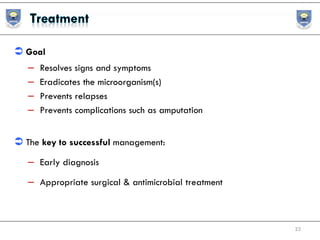
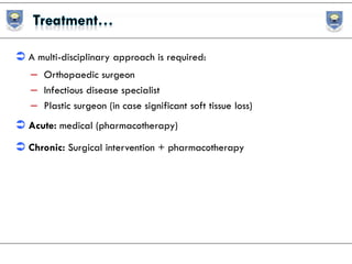
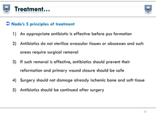
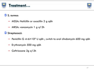
Lesson 7
Osteomyelitis
Tsegaye Melaku (BPharm, MSc)
Assistant Professor of Clinical Pharmacy
tsegayemlk@yahoo.com or tsegaye.melaku@ju.edu.et +251913765609December, 2020
 Session hits:
– Differentiating acute and chronic infections
– Most frequent pathogens in osteomyelitis
– Treatment protocol ( IV/Oral, duration)
– Monitoring plan
2
 It is a severe infection of the bone, bone marrow & surrounding soft tissue
 It is progressive infectious process that can involve one or multiple
components of bone
– The periosteum, medullary cavity, and cortical bone.
 Characterized by progressive inflammatory destruction of bone, by
necrosis, and by new bone formation.
3
 Acute or chronic infection
– Acute: first episode case or symptoms within
2 weeks
– Chronic: disease relapse or symptoms
persisting >2 months
– Characterized by presence of
necrotic bone
 Its Rx & Dx is challenging (complex nature)
5
6
 Uncommon disease
 Incidence: 0.4/1,000 children in US
 Vertebral osteomyelitis: in old age
(>50 years)
 Acute hematogenous osteomyelitis
– Bimodal distribution
– Younger than 2 years, and
8- 12 years
 Ethiopia
– TASH (COM): Biruk et al, 2006
– Mean age 18 years
– Trauma as cause: 27%
– Most affected Lower limb bones: Tibia, 36%, fibula 22%
and femur 21%
– Etiology: S.aureus
8
 Duration: Acute, subacute & chronic
– Differing prognoses and are treated in different ways
 Route of infection: hematogenous, contagious & inoculation
– Hematogenous
– Common in children & elderly
– Monomicrobial
– Contagious (with or without vascular insufficiency)
– Infection first begins in an area adjacent to bone spread
to bone
– Common in adult
– Polymicrobial
 Direct inoculation
– Trauma
– Open fracture 3-25%
– Surgery
 Contagious can be result of:
– Traumatic injury, penetrating injury, orthopedic surgery, or
– Diabetic or other forms of ischemic or neuropathic ulcer
10
 Can be classified into four stages based on extent of bone involvement:
– Medullary,
– Superficial (contagious focus/at base of soft tissue)
– Localized(discrete area)
– Diffuse (all regions)
11
 Essentially reflect their bacteremia incidence as a function of host age
 Can be comorbidity dependent
 Staphylococcus aureus: common cause
– 70% to 80% of bone infections
– ↑MRSA currently
 S. aureus -#1 pathogen
– Hematogenous osteomyelitis
– Device related infection
– Opportunistic pathogen
 Antibiotic resistance
– Methicillin resistant S. aureus (MRSA)
– 50% in some regions
– Vancomycin intermediate S. aureus (VISA)
– Vancomycin resistant S. aureus (VRSA)
 Biofilm formation
– Resistant abx/immunity/env’tal stress
13
 Hematogenous spread (usually 1 organism)
– Infant (<1 year)
– S. aureus, CoNS, GBS, E.coli
– Children & adults (>16 years)
– S. aureus, CoNS, E. Coli, Pseudomonas spp.,Serratia spp.
 Contiguous spread (polymicrobial)
– Microbiology depends on the primary site of infection
– S. Aureus, Streptococcus spp., Enterococcus
– Proteus mirabilis,Pseudomonas spp., Anaerobes
14
 It depends on mode of infection & patient factors
– Hematogenous /contiguous
– DM/PAD: MRSA, Enterobacteriaceae, & P. aeruginosa
– Sickle cell patients: Salmonella spp.
– Prosthetic implants: CoNS
– Neonates: E. coli or GBS
– Bed sores: polymicrobial
– IVDU: P. aeruginosa
– Fungus: chronically ill patients on long term intravenous therapy
15
 Bone is normally resistant to infection.
– However, when microorganisms are introduced into bone can cause
osteomyelitis.
 Determinant: microbial &patient-specific factors.
 Bone infected: hematogenous/inoculation/contagious(adjacent site) 
bacteria attachment to tissues & prosthetic material  
inflammation(leukocytes and cytokines)  local edema & ischemia
  bone necrosis.
16
 Dead bone(due to necrosis)   sequestra (bacteria nidus)  
chronic disease
– Difficult to treat(poor penetration)
– Surgical intervention
17
18
 Hematogenous
– Chills, fever, and malaise
– Pain and local swelling
– Localized back pain & tenderness (vertebrae infection)
– Increased ESR, CRP, procalcitonin
– Inflammation, erythema, edema,
– Decreased range of motion
19
 Contagious
– Increasing pain
– Mild fever and minimal drainage
– Imaging is often difficult to interpret
 A cardinal sign of chronic osteomyelitis
– The formation of a sinus tract (a channel from the infected site to
the skin) with purulent drainage
20
 CBC
– Usually normal WBC
 Bone biopsy: definitive dx
 Blood cultures: for hematogenous
 Superficial swabs
 History & physical examination
– Fever & malaise
– Pain & local tenderness
– Sweliing
– Compartment syndrome in children
 Normochromic normocytic anemia (anemia of chronic disease)
21
 Laboratory tests
– WBC, ESR, CRP
– Checked very 2- 3 days post treatment initiation
– Aspiration for suspected abscess
 Plain radiographs (usually used)
– Soft tissue swelling
– Periosteal reaction
– Bony destruction (10-12 days)
 Technetium-99m bone scan +/- MRI
– Confirm dx 24-48 hrs after onset
22
 Goal
– Resolves signs and symptoms
– Eradicates the microorganism(s)
– Prevents relapses
– Prevents complications such as amputation
 The key to successful management:
– Early diagnosis
– Appropriate surgical & antimicrobial treatment
23
 A multi-disciplinary approach is required:
– Orthopaedic surgeon
– Infectious disease specialist
– Plastic surgeon (in case significant soft tissue loss)
 Acute: medical (pharmacotherapy)
 Chronic: Surgical intervention + pharmacotherapy
 Nade’s 5 principles of treatment
1) An appropriate antibiotic is effective before pus formation
2) Antibiotics do not sterilize avascular tissues or abscesses and such
areas require surgical removal
3) If such removal is effective, antibiotics should prevent their
reformation and primary wound closure should be safe
4) Surgery should not damage already ischemic bone and soft tissue
5) Antibiotics should be continued after surgery
25
 Indication for surgery in acute case
– The presence of an abscess requiring drainage
– Failure of the patient to improve despite appropriate IV antibiotic
 Empiric antibiotic therapy is usually avoided.
– Treatment usually continues for 4- 8 weeks or longer
26
 S. aureus:
– MSSA: Nafcillin or oxacillin 2 g q6h
– MRSA: vancomycin 1 g q12h
 Streptococci:
– Penicillin G 4–6×10⁶ U q6h ; switch to oral clindamycin 600 mg q6h
– Erythromycin 500 mg q6h
– Ceftriaxone 2g q12h
27
 Enteric Gm(-) organisms:
– Oral ciprofloxacin 750 mg q12h
 Serratia or P. aeruginosa:
– Piperacillin–tazobactam 2–4 g q4h or
– Imipenem 500 mg q6h
 Anaerobes:
– Clindamycin 600mg q6h or
– Metronidazole 500mg q8h
– Amoxicillin–clavulanic acid 1.2g q6h–2.2g q8h
28
 Support for shorter duration therapy
– Adequate surgical debridement
– Diabetic foot osteomyelitis with good vascular supply,~6 weeks
– Prosthetic joint infection after implant removal, ~4 weeks
– Acute hematogenous osteomyelitis (non-vertebral), ~2-3 weeks
29
 Groups at risk for failure
– Recurrent infection – PJI requiring multiple surgeries
– Vertebral osteomyelitis
– Undrained abscess, endovascular infection, MRSA,
immunosuppression
– Lower recurrence rates in high risk patients treated with
≥6-8 weeks+
30
 Individualized duration
– Acute vs chronic infection
– Response to therapy
31
 Efficacy & safety
 Resolves signs and symptoms
 Symptoms resolution within 2 to 4 days of IV abx
 ↓CRP within 1 week of therapy
32
1. Septic arthritis
2. Sepsis and septic shock
3. HIV + OIs
4. TB
5. UTI
6. GI infections
7. Intra abdominal infections
8. Skin and soft tissue infections
9. Surgical prophylaxis
10. Parasitic infections, malaria & NTDs
11. Fungal infections(superficial & disseminated)
33
34

Osteomyelitis

  • 1.
    Lesson 7 Osteomyelitis Tsegaye Melaku(BPharm, MSc) Assistant Professor of Clinical Pharmacy tsegayemlk@yahoo.com or tsegaye.melaku@ju.edu.et +251913765609December, 2020
  • 2.
     Session hits: –Differentiating acute and chronic infections – Most frequent pathogens in osteomyelitis – Treatment protocol ( IV/Oral, duration) – Monitoring plan 2
  • 3.
     It isa severe infection of the bone, bone marrow & surrounding soft tissue  It is progressive infectious process that can involve one or multiple components of bone – The periosteum, medullary cavity, and cortical bone.  Characterized by progressive inflammatory destruction of bone, by necrosis, and by new bone formation. 3
  • 4.
     Acute orchronic infection – Acute: first episode case or symptoms within 2 weeks – Chronic: disease relapse or symptoms persisting >2 months – Characterized by presence of necrotic bone  Its Rx & Dx is challenging (complex nature)
  • 5.
  • 6.
  • 7.
     Uncommon disease Incidence: 0.4/1,000 children in US  Vertebral osteomyelitis: in old age (>50 years)  Acute hematogenous osteomyelitis – Bimodal distribution – Younger than 2 years, and 8- 12 years
  • 8.
     Ethiopia – TASH(COM): Biruk et al, 2006 – Mean age 18 years – Trauma as cause: 27% – Most affected Lower limb bones: Tibia, 36%, fibula 22% and femur 21% – Etiology: S.aureus 8
  • 9.
     Duration: Acute,subacute & chronic – Differing prognoses and are treated in different ways  Route of infection: hematogenous, contagious & inoculation – Hematogenous – Common in children & elderly – Monomicrobial – Contagious (with or without vascular insufficiency) – Infection first begins in an area adjacent to bone spread to bone – Common in adult – Polymicrobial
  • 10.
     Direct inoculation –Trauma – Open fracture 3-25% – Surgery  Contagious can be result of: – Traumatic injury, penetrating injury, orthopedic surgery, or – Diabetic or other forms of ischemic or neuropathic ulcer 10
  • 11.
     Can beclassified into four stages based on extent of bone involvement: – Medullary, – Superficial (contagious focus/at base of soft tissue) – Localized(discrete area) – Diffuse (all regions) 11
  • 12.
     Essentially reflecttheir bacteremia incidence as a function of host age  Can be comorbidity dependent  Staphylococcus aureus: common cause – 70% to 80% of bone infections – ↑MRSA currently
  • 13.
     S. aureus-#1 pathogen – Hematogenous osteomyelitis – Device related infection – Opportunistic pathogen  Antibiotic resistance – Methicillin resistant S. aureus (MRSA) – 50% in some regions – Vancomycin intermediate S. aureus (VISA) – Vancomycin resistant S. aureus (VRSA)  Biofilm formation – Resistant abx/immunity/env’tal stress 13
  • 14.
     Hematogenous spread(usually 1 organism) – Infant (<1 year) – S. aureus, CoNS, GBS, E.coli – Children & adults (>16 years) – S. aureus, CoNS, E. Coli, Pseudomonas spp.,Serratia spp.  Contiguous spread (polymicrobial) – Microbiology depends on the primary site of infection – S. Aureus, Streptococcus spp., Enterococcus – Proteus mirabilis,Pseudomonas spp., Anaerobes 14
  • 15.
     It dependson mode of infection & patient factors – Hematogenous /contiguous – DM/PAD: MRSA, Enterobacteriaceae, & P. aeruginosa – Sickle cell patients: Salmonella spp. – Prosthetic implants: CoNS – Neonates: E. coli or GBS – Bed sores: polymicrobial – IVDU: P. aeruginosa – Fungus: chronically ill patients on long term intravenous therapy 15
  • 16.
     Bone isnormally resistant to infection. – However, when microorganisms are introduced into bone can cause osteomyelitis.  Determinant: microbial &patient-specific factors.  Bone infected: hematogenous/inoculation/contagious(adjacent site)  bacteria attachment to tissues & prosthetic material   inflammation(leukocytes and cytokines)  local edema & ischemia   bone necrosis. 16
  • 17.
     Dead bone(dueto necrosis)   sequestra (bacteria nidus)   chronic disease – Difficult to treat(poor penetration) – Surgical intervention 17
  • 18.
  • 19.
     Hematogenous – Chills,fever, and malaise – Pain and local swelling – Localized back pain & tenderness (vertebrae infection) – Increased ESR, CRP, procalcitonin – Inflammation, erythema, edema, – Decreased range of motion 19
  • 20.
     Contagious – Increasingpain – Mild fever and minimal drainage – Imaging is often difficult to interpret  A cardinal sign of chronic osteomyelitis – The formation of a sinus tract (a channel from the infected site to the skin) with purulent drainage 20
  • 21.
     CBC – Usuallynormal WBC  Bone biopsy: definitive dx  Blood cultures: for hematogenous  Superficial swabs  History & physical examination – Fever & malaise – Pain & local tenderness – Sweliing – Compartment syndrome in children  Normochromic normocytic anemia (anemia of chronic disease) 21
  • 22.
     Laboratory tests –WBC, ESR, CRP – Checked very 2- 3 days post treatment initiation – Aspiration for suspected abscess  Plain radiographs (usually used) – Soft tissue swelling – Periosteal reaction – Bony destruction (10-12 days)  Technetium-99m bone scan +/- MRI – Confirm dx 24-48 hrs after onset 22
  • 23.
     Goal – Resolvessigns and symptoms – Eradicates the microorganism(s) – Prevents relapses – Prevents complications such as amputation  The key to successful management: – Early diagnosis – Appropriate surgical & antimicrobial treatment 23
  • 24.
     A multi-disciplinaryapproach is required: – Orthopaedic surgeon – Infectious disease specialist – Plastic surgeon (in case significant soft tissue loss)  Acute: medical (pharmacotherapy)  Chronic: Surgical intervention + pharmacotherapy
  • 25.
     Nade’s 5principles of treatment 1) An appropriate antibiotic is effective before pus formation 2) Antibiotics do not sterilize avascular tissues or abscesses and such areas require surgical removal 3) If such removal is effective, antibiotics should prevent their reformation and primary wound closure should be safe 4) Surgery should not damage already ischemic bone and soft tissue 5) Antibiotics should be continued after surgery 25
  • 26.
     Indication forsurgery in acute case – The presence of an abscess requiring drainage – Failure of the patient to improve despite appropriate IV antibiotic  Empiric antibiotic therapy is usually avoided. – Treatment usually continues for 4- 8 weeks or longer 26
  • 27.
     S. aureus: –MSSA: Nafcillin or oxacillin 2 g q6h – MRSA: vancomycin 1 g q12h  Streptococci: – Penicillin G 4–6×10⁶ U q6h ; switch to oral clindamycin 600 mg q6h – Erythromycin 500 mg q6h – Ceftriaxone 2g q12h 27
  • 28.
     Enteric Gm(-)organisms: – Oral ciprofloxacin 750 mg q12h  Serratia or P. aeruginosa: – Piperacillin–tazobactam 2–4 g q4h or – Imipenem 500 mg q6h  Anaerobes: – Clindamycin 600mg q6h or – Metronidazole 500mg q8h – Amoxicillin–clavulanic acid 1.2g q6h–2.2g q8h 28
  • 29.
     Support forshorter duration therapy – Adequate surgical debridement – Diabetic foot osteomyelitis with good vascular supply,~6 weeks – Prosthetic joint infection after implant removal, ~4 weeks – Acute hematogenous osteomyelitis (non-vertebral), ~2-3 weeks 29
  • 30.
     Groups atrisk for failure – Recurrent infection – PJI requiring multiple surgeries – Vertebral osteomyelitis – Undrained abscess, endovascular infection, MRSA, immunosuppression – Lower recurrence rates in high risk patients treated with ≥6-8 weeks+ 30
  • 31.
     Individualized duration –Acute vs chronic infection – Response to therapy 31
  • 32.
     Efficacy &safety  Resolves signs and symptoms  Symptoms resolution within 2 to 4 days of IV abx  ↓CRP within 1 week of therapy 32
  • 33.
    1. Septic arthritis 2.Sepsis and septic shock 3. HIV + OIs 4. TB 5. UTI 6. GI infections 7. Intra abdominal infections 8. Skin and soft tissue infections 9. Surgical prophylaxis 10. Parasitic infections, malaria & NTDs 11. Fungal infections(superficial & disseminated) 33
  • 34.