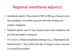
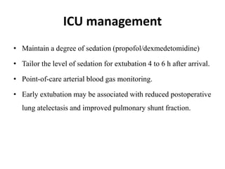
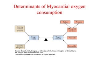
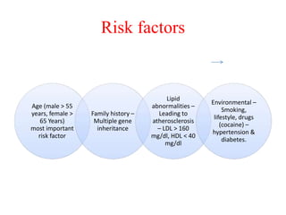
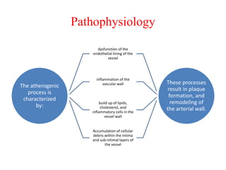
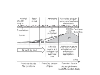
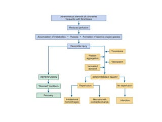
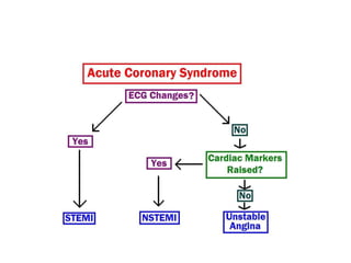
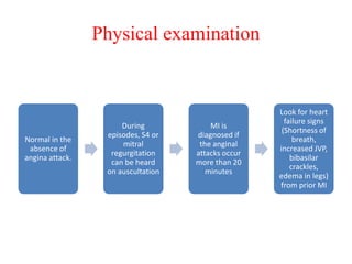
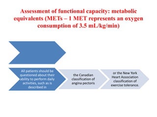
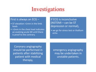
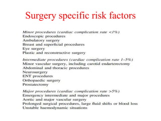
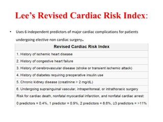
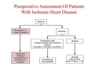
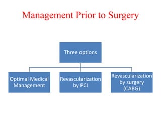
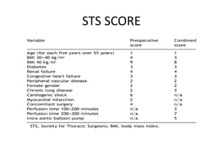
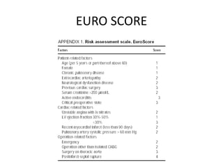
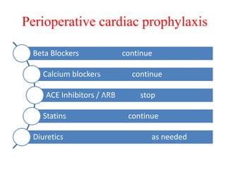
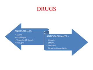
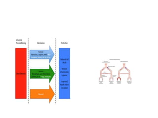
Ischaemic heart disease
(IHD)
DR. GAURAV GOGOI
PDT, CARDIAC ANESTHESIA
MODERATOR-DR.KAKALI GHOSH
Introduction
• IHD is due to imbalance between myocardial oxygen
demand and supply from the coronary arteries
• Coronary artery disease (CAD) is the number one cause
of death in United States
• Ischemia occurs secondary to coronary artery disease.
• Atherosclerosis number one cause of CAD
NORMAL ANATOMY
DETERMINANTS OF CORONARY BLOOD
FLOW
1. Physical
– Aortic blood pressure(aortic diast.
pressure-LVEDP)
– Heart rate
– Cardiac output
2. Chemical
– Metabolism
– Drugs
3. Neural
– Parasympathetic –coronary dilator
effect
– Sympathetic-coronary constriction
effect
4. Hormonal
– Thyroxin-cbf
– Vasopressin -CBF
5. REFLEX
– Gastrocoronary reflex
Determinants of Myocardial oxygen
consumption
Risk factors
Age (male > 55
years, female >
65 Years)
most important
risk factor
Family history –
Multiple gene
inheritance
Lipid
abnormalities –
Leading to
atherosclerosis
– LDL > 160
mg/dl, HDL < 40
mg/dl
Environmental –
Smoking,
lifestyle, drugs
(cocaine) –
hypertension &
diabetes.
Pathophysiology
The atherogenic
process is
characterized
by:
dysfunction of the
endothelial lining of the
vessel
inflammation of the
vascular wall
build up of lipids,
cholesterol, and
inflammatory cells in the
vessel wall
Accumulation of cellular
debris within the intima
and sub-intimal layers of
the vessel
These processes
result in plaque
formation, and
remodeling of
the arterial wall.
Coronary anatomy
(example of multiple lesions)
Coronary lesions are significant when there is obstruction of
one or more epicardial arteries with at least 70% stenosis
and/or that of LMD with at least 50% stenosis
Perioperative vs. non operative MI
• The pathophysiology of perioperative myocardial infarction differs
somewhat from that of myocardial infarction occurring in the non-operated
patient
• Rupture of a coronary arterial atherosclerotic plaque leads to platelet
aggregation and thrombus formation.
• In contrast the cause of perioperative myocardial are due to a prolonged
imbalance between myocardial oxygen supply and demand in the
setting of coronary artery disease.
CLINICAL MANIFESTATIONS
• The clinical manifestations of myocardial ischemia are
varied.
• Angina pectoris with or without signs of arrhythmias or
heart failure-assumed to be the classic manifestation.
• Angina is often classified according to its temporal
pattern and its relation to exertion because this loosely
reflects prognosis
Types of angina
Acute coronary syndromes(acs)
ACS- an operational term
used to describe a
constellation of symptoms
resulting from acute
myocardial ischemia.
ACS includes the diagnosis
of
ST-segment elevation
myocardial infarction
(STEMI).
non-ST-segment elevation
myocardial infarction
(NSTEMI), and
unstable angina (UA)
Physical examination
Normal in the
absence of
angina attack.
During
episodes, S4 or
mitral
regurgitation
can be heard
on auscultation
MI is
diagnosed if
the anginal
attacks occur
more than 20
minutes
Look for heart
failure signs
(Shortness of
breath,
increased JVP,
bibasilar
crackles,
edema in legs)
from prior MI
Assessment of functional capacity: metabolic
equivalents (METs – 1 MET represents an oxygen
consumption of 3.5 mL/kg/min)
All patients should be
questioned about their
ability to perform daily
activities, such as is
described in
the Canadian
classification of
angina pectoris
or the New York
Heart Association
classification of
exercise tolerance.
Investigations
First is always an ECG –
• ST elevation >1mm in the limb
leads
• >2mm in the chest lead indicates
an evolving acute MI until there
is proof to the contrary.
If ECG is inconclusive
(NSTEMI – can be ST
depression or normal),
• we go for stress test or thallium
echo.
Coronary angiography
should be performed in
patients after stabilizing
patient with medical
therapy,
emergency angiography
may be undertaken in
unstable patients.
• Do angiography – If you don’t find any
abnormal occlusion then Prinzmetal angina
can be suspected.
For Prinzmetal Angina
–ST elevation on ECG
is not specific.
• Wall motion abnormalities can help localize
obstructive coronary lesion
• Used to assess cardiac function
• Can be used to asses clot inside heart
Echocardiography
PET scan to
demonstrate regional
blood flow
&metabolism and
hence viability
Cardiac Markers
Myoglobin –
detected
from 1 to 5
hour of
chest pain.
• it is not
specific
No Troponin or
CK-MB will be
detected till 6-8
Hours
Troponin I –
• Start rising by 4th
hour,
• Peak at 16 hours
• Remain elevated for 7-
10 days (usually
drawn every 8 hours
three times till MI is
ruled out)
Creatine Kinase-
MB – Start rising
by 4th hours. Use
to detect re-
infarction, as
troponin level will
be high for up to
10 days. CK-MB
have sensitivity
and specificity of
95%
DIFFERENTIAL DIAGNOSIS
Anxiety and
hyperventilation
Musculoskeletal
chest wall pain
Cervical or thoracic
root pain
Pneumothorax,
pneumonia, or
pulmonary embolus
Esophageal problem
(inflammation/spasm
Pericarditis
Aortic dissection Mitral valve prolapse
Valvular heart
disease
POST MI COMPLICATIONS
Cardiac arrest
Tachyarrhythmia
Brady arrhythmia
Pericarditis
Left ventricular free wall rupture
Ventricular septal defect
Acute mitral regurgitation
Cardiogenic shock
Congestive heart failure
Dressler's syndrome
Left ventricular aneurysm
Risk Stratification
The ACC/AHA guidelines
integrate risk stratification
according to:
Clinical risk
factors & ECG
review.
Functional
Capacity.
Surgery specific
risk factors.
Clinical Predictors of Increased Perioperative
Cardiovascular risk
Major
• Unstable coronary
syndromes
• Acute / Recent MI
• Unstable/ Severe Angina
• Decompensated Heart failure
• High grade AV-block
• Symptomatic Ventricular
Dysrhythmias
• Severe Valvular Heart
Disease
Intermediate
Stable Angina
• Previous MI
(history/ECG)
• Compensated or previous
heart failure
• DM/Renal Insufficiency
Minor
• Age>70
• ECG: LVH,LBBB ST-T
abnormalities
• Low functional capacity
• H/O CVA/HTN
Functional capacity
• High-risk group : Ischemia at low-level exercise (<5
MET or heart rate <100 min)
• Low-risk group: > 7 MET (or heart rate >130 min)
without ischemia
• In the absence of valve pathology, the ejection fraction
of the left ventricle can be considered as an adequate
measurement of the myocardial functional reserve.
Surgery specific risk factors
Lee’s Revised Cardiac Risk Index:
• Uses 6 independent predictors of major cardiac complications for patients
undergoing elective non cardiac surgery.
Stepwise Assessment
Step 1: Assessment of urgency of surgical procedure.
Step 2: Assessment of presence of active cardiac conditions.
Step 3: Assessment of surgical risk. Low-risk surgery can be performed without additional cardiac testing.
Otherwise, further risk stratification is required.
Step 4: Assessment of functional capacity
Step 5: Re-assessment of surgical risk
Step 6: Assessment of cardiac risk factors
Step 7: Consideration of non-invasive testing
Preoperative Assessment Of Patients
With Ischemic Heart Disease
Management Prior to Surgery
Three options
Optimal Medical
Management
Revascularization
by PCI
Revascularization
by surgery
(CABG)
Medical Management
Any patient who comes with complains typical for angina –
Aspirin
Nitroglycerine (given sublingually or by spray)
STEMI patients
• Thrombolysis within 12
hrs. or ideally within
30mins of hospital arrival
• except recent stroke
history (6-12 Month) or
• stage 3 hypertension
(>180 mmHg)}
• Percutaneous coronary
intervention (PCI).-bare
metal or drug eluting
stents
NSTEMI patients
• Not a candidate for
immediate thrombolytic
• Low molecular weight
heparin
• If pain persists then may
be candidates for PCI
urgently or during
admission.
Indications of PCI and CABG
PCI -indicated if
• 1 coronary vessel is
occluded other
than main left
coronary artery.
CABG –
•most effective when 2
vessels with serious risk
factors like diabetes,
•3 vessels or
•left main coronary artery
occluded
In 2 or 3 vessel disease, if
right coronary artery (inferior
wall MI) is involved,
•STENTING EMERGENTLY in
RCA and then schedule
patient for CABG.
For long-term Management
Aspirin and prasiguel
Aspirin allergy – clopidegrel
Aspirin and clopidegrel fails –ticlopidine
High intensity statin therapy
Sublingual nitroglycerin
Beta-blockers (unless contraindicated) -first-line
Calcium channel blocker like verapamil-when b-blockers are contraindicated
If CCB is used as
monotherapy,
verapamil or diltiazem
should be used
beta-blockers should
not be prescribed
concurrently with
verapamil (risk of
complete heart block)
If used in combination
with a beta-blocker -
use a long-acting
CCB(e.g. modified-
release nifedipine).
If monotherapy not tolerated -consider one of
the following drugs: a long-acting nitrate,
ivabradine, nicorandil or ranolazine.
ACEi/ARBs benefits best with ejection fraction
below 40%.
TIMI(Thrombolysis in
Myocardial Infarction)
SCORE
• The TIMI score predicts
the risk of all
• mortality,
– MI and
– severe recurrent
ischemia requiring
urgent
revascularization
within 14 days after
admission as well as
benefit of enoxaparin. SCORE: 0-1= Low risk, 2-3 = Intermediate risk, 4 or
above =High risk
ACS management
Assessment for non cardiac surgery
Risk Assessment in Patients Scheduled for
Coronary Artery Bypass Grafting
The two risk score models most
commonly used to predict
outcomes for CABG are
the STS(society of thoracic
surgeons) score mostly used in
North America and
the European System for Cardiac
Operative Risk Evaluation (Euro
SCORE) in Europe.
Both models reliably predict
mortality associated with cardiac
surgery.
STS SCORE
EURO SCORE
Perioperative cardiac prophylaxis
Beta Blockers continue
Calcium blockers continue
ACE Inhibitors / ARB stop
Statins continue
Diuretics as needed
DRUGS
ANTIPLATELETS –
• Aspirin ,
• Clopidogrel,
• Ticagrelor (Brilanta),
• Prasugrel
ANTICOAGULANTS –
• Heparin,
• LMWH,
• Warfarin,
• Newer anticoagulants
Antiplatelet therapy
Aspirin is given
lifelong
Clopidogrel (DAPT) is
given for following
period as per the
cardiac status
Simple angioplasty
without stenting | 2-
4 weeks
PCI and bare metal
stents | 6 weeks
Acute coronary
syndrome | 3-6
months
PCI and drug eluting
stents | Minimum of
12 months
Anticoagulant therapy
The last dose of LMWH should be administered no later
than 12 hours before surgery
Patients on oral vitamin K antagonists can undergo non-
cardiac surgery when the INR is <1.5. (Vit.K or FFP)
Risk of perioperative bleeding weighed against
thromboembolic risk..
• For patients receiving non-vitamin K antagonist
direct oral anticoagulants (NOACs) like
dabigatran, rivaroxaban –
– discontinue NOACs for 2-3 times their respective half-
lives before surgery with average risk of bleeding,
– and for 4-5 times their biological half-lives before
surgery with high risk of bleeding.
Bridging therapy
After stopping of APT
anticoagulant therapy
Started with heparin or low
molecular heparin.
Heparin has no antiplatelet
action
Short acting platelet
glycoprotein inhibitor like
eptifibatide, tirofiban
substitute for clopidogrel
while aspirin is continued
Cont..
If low risk stop
warfarin 5 days before
surgery so that INR drops
to < 1.5
If high risk stop
warfarin start heparin
(unfractionated heparin
infusion or low
molecular weight
heparin)
during the period
when the INR is
below the
therapeutic range.
Urgent surgery
• Patient on Warfarin – rapid reversal of anticoagulants
by -
– Fresh frozen plasma
– Prothrombin complex concentrates, recombinant factor
VIIa
• If surgery can be delayed by 18-24 hours,
– IV Vitamin K 2.5-5 mg given without the need for blood
products.
PTs on antiplatelets
• No pharmacologic agent can reverse effects of
aspirin,clopidogrel or ticlopidine
• Platelet transfusion can be given if bleeding
• Prohemostatic agents- E aminocaproic acid,
tranexamic acid
Intra-op monitoring for myocardial ischemia
Detection of
intraoperative ischemia
critically important
because
• intraoperative ischemia is an independent
predictor of postoperative myocardial infarction
• The presence ofnew persistent Q waves of at
least 0.03-s duration, broadening of preexisting Q
waves, or new QS deflections on the
postoperative ECG have been considered
evidence of perioperative
AMI.
Intra-op monitoring for myocardial ischemia
• Typical monitoring for CABG surgery includes
– the standard American Society of Anesthesiology
(ASA) monitors and
– invasive arterial blood pressure monitoring.
– TEE used more frequently
in current practice, as a
category II indication
– Pulmonary arterial (PA)
catheters for routine
CABG is controversial.
– ECG
– Cardiac output monitoring
– Neurophysiologic
monitoring
– Temperature monitoring
– Foley placement for all
patients
Goals of anesthetic management
• The primary aim during anesthetic management of the patient
with coronary artery disease is
– prevention of imbalance between myocardial oxygen
supply and demand.
Induction and Maintenance of General
Anesthesia
The main considerations in
choosing an induction
technique are
• LV function and
• coronary pathology.
No single approach to
anesthesia for is suitable for
all patients.
Fast-track cardiac anesthesia
techniques to facilitate early
extubation, awakening, and
safe transfer from intensive
care environments.
• Fast-track cardiac anesthesia
typically involves a “balanced”
technique utilizing a
combination of inhalational
anesthetic, modest amounts of
opioid, and intermediate-
duration muscle relaxation.
Coronary steal-phenomenon in which dilation of
normal vascular beds diverts blood away from
other beds that are ischemic
Preconditioning- Volatile anesthetics may confer
a degree of preconditioning-like protective
effect against ischemia-reperfusion injury in
human myocardial tissue and improve late
cardiac events following cardiac surgery
• Intraoperative extubation can be done In the absence of
ventricular impairment, hemodynamic instability,
hypothermia, or significant coagulopathy
• Patients remaining intubated need adequate
sedation/anesthesia to last through the transport and for at
least 30 min beyond
Regional anesthesia adjuncts
• Intrathecal opioid -Placement of 200 to 400 μg of preservative-
free morphine via lumbar puncture provides lasting, pre-
emptive analgesia
• Epidural opioid -up to 5 mg of preservative-free morphine can
provide preemptive analgesia.
• Precautions -Potent antiplatelet therapy (e.g., clopidogrel) not
halted at least 7 days before the day of surgery raises concerns
in neuraxial procedure
ICU management
• Maintain a degree of sedation (propofol/dexmedetomidine)
• Tailor the level of sedation for extubation 4 to 6 h after arrival.
• Point-of-care arterial blood gas monitoring.
• Early extubation may be associated with reduced postoperative
lung atelectasis and improved pulmonary shunt fraction.
Post Op Cabg Complication
References
Kaplan Cardiac Anesthesia
Braunwald Textbook Of Cardiovascular
Medicine
Fundamentals Of Cardiology For USMLE
Hensley Martin Practical Approach To Cardiac
Anesthesia
WWW
Ishemic heart disease

Ishemic heart disease

  • 1.
    Ischaemic heart disease (IHD) DR.GAURAV GOGOI PDT, CARDIAC ANESTHESIA MODERATOR-DR.KAKALI GHOSH
  • 2.
    Introduction • IHD isdue to imbalance between myocardial oxygen demand and supply from the coronary arteries • Coronary artery disease (CAD) is the number one cause of death in United States • Ischemia occurs secondary to coronary artery disease. • Atherosclerosis number one cause of CAD
  • 3.
  • 4.
    DETERMINANTS OF CORONARYBLOOD FLOW 1. Physical – Aortic blood pressure(aortic diast. pressure-LVEDP) – Heart rate – Cardiac output 2. Chemical – Metabolism – Drugs 3. Neural – Parasympathetic –coronary dilator effect – Sympathetic-coronary constriction effect 4. Hormonal – Thyroxin-cbf – Vasopressin -CBF 5. REFLEX – Gastrocoronary reflex
  • 5.
    Determinants of Myocardialoxygen consumption
  • 6.
    Risk factors Age (male> 55 years, female > 65 Years) most important risk factor Family history – Multiple gene inheritance Lipid abnormalities – Leading to atherosclerosis – LDL > 160 mg/dl, HDL < 40 mg/dl Environmental – Smoking, lifestyle, drugs (cocaine) – hypertension & diabetes.
  • 7.
    Pathophysiology The atherogenic process is characterized by: dysfunctionof the endothelial lining of the vessel inflammation of the vascular wall build up of lipids, cholesterol, and inflammatory cells in the vessel wall Accumulation of cellular debris within the intima and sub-intimal layers of the vessel These processes result in plaque formation, and remodeling of the arterial wall.
  • 10.
    Coronary anatomy (example ofmultiple lesions) Coronary lesions are significant when there is obstruction of one or more epicardial arteries with at least 70% stenosis and/or that of LMD with at least 50% stenosis
  • 11.
    Perioperative vs. nonoperative MI • The pathophysiology of perioperative myocardial infarction differs somewhat from that of myocardial infarction occurring in the non-operated patient • Rupture of a coronary arterial atherosclerotic plaque leads to platelet aggregation and thrombus formation. • In contrast the cause of perioperative myocardial are due to a prolonged imbalance between myocardial oxygen supply and demand in the setting of coronary artery disease.
  • 12.
    CLINICAL MANIFESTATIONS • Theclinical manifestations of myocardial ischemia are varied. • Angina pectoris with or without signs of arrhythmias or heart failure-assumed to be the classic manifestation. • Angina is often classified according to its temporal pattern and its relation to exertion because this loosely reflects prognosis
  • 13.
  • 14.
    Acute coronary syndromes(acs) ACS-an operational term used to describe a constellation of symptoms resulting from acute myocardial ischemia. ACS includes the diagnosis of ST-segment elevation myocardial infarction (STEMI). non-ST-segment elevation myocardial infarction (NSTEMI), and unstable angina (UA)
  • 16.
    Physical examination Normal inthe absence of angina attack. During episodes, S4 or mitral regurgitation can be heard on auscultation MI is diagnosed if the anginal attacks occur more than 20 minutes Look for heart failure signs (Shortness of breath, increased JVP, bibasilar crackles, edema in legs) from prior MI
  • 17.
    Assessment of functionalcapacity: metabolic equivalents (METs – 1 MET represents an oxygen consumption of 3.5 mL/kg/min) All patients should be questioned about their ability to perform daily activities, such as is described in the Canadian classification of angina pectoris or the New York Heart Association classification of exercise tolerance.
  • 20.
    Investigations First is alwaysan ECG – • ST elevation >1mm in the limb leads • >2mm in the chest lead indicates an evolving acute MI until there is proof to the contrary. If ECG is inconclusive (NSTEMI – can be ST depression or normal), • we go for stress test or thallium echo. Coronary angiography should be performed in patients after stabilizing patient with medical therapy, emergency angiography may be undertaken in unstable patients.
  • 21.
    • Do angiography– If you don’t find any abnormal occlusion then Prinzmetal angina can be suspected. For Prinzmetal Angina –ST elevation on ECG is not specific. • Wall motion abnormalities can help localize obstructive coronary lesion • Used to assess cardiac function • Can be used to asses clot inside heart Echocardiography PET scan to demonstrate regional blood flow &metabolism and hence viability
  • 22.
    Cardiac Markers Myoglobin – detected from1 to 5 hour of chest pain. • it is not specific No Troponin or CK-MB will be detected till 6-8 Hours Troponin I – • Start rising by 4th hour, • Peak at 16 hours • Remain elevated for 7- 10 days (usually drawn every 8 hours three times till MI is ruled out) Creatine Kinase- MB – Start rising by 4th hours. Use to detect re- infarction, as troponin level will be high for up to 10 days. CK-MB have sensitivity and specificity of 95%
  • 24.
    DIFFERENTIAL DIAGNOSIS Anxiety and hyperventilation Musculoskeletal chestwall pain Cervical or thoracic root pain Pneumothorax, pneumonia, or pulmonary embolus Esophageal problem (inflammation/spasm Pericarditis Aortic dissection Mitral valve prolapse Valvular heart disease
  • 25.
    POST MI COMPLICATIONS Cardiacarrest Tachyarrhythmia Brady arrhythmia Pericarditis Left ventricular free wall rupture Ventricular septal defect Acute mitral regurgitation Cardiogenic shock Congestive heart failure Dressler's syndrome Left ventricular aneurysm
  • 26.
    Risk Stratification The ACC/AHAguidelines integrate risk stratification according to: Clinical risk factors & ECG review. Functional Capacity. Surgery specific risk factors.
  • 27.
    Clinical Predictors ofIncreased Perioperative Cardiovascular risk Major • Unstable coronary syndromes • Acute / Recent MI • Unstable/ Severe Angina • Decompensated Heart failure • High grade AV-block • Symptomatic Ventricular Dysrhythmias • Severe Valvular Heart Disease Intermediate Stable Angina • Previous MI (history/ECG) • Compensated or previous heart failure • DM/Renal Insufficiency Minor • Age>70 • ECG: LVH,LBBB ST-T abnormalities • Low functional capacity • H/O CVA/HTN
  • 28.
    Functional capacity • High-riskgroup : Ischemia at low-level exercise (<5 MET or heart rate <100 min) • Low-risk group: > 7 MET (or heart rate >130 min) without ischemia • In the absence of valve pathology, the ejection fraction of the left ventricle can be considered as an adequate measurement of the myocardial functional reserve.
  • 29.
  • 30.
    Lee’s Revised CardiacRisk Index: • Uses 6 independent predictors of major cardiac complications for patients undergoing elective non cardiac surgery.
  • 31.
    Stepwise Assessment Step 1:Assessment of urgency of surgical procedure. Step 2: Assessment of presence of active cardiac conditions. Step 3: Assessment of surgical risk. Low-risk surgery can be performed without additional cardiac testing. Otherwise, further risk stratification is required. Step 4: Assessment of functional capacity Step 5: Re-assessment of surgical risk Step 6: Assessment of cardiac risk factors Step 7: Consideration of non-invasive testing
  • 32.
    Preoperative Assessment OfPatients With Ischemic Heart Disease
  • 33.
    Management Prior toSurgery Three options Optimal Medical Management Revascularization by PCI Revascularization by surgery (CABG)
  • 34.
    Medical Management Any patientwho comes with complains typical for angina – Aspirin Nitroglycerine (given sublingually or by spray)
  • 35.
    STEMI patients • Thrombolysiswithin 12 hrs. or ideally within 30mins of hospital arrival • except recent stroke history (6-12 Month) or • stage 3 hypertension (>180 mmHg)} • Percutaneous coronary intervention (PCI).-bare metal or drug eluting stents NSTEMI patients • Not a candidate for immediate thrombolytic • Low molecular weight heparin • If pain persists then may be candidates for PCI urgently or during admission.
  • 36.
    Indications of PCIand CABG PCI -indicated if • 1 coronary vessel is occluded other than main left coronary artery. CABG – •most effective when 2 vessels with serious risk factors like diabetes, •3 vessels or •left main coronary artery occluded In 2 or 3 vessel disease, if right coronary artery (inferior wall MI) is involved, •STENTING EMERGENTLY in RCA and then schedule patient for CABG.
  • 37.
    For long-term Management Aspirinand prasiguel Aspirin allergy – clopidegrel Aspirin and clopidegrel fails –ticlopidine High intensity statin therapy Sublingual nitroglycerin Beta-blockers (unless contraindicated) -first-line Calcium channel blocker like verapamil-when b-blockers are contraindicated
  • 38.
    If CCB isused as monotherapy, verapamil or diltiazem should be used beta-blockers should not be prescribed concurrently with verapamil (risk of complete heart block) If used in combination with a beta-blocker - use a long-acting CCB(e.g. modified- release nifedipine).
  • 39.
    If monotherapy nottolerated -consider one of the following drugs: a long-acting nitrate, ivabradine, nicorandil or ranolazine. ACEi/ARBs benefits best with ejection fraction below 40%.
  • 40.
    TIMI(Thrombolysis in Myocardial Infarction) SCORE •The TIMI score predicts the risk of all • mortality, – MI and – severe recurrent ischemia requiring urgent revascularization within 14 days after admission as well as benefit of enoxaparin. SCORE: 0-1= Low risk, 2-3 = Intermediate risk, 4 or above =High risk
  • 41.
  • 42.
    Assessment for noncardiac surgery
  • 43.
    Assessment for noncardiac surgery
  • 46.
    Risk Assessment inPatients Scheduled for Coronary Artery Bypass Grafting The two risk score models most commonly used to predict outcomes for CABG are the STS(society of thoracic surgeons) score mostly used in North America and the European System for Cardiac Operative Risk Evaluation (Euro SCORE) in Europe. Both models reliably predict mortality associated with cardiac surgery.
  • 47.
  • 48.
  • 49.
    Perioperative cardiac prophylaxis BetaBlockers continue Calcium blockers continue ACE Inhibitors / ARB stop Statins continue Diuretics as needed
  • 50.
    DRUGS ANTIPLATELETS – • Aspirin, • Clopidogrel, • Ticagrelor (Brilanta), • Prasugrel ANTICOAGULANTS – • Heparin, • LMWH, • Warfarin, • Newer anticoagulants
  • 51.
    Antiplatelet therapy Aspirin isgiven lifelong Clopidogrel (DAPT) is given for following period as per the cardiac status Simple angioplasty without stenting | 2- 4 weeks PCI and bare metal stents | 6 weeks Acute coronary syndrome | 3-6 months PCI and drug eluting stents | Minimum of 12 months
  • 52.
    Anticoagulant therapy The lastdose of LMWH should be administered no later than 12 hours before surgery Patients on oral vitamin K antagonists can undergo non- cardiac surgery when the INR is <1.5. (Vit.K or FFP) Risk of perioperative bleeding weighed against thromboembolic risk..
  • 53.
    • For patientsreceiving non-vitamin K antagonist direct oral anticoagulants (NOACs) like dabigatran, rivaroxaban – – discontinue NOACs for 2-3 times their respective half- lives before surgery with average risk of bleeding, – and for 4-5 times their biological half-lives before surgery with high risk of bleeding.
  • 54.
    Bridging therapy After stoppingof APT anticoagulant therapy Started with heparin or low molecular heparin. Heparin has no antiplatelet action Short acting platelet glycoprotein inhibitor like eptifibatide, tirofiban substitute for clopidogrel while aspirin is continued
  • 55.
    Cont.. If low riskstop warfarin 5 days before surgery so that INR drops to < 1.5 If high risk stop warfarin start heparin (unfractionated heparin infusion or low molecular weight heparin) during the period when the INR is below the therapeutic range.
  • 56.
    Urgent surgery • Patienton Warfarin – rapid reversal of anticoagulants by - – Fresh frozen plasma – Prothrombin complex concentrates, recombinant factor VIIa • If surgery can be delayed by 18-24 hours, – IV Vitamin K 2.5-5 mg given without the need for blood products.
  • 57.
    PTs on antiplatelets •No pharmacologic agent can reverse effects of aspirin,clopidogrel or ticlopidine • Platelet transfusion can be given if bleeding • Prohemostatic agents- E aminocaproic acid, tranexamic acid
  • 58.
    Intra-op monitoring formyocardial ischemia Detection of intraoperative ischemia critically important because • intraoperative ischemia is an independent predictor of postoperative myocardial infarction • The presence ofnew persistent Q waves of at least 0.03-s duration, broadening of preexisting Q waves, or new QS deflections on the postoperative ECG have been considered evidence of perioperative AMI.
  • 59.
    Intra-op monitoring formyocardial ischemia • Typical monitoring for CABG surgery includes – the standard American Society of Anesthesiology (ASA) monitors and – invasive arterial blood pressure monitoring.
  • 60.
    – TEE usedmore frequently in current practice, as a category II indication – Pulmonary arterial (PA) catheters for routine CABG is controversial. – ECG – Cardiac output monitoring – Neurophysiologic monitoring – Temperature monitoring – Foley placement for all patients
  • 61.
    Goals of anestheticmanagement • The primary aim during anesthetic management of the patient with coronary artery disease is – prevention of imbalance between myocardial oxygen supply and demand.
  • 62.
    Induction and Maintenanceof General Anesthesia The main considerations in choosing an induction technique are • LV function and • coronary pathology. No single approach to anesthesia for is suitable for all patients. Fast-track cardiac anesthesia techniques to facilitate early extubation, awakening, and safe transfer from intensive care environments.
  • 63.
    • Fast-track cardiacanesthesia typically involves a “balanced” technique utilizing a combination of inhalational anesthetic, modest amounts of opioid, and intermediate- duration muscle relaxation.
  • 64.
    Coronary steal-phenomenon inwhich dilation of normal vascular beds diverts blood away from other beds that are ischemic Preconditioning- Volatile anesthetics may confer a degree of preconditioning-like protective effect against ischemia-reperfusion injury in human myocardial tissue and improve late cardiac events following cardiac surgery
  • 66.
    • Intraoperative extubationcan be done In the absence of ventricular impairment, hemodynamic instability, hypothermia, or significant coagulopathy • Patients remaining intubated need adequate sedation/anesthesia to last through the transport and for at least 30 min beyond
  • 67.
    Regional anesthesia adjuncts •Intrathecal opioid -Placement of 200 to 400 μg of preservative- free morphine via lumbar puncture provides lasting, pre- emptive analgesia • Epidural opioid -up to 5 mg of preservative-free morphine can provide preemptive analgesia. • Precautions -Potent antiplatelet therapy (e.g., clopidogrel) not halted at least 7 days before the day of surgery raises concerns in neuraxial procedure
  • 68.
    ICU management • Maintaina degree of sedation (propofol/dexmedetomidine) • Tailor the level of sedation for extubation 4 to 6 h after arrival. • Point-of-care arterial blood gas monitoring. • Early extubation may be associated with reduced postoperative lung atelectasis and improved pulmonary shunt fraction.
  • 69.
    Post Op CabgComplication
  • 70.
    References Kaplan Cardiac Anesthesia BraunwaldTextbook Of Cardiovascular Medicine Fundamentals Of Cardiology For USMLE Hensley Martin Practical Approach To Cardiac Anesthesia WWW