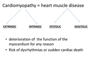
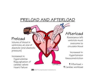
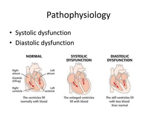
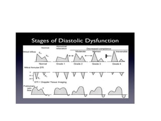
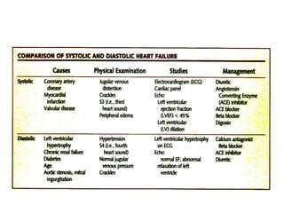
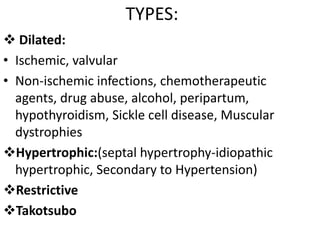
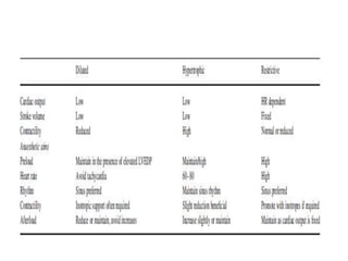
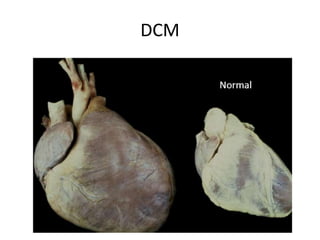
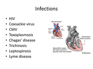
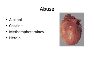
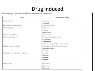
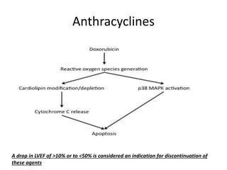
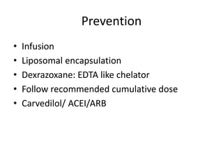
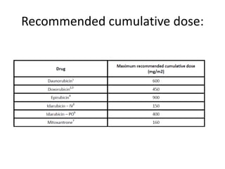
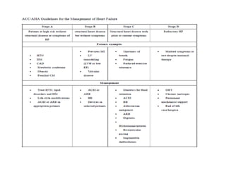
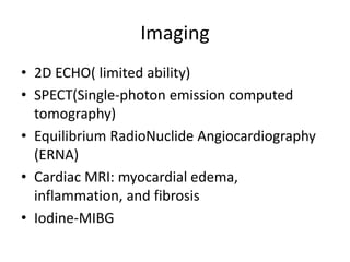
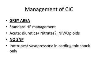
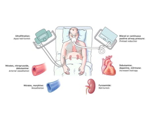
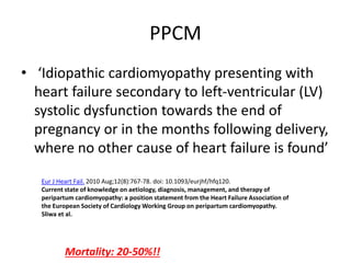
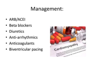
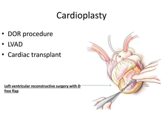
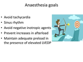
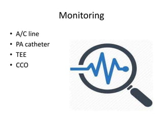
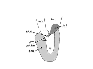
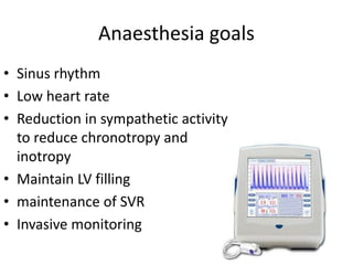
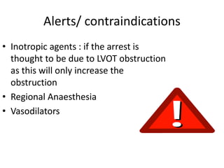
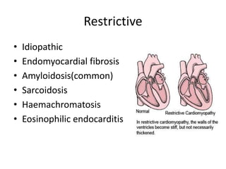
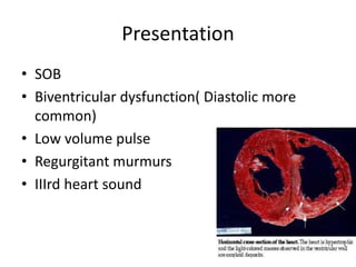
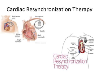
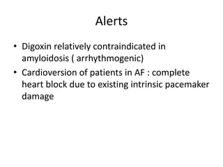
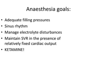
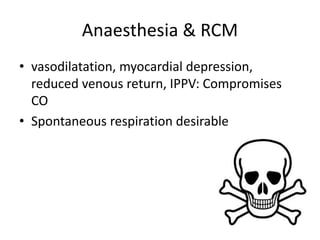
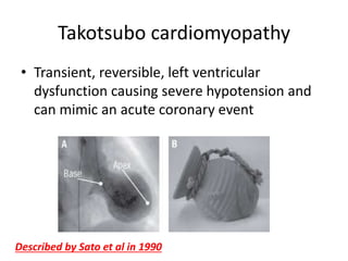
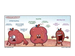
CARDIOMYOPATHY &
ANAESTHESIA
DR. ABHIJIT S. NAIR
Consultant Anaesthesiologist,
Basavatarakam Indo-American Cancer Hospital &
Research Institute,
Hyderabad.
Cardiomyopathy = heart muscle disease
• deterioration of the function of the
myocardium for any reason
• Risk of dysrhythmias or sudden cardiac death
EXTRINSIC INTRINSIC SYSTOLIC DIASTOLIC
Pathophysiology
• Systolic dysfunction
• Diastolic dysfunction
TYPES:
 Dilated:
• Ischemic, valvular
• Non-ischemic infections, chemotherapeutic
agents, drug abuse, alcohol, peripartum,
hypothyroidism, Sickle cell disease, Muscular
dystrophies
Hypertrophic:(septal hypertrophy-idiopathic
hypertrophic, Secondary to Hypertension)
Restrictive
Takotsubo
DCM
Infections
• HIV
• Coxsackie virus
• CMV
• Toxoplasmosis
• Chagas’ disease
• Trichinosis
• Leptospirosis
• Lyme disease
Abuse
• Alcohol
• Cocaine
• Methamphetamines
• Heroin
Drug induced
Anthracyclines
A drop in LVEF of >10% or to <50% is considered an indication for discontinuation of
these agents
Prevention
• Infusion
• Liposomal encapsulation
• Dexrazoxane: EDTA like chelator
• Follow recommended cumulative dose
• Carvedilol/ ACEI/ARB
Recommended cumulative dose:
Imaging
• 2D ECHO( limited ability)
• SPECT(Single-photon emission computed
tomography)
• Equilibrium RadioNuclide Angiocardiography
(ERNA)
• Cardiac MRI: myocardial edema,
inflammation, and fibrosis
• Iodine-MIBG
Management of CIC
• GREY AREA
• Standard HF management
• Acute: diuretics+ Nitrates?, NIV/Opioids
• NO SNP
• Inotropes/ vasopressors: in cardiogenic shock
only
PPCM
• ‘Idiopathic cardiomyopathy presenting with
heart failure secondary to left-ventricular (LV)
systolic dysfunction towards the end of
pregnancy or in the months following delivery,
where no other cause of heart failure is found’
Eur J Heart Fail. 2010 Aug;12(8):767-78. doi: 10.1093/eurjhf/hfq120.
Current state of knowledge on aetiology, diagnosis, management, and therapy of
peripartum cardiomyopathy: a position statement from the Heart Failure Association of
the European Society of Cardiology Working Group on peripartum cardiomyopathy.
Sliwa et al.
Mortality: 20-50%!!
Peripartum( 1:100-1:1000)
• Last trimester up to 5 months
Causes:
• Obesity
• myocarditis
• Drugs?
• Smoking
• Alcoholism
• Multiple pregnancies
• African-American descent
• Poor nourishment
Management:
• Beta blockers/ACEI/ARB/Diuretics
• Bromocriptine/ pentoxyphylline
• AICD
• Anticoagulation
S & S of DCM:
• Tachycardia
• SOB
• Ascites/ oedema
• Regurgitation/ arrhythmias/
Thrombus
• Biventricular dysfunction
Management:
• ARB/ACEI
• Beta blockers
• Diuretics
• Anti-arrhythmics
• Anticoagulants
• Biventricular pacing
Cardioplasty
• DOR procedure
• LVAD
• Cardiac transplant
Left ventricular reconstructive surgery with D
free flap
Anaesthesia goals
• Avoid tachycardia
• Sinus rhythm
• Avoid negative inotropic agents
• Prevent increases in afterload
• Maintain adequate preload in
the presence of elevated LVEDP
Monitoring
• A/C line
• PA catheter
• TEE
• CCO
HCM
• HOCM ( Obstructive)
• HHCM ( Hypertensive)
• IHSS (idiopathic hypertrophic subaortic
stenosis)
HCM
• Autosomal dominant
• Young adults
• SCD
• Family history of sudden death
• Significant LV hypertrophy(> 30
mm)
• Unexplained syncope
• Non-sustained VT
HOCM
Management
• Beta blockers
• Anti-Arrhythmics
• AICD
• Myomectomy
LVOTO
• Common at IVS
• dynamic LVOTO in sub-aortic region during
systole
• exacerbated when there is under filling of LV with
forceful sub-aortic narrowing
• velocity of blood in the outflow tract draws the
anterior mitral valve leaflet towards IVS
• SAM: Anterior leaflet pulled from posterior MV
leaflet—> obstruction
Anaesthesia goals
• Sinus rhythm
• Low heart rate
• Reduction in sympathetic activity
to reduce chronotropy and
inotropy
• Maintain LV filling
• maintenance of SVR
• Invasive monitoring
• “ Defibrillator paddles throughout surgery”
Alerts/ contraindications
• Inotropic agents : if the arrest is
thought to be due to LVOT obstruction
as this will only increase the
obstruction
• Regional Anaesthesia
• Vasodilators
Restrictive
• Idiopathic
• Endomyocardial fibrosis
• Amyloidosis(common)
• Sarcoidosis
• Haemachromatosis
• Eosinophilic endocarditis
Presentation
• SOB
• Biventricular dysfunction( Diastolic more
common)
• Low volume pulse
• Regurgitant murmurs
• IIIrd heart sound
Management
• Lower the elevated filling pressures caused by
low ventricular compliance without reducing
cardiac output
• ACEI or β-blockade/ diuretics
• Anti-arrhythmics
• Anticoagulants
• Disease modifying drugs
• Heart transplantation!
Cardiac Resynchronization Therapy
Alerts
• Digoxin relatively contraindicated in
amyloidosis ( arrhythmogenic)
• Cardioversion of patients in AF : complete
heart block due to existing intrinsic pacemaker
damage
Anaesthesia goals:
• Adequate filling pressures
• Sinus rhythm
• Manage electrolyte disturbances
• Maintain SVR in the presence of
relatively fixed cardiac output
• KETAMINE!
Anaesthesia & RCM
• vasodilatation, myocardial depression,
reduced venous return, IPPV: Compromises
CO
• Spontaneous respiration desirable
Takotsubo cardiomyopathy
• Transient, reversible, left ventricular
dysfunction causing severe hypotension and
can mimic an acute coronary event
Described by Sato et al in 1990
CAG: Normal
Management:
• No evidence-based guidelines
• Supportive
• Beta blockers, ACE inhibitors, diuretics
• Anticoagulation
• Possibility of recurrence in similar situation
Conclusion
• Understand the pathology
• Avoid worsening of heart function
• Sinus rhythm
• Invasive monitoring
• Electrolyte imbalance/ arrhythmia
• Judicious use of vasopressor / inotrope
• Multimodal analgesia
• Look for DIASTOLIC DYSFUNCTION

Cardiomyopathy

  • 1.
    CARDIOMYOPATHY & ANAESTHESIA DR. ABHIJITS. NAIR Consultant Anaesthesiologist, Basavatarakam Indo-American Cancer Hospital & Research Institute, Hyderabad.
  • 2.
    Cardiomyopathy = heartmuscle disease • deterioration of the function of the myocardium for any reason • Risk of dysrhythmias or sudden cardiac death EXTRINSIC INTRINSIC SYSTOLIC DIASTOLIC
  • 4.
  • 7.
    TYPES:  Dilated: • Ischemic,valvular • Non-ischemic infections, chemotherapeutic agents, drug abuse, alcohol, peripartum, hypothyroidism, Sickle cell disease, Muscular dystrophies Hypertrophic:(septal hypertrophy-idiopathic hypertrophic, Secondary to Hypertension) Restrictive Takotsubo
  • 9.
  • 10.
    Infections • HIV • Coxsackievirus • CMV • Toxoplasmosis • Chagas’ disease • Trichinosis • Leptospirosis • Lyme disease
  • 11.
    Abuse • Alcohol • Cocaine •Methamphetamines • Heroin
  • 12.
  • 13.
    Anthracyclines A drop inLVEF of >10% or to <50% is considered an indication for discontinuation of these agents
  • 14.
    Prevention • Infusion • Liposomalencapsulation • Dexrazoxane: EDTA like chelator • Follow recommended cumulative dose • Carvedilol/ ACEI/ARB
  • 15.
  • 17.
    Imaging • 2D ECHO(limited ability) • SPECT(Single-photon emission computed tomography) • Equilibrium RadioNuclide Angiocardiography (ERNA) • Cardiac MRI: myocardial edema, inflammation, and fibrosis • Iodine-MIBG
  • 18.
    Management of CIC •GREY AREA • Standard HF management • Acute: diuretics+ Nitrates?, NIV/Opioids • NO SNP • Inotropes/ vasopressors: in cardiogenic shock only
  • 20.
    PPCM • ‘Idiopathic cardiomyopathypresenting with heart failure secondary to left-ventricular (LV) systolic dysfunction towards the end of pregnancy or in the months following delivery, where no other cause of heart failure is found’ Eur J Heart Fail. 2010 Aug;12(8):767-78. doi: 10.1093/eurjhf/hfq120. Current state of knowledge on aetiology, diagnosis, management, and therapy of peripartum cardiomyopathy: a position statement from the Heart Failure Association of the European Society of Cardiology Working Group on peripartum cardiomyopathy. Sliwa et al. Mortality: 20-50%!!
  • 21.
    Peripartum( 1:100-1:1000) • Lasttrimester up to 5 months
  • 22.
    Causes: • Obesity • myocarditis •Drugs? • Smoking • Alcoholism • Multiple pregnancies • African-American descent • Poor nourishment
  • 23.
    Management: • Beta blockers/ACEI/ARB/Diuretics •Bromocriptine/ pentoxyphylline • AICD • Anticoagulation
  • 24.
    S & Sof DCM: • Tachycardia • SOB • Ascites/ oedema • Regurgitation/ arrhythmias/ Thrombus • Biventricular dysfunction
  • 25.
    Management: • ARB/ACEI • Betablockers • Diuretics • Anti-arrhythmics • Anticoagulants • Biventricular pacing
  • 26.
    Cardioplasty • DOR procedure •LVAD • Cardiac transplant Left ventricular reconstructive surgery with D free flap
  • 27.
    Anaesthesia goals • Avoidtachycardia • Sinus rhythm • Avoid negative inotropic agents • Prevent increases in afterload • Maintain adequate preload in the presence of elevated LVEDP
  • 28.
    Monitoring • A/C line •PA catheter • TEE • CCO
  • 29.
    HCM • HOCM (Obstructive) • HHCM ( Hypertensive) • IHSS (idiopathic hypertrophic subaortic stenosis)
  • 30.
    HCM • Autosomal dominant •Young adults • SCD • Family history of sudden death • Significant LV hypertrophy(> 30 mm) • Unexplained syncope • Non-sustained VT
  • 31.
  • 32.
    Management • Beta blockers •Anti-Arrhythmics • AICD • Myomectomy
  • 33.
    LVOTO • Common atIVS • dynamic LVOTO in sub-aortic region during systole • exacerbated when there is under filling of LV with forceful sub-aortic narrowing • velocity of blood in the outflow tract draws the anterior mitral valve leaflet towards IVS • SAM: Anterior leaflet pulled from posterior MV leaflet—> obstruction
  • 35.
    Anaesthesia goals • Sinusrhythm • Low heart rate • Reduction in sympathetic activity to reduce chronotropy and inotropy • Maintain LV filling • maintenance of SVR • Invasive monitoring
  • 36.
    • “ Defibrillatorpaddles throughout surgery”
  • 37.
    Alerts/ contraindications • Inotropicagents : if the arrest is thought to be due to LVOT obstruction as this will only increase the obstruction • Regional Anaesthesia • Vasodilators
  • 39.
    Restrictive • Idiopathic • Endomyocardialfibrosis • Amyloidosis(common) • Sarcoidosis • Haemachromatosis • Eosinophilic endocarditis
  • 40.
    Presentation • SOB • Biventriculardysfunction( Diastolic more common) • Low volume pulse • Regurgitant murmurs • IIIrd heart sound
  • 41.
    Management • Lower theelevated filling pressures caused by low ventricular compliance without reducing cardiac output • ACEI or β-blockade/ diuretics • Anti-arrhythmics • Anticoagulants • Disease modifying drugs • Heart transplantation!
  • 42.
  • 43.
    Alerts • Digoxin relativelycontraindicated in amyloidosis ( arrhythmogenic) • Cardioversion of patients in AF : complete heart block due to existing intrinsic pacemaker damage
  • 44.
    Anaesthesia goals: • Adequatefilling pressures • Sinus rhythm • Manage electrolyte disturbances • Maintain SVR in the presence of relatively fixed cardiac output • KETAMINE!
  • 45.
    Anaesthesia & RCM •vasodilatation, myocardial depression, reduced venous return, IPPV: Compromises CO • Spontaneous respiration desirable
  • 46.
    Takotsubo cardiomyopathy • Transient,reversible, left ventricular dysfunction causing severe hypotension and can mimic an acute coronary event Described by Sato et al in 1990
  • 47.
  • 49.
    Management: • No evidence-basedguidelines • Supportive • Beta blockers, ACE inhibitors, diuretics • Anticoagulation • Possibility of recurrence in similar situation
  • 51.
    Conclusion • Understand thepathology • Avoid worsening of heart function • Sinus rhythm • Invasive monitoring • Electrolyte imbalance/ arrhythmia • Judicious use of vasopressor / inotrope • Multimodal analgesia • Look for DIASTOLIC DYSFUNCTION