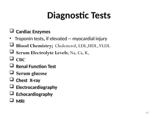
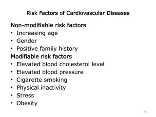
1
Nursing Health Assessment of
CVS
2
Outline
3
Purpose of cardiovascular examination
• To assess the patient appearance
• To assess effectiveness of the heart as a
pump
• To assess filling volume and pressure
• To assess the cardiac out put
• To identify the presence of compensatory
mechanism that help to maintain cardiac out
put
4
Subjective data
Ask for any
• Chest pain, dyspnea, orthopnea,
• Cough, fatigue,
• Cyanosis or pallor, edema, nocturia,
• Past history, family history, personal
habit,(nutrition, smoking, alcohol,
exercise)
5
Objective data
The neck vessels
1. The carotid arteries
• palpate each carotid arteries medial to the
sternomastiod muscle in the lower third of
the neck.
6
• Palpate only one carotid at a time
• Feel the contour and amplitude of the
pulse.
• Normally the contour is smooth and the
normal stroke is 2+ or moderate.
• Diminished pulse fells small and weak
occurs with decreased stroke volume.
7
Auscultate the carotid artery
• For persons who show signs of cardiovascular
disease, auscultate each carotid artery for the
presence of bruits.
• This is blowing, swishing sound indicating
blood flow turbulence; normally there is none.
• Ask the person to hold his or her breath while
you listen?
8
2. The jugular vein
• From the jugular vein you can asses CVP and
thus the heart efficiency as pump.
• You can not see the internal jugular vein it self
but you can see its pulsation.
9
• Position the person any where from a 30-
45 degree angle, where ever you can best
see the pulsations.
• Turn the person’s head slightly away from
the examined side.
• Note the external jugular vein overlying
the sternomastoid muscle.
10
• Full distention of external jugular veins
above 45 degree signify increased CVP.
11
• Now look for pulsation’s of the internal jugular
vein in the area of the supra sternal notch or
around the origin of the sternomastoid muscle
around the clavicle.
• You must be able to distinguish internal jugular
vein pulsation from that of the carotid artery.
12
The pericardium
Inspect the anterior chest.
• You may or may not see the apical
impulse (pulsation of left ventricle,) when
visible it occupies the fourth or fifith
intercostals space at the mid-clavicular
line.
• Easier to see in children or those with
thinner chest walls.
13
14
Assessment
• Position client supine
• Then head elevated at 45 degrees
INSPECTION:
• Lifts, heaves
• PMI (assess location)
15
Inspection
• Chest for visible cardiac motion
• Estimate Jugular venous pressure
• Patient supine and head elevated to 15-30
degrees.
• JVP is the distance b/w highest point at which
pulsation can be seen and the sternal angle
16
Jugular Venous Pressure
• An indirect measure of right atrial pressure.
• Measured in centimeters from the sternal angle
and is best visualized with the patient's head
rotated to the left.
• Described for its quality and character, effects of
respiration, and patient position-induced
changes.
17
Palpation
18
Physical Landmarks
• Suprasternal
notch
• Sternum
• Manubriosternal
angle – Angle of
Louis
• Intercostals
Spaces
19
Palpations
• Palpate for PMI; easiest if patient sits up and
leans forward
• has a diameter of  2cm and located with 10
cm of the midsternal line
• Palpate for general cardiac motion with
fingertips and patient in supine position
• Palpate for radial, carotid, brachial, femoral,
popliteal, posterior tibial And dorsalis pedis
peripheral pulses
20
radial pulse Brachial pulse
popliteal pulse
21
Dorsalis pedis pulse Posterior tibial pulse
22
Palpations…
• Rate strength of the pulse normal, diminished,
or absent on a scale of 0 to +4, where 2+ is
normal.
23
Auscultation:
Auscultatory Sites
24
Auscultation
• With a stethoscope
• Use diaphragm to assess higher pitched
sounds
• Needs a lot of practice and experience
• Listen in a quiet area or to close eyes to
reduce conflicting stimuli
• See also figure 4-10 for auscultatory Sites
25
Auscultatory Sites: Cont.
26
Auscultatory Sites
• The auscultatory Sites are close to but not the
same as the anatomic locations of the valves.
• Aortic area2nd
ICS at the right sternal border
• Pulmonic  2nd
ICS at the left sternal border
• Tricuspid  lt lower sternal border
• Mitral cardiac apex
27
Heart Sounds
• Heart sounds are characterized by location,
pitch, intensity, duration, and timing within the
cardiac cycle
28
Heart Sounds
• High-pitched sounds such as S1 and S2,
murmurs of aortic and mitral regurgitation,
and pericardial friction rubs are best heard
with the diaphragm.
• The bell is preferred for low-pitched
sounds such as S3 and S4.
29
Heart Sounds – S1…(Lub)…
• S1: Closure of AV valves
(mitral and tricuspid
valves: M1 before T1)
• Correlates with the
carotid pulse
• Loudest at the cardiac
apex
• Can be split but not often
30
Heart Sounds – S2…(Dub)…
• S2: Closure of
Semilunar valves
(aortic & pulmonic)
• Loudest at the base
of the heart
• May have a split
sound (A2 before
P2)
31
Heart Sounds – S2…(Dub)…
• S1 and S2 assessed in all four sites in upright
and supine position
• S1 precedes and the S2 follows the carotid
pulse
32
Heart Sounds…
• Base (R/L 2nd ICS)
– S2 louder than S1
• Apex
– S1 louder than S2
• Normal physiologic S2 Split
– Best heard at pulmonic area
during inspiration
• Fixed split (no variation
with inspiration)
Slide 19-33
Sample Charting
Slide 19-34
Sample Charting
(cont.)
35
Extra Heart Sounds
S3…
• Due to volume overload
• Due to Rapid ventricular
filling: ventricular gallop
• S1 -- S2-S3 (Ken--tuc-ky)
S4…
• Due to pressure overload
• Due to slow ventricular
contraction: atrial gallop
• S4-S1 — S2 (Ten-nes—see)
36
Extra Heart Sounds
S3…
• Low-pitched sound
• Usually heard at the
apex of the heart.
• Caused by rapid filling
and stretching of the left
ventricle when the left
ventricle is somewhat
noncompliant.
• Characteristic of volume
overloading, such as in
CHF (especially left-
sided heart failure),
tricuspid or mitral valve
insufficiency.
S4…
• A dull, low-pitched
postsystolic atrial gallop
• Usually caused by reduced
ventricular compliance.
• Best heard at the apex in the
left lateral position.
• Occurs with reduced
ventricular compliance and is
present in conditions such as
aortic stenosis, hypertension,
hypertrophic
cardiomyopathies, and
coronary artery disease.
• Less specific for CHF than S3.
37
Murmurs
• Turbulent blood flow across a valve or a disease
such as anemia or hyperthyroidism
• Listen for murmurs in the same auscultatory sites
APETM
• Systolic b/n S1 & S2
• Diastolic b/n S2 & S1
38
Characteristics of Heart Sounds
Type of
Murmur Examples Location Pitch Radiation Quality
Midsystolic
Aortic
stenosis 2nd RICS Medium Neck, left
sternal border Harsh
Pulmonic
stenosis
2nd and 3rd
LICS Medium Left shoulder
and neck Harsh
Hypertrophic
cardiomyopat
hy
3rd and 4th
LICS Medium
Left sternal
border to
apex
Harsh
Pansystolic
Mitral
regurgitation Apex Medium to
high Left axilla Blowing
Tricuspid
regurgitation
Lower left
sternal border Medium
Right
sternum,
xiphoid
Blowing
Ventricular
septal defect
3rd, 4th, and
5th LICS High Often harsh
Diastolic
Aortic
regurgitation
2nd to 4th
LICS High Apex Blowing
Mitral stenosis Apex Low Little or none
39
Murmurs
• They are classified by
– timing and duration within the cardiac cycle (systolic,
diastolic, and continuous),
– location,
– intensity,
– shape (configuration or pattern),
– pitch (frequency),
– quality, and radiation
40
Murmurs
• Grade I: barely audible
• Gr II: audible but quiet and soft
• Gr III: moderated loud, without thrust or thrill
• Gr IV: loud, with thrill
• Gr V: louder with thrill, steth on chest wall
• Gr VI: loud enough to be heard before steth on
chest
41
Murmurs
• Thrill:
– a palpable murmur
• Bruits:
– Vascular murmur
– sounds made by turbulent blood flow
– Heard over blood vessels with constricted lumens.
– Carotid and femoral are routinely assessed for
bruits
– Sometimes found over the vertebral, subclavian
and abdominal arteries
Slide 19-42
Abnormal Findings
Murmurs Caused by Valvular Defects
• Midsystolic ejection
murmurs
– Aortic stenosis
– Pulmonic stenosis
• Pansystolic regurgitant
murmurs
– Mitral regurgitation
– Tricuspid regurgitation
• Diastolic rumbles of
atrioventricular valves
– Mitral stenosis
– Tricuspid stenosis
• Early diastolic
murmurs
– Aortic regurgitation
– Pulmonic
regurgitation
43
Diagnostic Tests
 Cardiac Enzymes
• Troponin tests, if elevated -- myocardial injury
 Blood Chemistry; Cholesterol, LDL,HDL,VLDL
 Serum Electrolyte Levels; Na, Ca, K,
 CBC
 Renal Function Test
 Serum glucose
 Chest X-ray
 Electrocardiography
 Echocardiography
 MRI
44
Risk Factors of Cardiovascular Diseases
Non-modifiable risk factors
• Increasing age
• Gender
• Positive family history
Modifiable risk factors
• Elevated blood cholesterol level
• Elevated blood pressure
• Cigarette smoking
• Physical inactivity
• Stress
• Obesity
45

2-Assessment Cardiovascular system .pptx

  • 1.
  • 2.
  • 3.
    3 Purpose of cardiovascularexamination • To assess the patient appearance • To assess effectiveness of the heart as a pump • To assess filling volume and pressure • To assess the cardiac out put • To identify the presence of compensatory mechanism that help to maintain cardiac out put
  • 4.
    4 Subjective data Ask forany • Chest pain, dyspnea, orthopnea, • Cough, fatigue, • Cyanosis or pallor, edema, nocturia, • Past history, family history, personal habit,(nutrition, smoking, alcohol, exercise)
  • 5.
    5 Objective data The neckvessels 1. The carotid arteries • palpate each carotid arteries medial to the sternomastiod muscle in the lower third of the neck.
  • 6.
    6 • Palpate onlyone carotid at a time • Feel the contour and amplitude of the pulse. • Normally the contour is smooth and the normal stroke is 2+ or moderate. • Diminished pulse fells small and weak occurs with decreased stroke volume.
  • 7.
    7 Auscultate the carotidartery • For persons who show signs of cardiovascular disease, auscultate each carotid artery for the presence of bruits. • This is blowing, swishing sound indicating blood flow turbulence; normally there is none. • Ask the person to hold his or her breath while you listen?
  • 8.
    8 2. The jugularvein • From the jugular vein you can asses CVP and thus the heart efficiency as pump. • You can not see the internal jugular vein it self but you can see its pulsation.
  • 9.
    9 • Position theperson any where from a 30- 45 degree angle, where ever you can best see the pulsations. • Turn the person’s head slightly away from the examined side. • Note the external jugular vein overlying the sternomastoid muscle.
  • 10.
    10 • Full distentionof external jugular veins above 45 degree signify increased CVP.
  • 11.
    11 • Now lookfor pulsation’s of the internal jugular vein in the area of the supra sternal notch or around the origin of the sternomastoid muscle around the clavicle. • You must be able to distinguish internal jugular vein pulsation from that of the carotid artery.
  • 12.
    12 The pericardium Inspect theanterior chest. • You may or may not see the apical impulse (pulsation of left ventricle,) when visible it occupies the fourth or fifith intercostals space at the mid-clavicular line. • Easier to see in children or those with thinner chest walls.
  • 13.
  • 14.
    14 Assessment • Position clientsupine • Then head elevated at 45 degrees INSPECTION: • Lifts, heaves • PMI (assess location)
  • 15.
    15 Inspection • Chest forvisible cardiac motion • Estimate Jugular venous pressure • Patient supine and head elevated to 15-30 degrees. • JVP is the distance b/w highest point at which pulsation can be seen and the sternal angle
  • 16.
    16 Jugular Venous Pressure •An indirect measure of right atrial pressure. • Measured in centimeters from the sternal angle and is best visualized with the patient's head rotated to the left. • Described for its quality and character, effects of respiration, and patient position-induced changes.
  • 17.
  • 18.
    18 Physical Landmarks • Suprasternal notch •Sternum • Manubriosternal angle – Angle of Louis • Intercostals Spaces
  • 19.
    19 Palpations • Palpate forPMI; easiest if patient sits up and leans forward • has a diameter of  2cm and located with 10 cm of the midsternal line • Palpate for general cardiac motion with fingertips and patient in supine position • Palpate for radial, carotid, brachial, femoral, popliteal, posterior tibial And dorsalis pedis peripheral pulses
  • 20.
    20 radial pulse Brachialpulse popliteal pulse
  • 21.
    21 Dorsalis pedis pulsePosterior tibial pulse
  • 22.
    22 Palpations… • Rate strengthof the pulse normal, diminished, or absent on a scale of 0 to +4, where 2+ is normal.
  • 23.
  • 24.
    24 Auscultation • With astethoscope • Use diaphragm to assess higher pitched sounds • Needs a lot of practice and experience • Listen in a quiet area or to close eyes to reduce conflicting stimuli • See also figure 4-10 for auscultatory Sites
  • 25.
  • 26.
    26 Auscultatory Sites • Theauscultatory Sites are close to but not the same as the anatomic locations of the valves. • Aortic area2nd ICS at the right sternal border • Pulmonic  2nd ICS at the left sternal border • Tricuspid  lt lower sternal border • Mitral cardiac apex
  • 27.
    27 Heart Sounds • Heartsounds are characterized by location, pitch, intensity, duration, and timing within the cardiac cycle
  • 28.
    28 Heart Sounds • High-pitchedsounds such as S1 and S2, murmurs of aortic and mitral regurgitation, and pericardial friction rubs are best heard with the diaphragm. • The bell is preferred for low-pitched sounds such as S3 and S4.
  • 29.
    29 Heart Sounds –S1…(Lub)… • S1: Closure of AV valves (mitral and tricuspid valves: M1 before T1) • Correlates with the carotid pulse • Loudest at the cardiac apex • Can be split but not often
  • 30.
    30 Heart Sounds –S2…(Dub)… • S2: Closure of Semilunar valves (aortic & pulmonic) • Loudest at the base of the heart • May have a split sound (A2 before P2)
  • 31.
    31 Heart Sounds –S2…(Dub)… • S1 and S2 assessed in all four sites in upright and supine position • S1 precedes and the S2 follows the carotid pulse
  • 32.
    32 Heart Sounds… • Base(R/L 2nd ICS) – S2 louder than S1 • Apex – S1 louder than S2 • Normal physiologic S2 Split – Best heard at pulmonic area during inspiration • Fixed split (no variation with inspiration)
  • 33.
  • 34.
  • 35.
    35 Extra Heart Sounds S3… •Due to volume overload • Due to Rapid ventricular filling: ventricular gallop • S1 -- S2-S3 (Ken--tuc-ky) S4… • Due to pressure overload • Due to slow ventricular contraction: atrial gallop • S4-S1 — S2 (Ten-nes—see)
  • 36.
    36 Extra Heart Sounds S3… •Low-pitched sound • Usually heard at the apex of the heart. • Caused by rapid filling and stretching of the left ventricle when the left ventricle is somewhat noncompliant. • Characteristic of volume overloading, such as in CHF (especially left- sided heart failure), tricuspid or mitral valve insufficiency. S4… • A dull, low-pitched postsystolic atrial gallop • Usually caused by reduced ventricular compliance. • Best heard at the apex in the left lateral position. • Occurs with reduced ventricular compliance and is present in conditions such as aortic stenosis, hypertension, hypertrophic cardiomyopathies, and coronary artery disease. • Less specific for CHF than S3.
  • 37.
    37 Murmurs • Turbulent bloodflow across a valve or a disease such as anemia or hyperthyroidism • Listen for murmurs in the same auscultatory sites APETM • Systolic b/n S1 & S2 • Diastolic b/n S2 & S1
  • 38.
    38 Characteristics of HeartSounds Type of Murmur Examples Location Pitch Radiation Quality Midsystolic Aortic stenosis 2nd RICS Medium Neck, left sternal border Harsh Pulmonic stenosis 2nd and 3rd LICS Medium Left shoulder and neck Harsh Hypertrophic cardiomyopat hy 3rd and 4th LICS Medium Left sternal border to apex Harsh Pansystolic Mitral regurgitation Apex Medium to high Left axilla Blowing Tricuspid regurgitation Lower left sternal border Medium Right sternum, xiphoid Blowing Ventricular septal defect 3rd, 4th, and 5th LICS High Often harsh Diastolic Aortic regurgitation 2nd to 4th LICS High Apex Blowing Mitral stenosis Apex Low Little or none
  • 39.
    39 Murmurs • They areclassified by – timing and duration within the cardiac cycle (systolic, diastolic, and continuous), – location, – intensity, – shape (configuration or pattern), – pitch (frequency), – quality, and radiation
  • 40.
    40 Murmurs • Grade I:barely audible • Gr II: audible but quiet and soft • Gr III: moderated loud, without thrust or thrill • Gr IV: loud, with thrill • Gr V: louder with thrill, steth on chest wall • Gr VI: loud enough to be heard before steth on chest
  • 41.
    41 Murmurs • Thrill: – apalpable murmur • Bruits: – Vascular murmur – sounds made by turbulent blood flow – Heard over blood vessels with constricted lumens. – Carotid and femoral are routinely assessed for bruits – Sometimes found over the vertebral, subclavian and abdominal arteries
  • 42.
    Slide 19-42 Abnormal Findings MurmursCaused by Valvular Defects • Midsystolic ejection murmurs – Aortic stenosis – Pulmonic stenosis • Pansystolic regurgitant murmurs – Mitral regurgitation – Tricuspid regurgitation • Diastolic rumbles of atrioventricular valves – Mitral stenosis – Tricuspid stenosis • Early diastolic murmurs – Aortic regurgitation – Pulmonic regurgitation
  • 43.
    43 Diagnostic Tests  CardiacEnzymes • Troponin tests, if elevated -- myocardial injury  Blood Chemistry; Cholesterol, LDL,HDL,VLDL  Serum Electrolyte Levels; Na, Ca, K,  CBC  Renal Function Test  Serum glucose  Chest X-ray  Electrocardiography  Echocardiography  MRI
  • 44.
    44 Risk Factors ofCardiovascular Diseases Non-modifiable risk factors • Increasing age • Gender • Positive family history Modifiable risk factors • Elevated blood cholesterol level • Elevated blood pressure • Cigarette smoking • Physical inactivity • Stress • Obesity
  • 45.