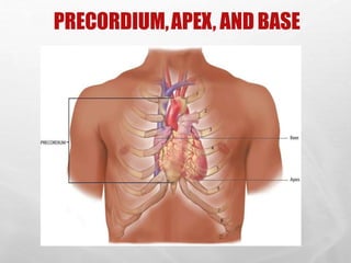
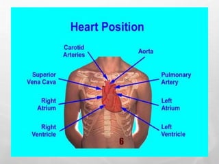
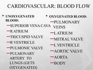
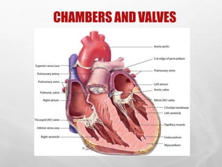
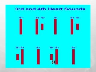
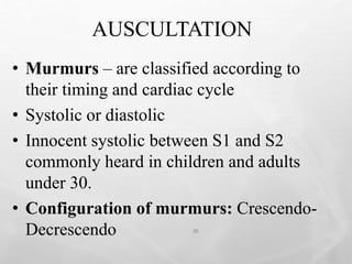
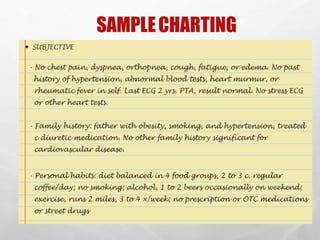
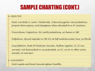
UNIT 08:
ASSESSMENT OF
CARDIOVASCULAR SYSTEM
By: Nosheen Hussain
OBJECTIVES
2
• By the end of the unit, learners will be able to:
• Describe the components of health history that should be
elicited during the assessment of cardiovascular system.
• Identify the landmarks of the chest.
• Describe the following:
– Pulse rate, rhythm and pulsation characteristics
– PMI
– Heart sounds
• Discuss systolic and diastolic murmurs.
• Assess the cardiovascular system systematically.
• Document findings.
• List the changes in cardiovascular system that is
characteristics of aging process.
CARDIOVASCULAR SYSTEM
3
• Cardiovascular system consists of heart
(a muscular pump) and blood vessels
• Blood vessels are arranged in two
continuous loops
– Pulmonary circulation
– Systemic circulation
• When the heart contracts, it pumps blood
simultaneously into both loops
CARDIOVASCULARANATOMY &
PHYSIOLOGY
4
• Heart is shaped like “Cone”
• “top” of the heart is the base
• “bottom” is the apex
• Heart size = clenched fist
• Precordium - area on anterior chest that covers
heart and great vessels
• Atria are tilted slightly toward the back and
ventricles extend to left and toward anterior
chest wall.
PRECORDIUM,APEX, AND BASE
5
6
CARDIOVASCULAR: BLOOD FLOW
• UNOXYGENATED
BLOOD:
–SUPERIOR VENACAVA
–RATRIUM
–TRICUSPID VALVE
–R VENTRICLE
–PULMONIC VALVE
–PULMONARY
ARTERY TO
LUNGS (GETS
OXYGENATED)
• OXYGENATED BLOOD:
–PULMONARY
VEINS
–LATRIUM
–MITRAL VALVE
–LVENTRICLE
–AORTIC VALVE
–AORTA
–BODY
7
CHAMBERS ANDVALVES
8
CARDIOVASCULAR:
BLOOD FLOW
9
• There are two main coronary arteries, the left
(LCA) and the right (RCA)
• Coronary artery blood flow to the
myocardium occurs primarily during
diastole, when coronary vascular
resistance is minimized.
• To maintain adequate blood flow through the
coronary arteries, the diastolic pressure must
be at least 60 mmHg.
CARDIOVASCULAR:
CARDIAC CYCLE
10
• 2 phases:
–DIASTOLE: ventricles relax and fill
with blood
–SYSTOLE : ventricles contract pump
blood into pulmonary and systemic
arteries
CARDIOVASCULAR: HEART SOUNDS
11
• Heart sounds: lub dub
• SYSTOLE: lub= S1 (closing ofAVvalves)
• DIASTOLE: dub = S2 (closing of semilunar
valves)
• During the cardiac cycle, valves are opening
and closing, causing different heart sounds (S1
and S2).
• Sometimes abnormal heart sounds are heard
due to improper opening or closing of the
valves.(murmurs)
12
CARDIOVASCULAR:
HEART SOUNDS
13
• Characteristics of Heart Sounds
• Frequency (pitch): high or low
• Intensity (loudness): loud or soft
• Duration: very short hear sounds or
longer periods of silence
• Timing: systole or diastole
CARDIOVASCULAR:
CONDUCTION
14
• Heart contracts by itself through its
own conduction system:
• Sinoatrial (SA)node – (pacemaker)
initiates electrical impulse
• A
V node
• Bundle of HIS (L & R Bundle
Branches)
• Purkinje fibers
CARDIOVASCULAR:
CONDUCTION
15
• Electrical impulses shown on ECG
(EKG)
• PQRST wave correlates to impulses
traveling through the heart.
• SAtoA
V = P wave, (atrial stimulation)
• Stimulus spreads through bundle of His =
QRS complex
• Repolarization of ventricles =T wave on
16
CARDIOVASCULAR: PUMPING ABILITY
• Cardiac Output (C.O.) = volume of blood
in liters ejected by the heart each minute.
• Adult = 4-7 liters/minute
• C.O. = HR x SV
• Heart Rate (HR) = number of
times ventricles contract each
minute.
• Stroke Volume (SV) = The amount of
blood ejected by the left ventricle during 17
CARDIOVASCULAR
18
• Preload = degree of stretch of myocardial fibers
at end of DIASTOLE. The more the heart is
filled (within limits, i.e., not over- filled), the
more forcefully it contracts.
• Afterload = pressure or resistance the ventricles
must overcome to pump out blood. The amount
of resistance is directly related
to arterial blood pressure and the
diameter of the vessels.
ASSESSMENT: SUBJECTIVE
19
• Personal and family history
• Diet history: 24 hr. sample diet
Opportunity for teaching food
selection and preparation
• Socioeconomic status – ability to purchase
proper foods, medicines. Employment and
its effects on health?
• Cigarette smoking : # packs /day and
also # years smoked
ASSESSMENT: SUBJECTIVE
20
• PhysicalActivity/Inactivity – 30 minutes
daily of light to moderate exercise
recommended byAmerican HeartAssoc.
• Obesity – associated with HTN,
hyperlipidemia, and diabetes and all
contribute to CV disease.
• TypeApersonality – not conclusive proof
• Current Health Problems – describe health
concerns.
ASSESSMENT: SUBJECTIVE
21
• Chest pain: or discomfort, a symptom of
cardiac disease, can result from ischemic
heart disease, pericarditis and aortic
dissection.
• Chest pain: can also be due to non-
cardiac causes; pleurisy, pulmonary
embolus, hiatal hernia and anxiety.
ASSESSMENT- CHEST PAIN
22
• Onset
• Duration
• Frequency
• Precipitating factors
• Location
• Radiation
• Quality
• Intensity
ASSESSMENT: SUBJECTIVE
23
• Paroxysmal Nocturnal Dyspnea – client
has been recumbent for several hours,
increase in venous return leads to
pulmonary congestion.
• Fatigue- resulting from decreased cardiac
output is usually worse in evening.Ask
pt. if can they perform same activities as
a year ago
ASSESSMENT: SUBJECTIVE
24
• Palpitations- fluttering or unpleasant awareness of
heartbeat. Non- cardiac- causes-
fatigue, caffeine, nicotine, alcohol
• Weight gain- a sudden increase in wt. of
• 2.2 pounds (1 kg) can be result of
accumulation of fluid (1L) in interstitial
• spaces, known as edema.
• Syncope- transient loss of consciousness,
decrease in perfusion to brain.
ASSESSMENT: OBJECTIVE
25
• General appearance: Build, skin
color, LOC, presence of SOB, DOE
• Skin- color and temperature – look
for symmetry in color, temp, any
cyanosis?
• Extremities – assess skin changes,
vascular changes, clubbing,
capillary fillingand edema.
• Orthostatic BP– postural hypotension
CYANOSIS WITH ARTERIAL INSUFFICIENCY
SHAHZAD BASHIR,
NLCON, KARACHI.
26
ARTERIAL INSUFFICIENCY
27
CAPILLARY REFILLS
SHAHZAD BASHIR,
NLCON, KARACHI.
2/26/2016 28
PITTINGEDEMA
SHAHZAD BASHIR,
NLCON, KARACHI.
2/26/2016 29
ASSESSMENT: OBJECTIVE
30
• BP: supine – change position 1-2 minutes, check
again.
• Normally, systolic drops slightly or remains
unchanged and diastolic increases slightly.
• Peripheral pulses are assessed for:
– Presence
– Amplitude
– Rhythm
– Rate
– Equality
ASSESSMENT: OBJECTIVE
31
• Specific assessments for particular
populations:
• Assessment for Infants
• Assessment for Children
• Assessment for Pregnant Females
• Assessment for Elderly,
ASSESSMENT: OBJECTIVE
32
• PrecordiumAssessment- area over heart, done
by:
– Inspection (I)
– Palpation (P)
– Percussion (P)
– Auscultation (A)
PHYSICALASSESSMENT
33
• Inspection- side to side, at right angle and
downward over precordium where vibrations are
visible.
• Point of Maximal Intensity (PMI) – located at 5th
intercostal (IC) space at midclavicular line (MCL)
– mitral area
• Right Ventricular (RV area)
• Epigastric area
• Pulmonic area
PHYSICALASSESSMENT
• Palpation: fingers and most sensitive part
of palm of hand to detect any precordial
motion or thrills.
• Palpate apical impulse
• Percussion: estimate heart size, most
accurately done by chest x-ray
• Auscultation:– evaluates heart rate,
rhythm, cardiac cycle and valvular
function. 34
PHYSICALASSESSMENT:AUSCULTATION
35
• Diaphragm of stethoscope – 1st and 2nd
heart sounds and high frequency
murmurs. lub-dub
• Use bell of stethoscope – low frequency
• gallops and murmurs.
• Paradoxical splitting of S2 – severe
myocardial depression, may be seen with
an MI, aortic stenosis or other causes.
AUSCULTATION:
36
• 3rd & 4th Heart Sounds
• S3 (Gallops): rapid, passive filling phase during
diastole into noncompliant ventricle.
• S4: pathologic, may be heard with advancing age
because of stiffened ventricle.
• Both S3 and S4 = Summation Gallop:
indication of severe heart failure.
• Murmurs – Turbulent blood flow through normal
or abnormal valves.
37
AUSCULTATION
38
• Murmurs – are classified according to
their timing and cardiac cycle
• Systolic or diastolic
• Innocent systolic between S1 and S2
commonly heard in children and adults
under 30.
• Configuration of murmurs: Crescendo-
Decrescendo
AUSCULTATION
39
• Intensity of murmur:
• Grade:
– 1: faint
– 2: soft
– 3: moderately loud
– 4: loud with thrill
– 5: very loud
• (stethoscope partially off chest)
– 6: stethoscope off chest, thrill
AUSCULTATION
40
• Pericardial Friction Rubs- results from
inflammation of pericardial membrane.
• Ejection Click- Early systole, stiff
deformed valve, high pitch, apex,
diaphragm.
• Opening snap – Immediately after S2
stenotic mitral or tricuspid valve leaflets
recoil abruptly during diastole.
AUSCULTATION
TECHNIQUES
41
• Listen for: S1, S2, extra sounds in S1 and
S2, murmurs.
• Listen R. 2nd ICS close to sternum (aortic area)
• Listen L. 2nd ICS (pulmonic)
• Listen L. 3rd ICS (Erb’s point)
• Listen L. 5th ICS (tricuspid area)
• Listen L. 5th ICS medial midclavicular line (mitral)
• Listen with diaphragm and bell in each area.
• Position pt. Supine, L. side lying and sitting,
leaning forward.
42
AUSCULTATORYAREAS
SHAHZAD BASHIR,
NLCON, KARACHI.
43
CONTI
…
44
• Upon completion of auscultation of the
precordium:
• Assessment of Cardiovascular system
continues with the assessment of the
peripheral vascular system…..
SUMMARY:
CARDIOVASCULAR
45
• Physical assessment Includes:
• Neck vessels
• Precordium
• Inspection and palpation of peripheral
system with auscultation of the carotids
SAMPLECHARTING
SHAHZAD BASHIR,
NLCON, KARACHI.
2/26/2016 46
SAMPLE CHARTING(CONT.)
SHAHZAD BASHIR,
NLCON, KARACHI.
47

unit8 cardiovascular.pptx

  • 1.
    UNIT 08: ASSESSMENT OF CARDIOVASCULARSYSTEM By: Nosheen Hussain
  • 2.
    OBJECTIVES 2 • By theend of the unit, learners will be able to: • Describe the components of health history that should be elicited during the assessment of cardiovascular system. • Identify the landmarks of the chest. • Describe the following: – Pulse rate, rhythm and pulsation characteristics – PMI – Heart sounds • Discuss systolic and diastolic murmurs. • Assess the cardiovascular system systematically. • Document findings. • List the changes in cardiovascular system that is characteristics of aging process.
  • 3.
    CARDIOVASCULAR SYSTEM 3 • Cardiovascularsystem consists of heart (a muscular pump) and blood vessels • Blood vessels are arranged in two continuous loops – Pulmonary circulation – Systemic circulation • When the heart contracts, it pumps blood simultaneously into both loops
  • 4.
    CARDIOVASCULARANATOMY & PHYSIOLOGY 4 • Heartis shaped like “Cone” • “top” of the heart is the base • “bottom” is the apex • Heart size = clenched fist • Precordium - area on anterior chest that covers heart and great vessels • Atria are tilted slightly toward the back and ventricles extend to left and toward anterior chest wall.
  • 5.
  • 6.
  • 7.
    CARDIOVASCULAR: BLOOD FLOW •UNOXYGENATED BLOOD: –SUPERIOR VENACAVA –RATRIUM –TRICUSPID VALVE –R VENTRICLE –PULMONIC VALVE –PULMONARY ARTERY TO LUNGS (GETS OXYGENATED) • OXYGENATED BLOOD: –PULMONARY VEINS –LATRIUM –MITRAL VALVE –LVENTRICLE –AORTIC VALVE –AORTA –BODY 7
  • 8.
  • 9.
    CARDIOVASCULAR: BLOOD FLOW 9 • Thereare two main coronary arteries, the left (LCA) and the right (RCA) • Coronary artery blood flow to the myocardium occurs primarily during diastole, when coronary vascular resistance is minimized. • To maintain adequate blood flow through the coronary arteries, the diastolic pressure must be at least 60 mmHg.
  • 10.
    CARDIOVASCULAR: CARDIAC CYCLE 10 • 2phases: –DIASTOLE: ventricles relax and fill with blood –SYSTOLE : ventricles contract pump blood into pulmonary and systemic arteries
  • 11.
    CARDIOVASCULAR: HEART SOUNDS 11 •Heart sounds: lub dub • SYSTOLE: lub= S1 (closing ofAVvalves) • DIASTOLE: dub = S2 (closing of semilunar valves) • During the cardiac cycle, valves are opening and closing, causing different heart sounds (S1 and S2). • Sometimes abnormal heart sounds are heard due to improper opening or closing of the valves.(murmurs)
  • 12.
  • 13.
    CARDIOVASCULAR: HEART SOUNDS 13 • Characteristicsof Heart Sounds • Frequency (pitch): high or low • Intensity (loudness): loud or soft • Duration: very short hear sounds or longer periods of silence • Timing: systole or diastole
  • 14.
    CARDIOVASCULAR: CONDUCTION 14 • Heart contractsby itself through its own conduction system: • Sinoatrial (SA)node – (pacemaker) initiates electrical impulse • A V node • Bundle of HIS (L & R Bundle Branches) • Purkinje fibers
  • 15.
    CARDIOVASCULAR: CONDUCTION 15 • Electrical impulsesshown on ECG (EKG) • PQRST wave correlates to impulses traveling through the heart. • SAtoA V = P wave, (atrial stimulation) • Stimulus spreads through bundle of His = QRS complex • Repolarization of ventricles =T wave on
  • 16.
  • 17.
    CARDIOVASCULAR: PUMPING ABILITY •Cardiac Output (C.O.) = volume of blood in liters ejected by the heart each minute. • Adult = 4-7 liters/minute • C.O. = HR x SV • Heart Rate (HR) = number of times ventricles contract each minute. • Stroke Volume (SV) = The amount of blood ejected by the left ventricle during 17
  • 18.
    CARDIOVASCULAR 18 • Preload =degree of stretch of myocardial fibers at end of DIASTOLE. The more the heart is filled (within limits, i.e., not over- filled), the more forcefully it contracts. • Afterload = pressure or resistance the ventricles must overcome to pump out blood. The amount of resistance is directly related to arterial blood pressure and the diameter of the vessels.
  • 19.
    ASSESSMENT: SUBJECTIVE 19 • Personaland family history • Diet history: 24 hr. sample diet Opportunity for teaching food selection and preparation • Socioeconomic status – ability to purchase proper foods, medicines. Employment and its effects on health? • Cigarette smoking : # packs /day and also # years smoked
  • 20.
    ASSESSMENT: SUBJECTIVE 20 • PhysicalActivity/Inactivity– 30 minutes daily of light to moderate exercise recommended byAmerican HeartAssoc. • Obesity – associated with HTN, hyperlipidemia, and diabetes and all contribute to CV disease. • TypeApersonality – not conclusive proof • Current Health Problems – describe health concerns.
  • 21.
    ASSESSMENT: SUBJECTIVE 21 • Chestpain: or discomfort, a symptom of cardiac disease, can result from ischemic heart disease, pericarditis and aortic dissection. • Chest pain: can also be due to non- cardiac causes; pleurisy, pulmonary embolus, hiatal hernia and anxiety.
  • 22.
    ASSESSMENT- CHEST PAIN 22 •Onset • Duration • Frequency • Precipitating factors • Location • Radiation • Quality • Intensity
  • 23.
    ASSESSMENT: SUBJECTIVE 23 • ParoxysmalNocturnal Dyspnea – client has been recumbent for several hours, increase in venous return leads to pulmonary congestion. • Fatigue- resulting from decreased cardiac output is usually worse in evening.Ask pt. if can they perform same activities as a year ago
  • 24.
    ASSESSMENT: SUBJECTIVE 24 • Palpitations-fluttering or unpleasant awareness of heartbeat. Non- cardiac- causes- fatigue, caffeine, nicotine, alcohol • Weight gain- a sudden increase in wt. of • 2.2 pounds (1 kg) can be result of accumulation of fluid (1L) in interstitial • spaces, known as edema. • Syncope- transient loss of consciousness, decrease in perfusion to brain.
  • 25.
    ASSESSMENT: OBJECTIVE 25 • Generalappearance: Build, skin color, LOC, presence of SOB, DOE • Skin- color and temperature – look for symmetry in color, temp, any cyanosis? • Extremities – assess skin changes, vascular changes, clubbing, capillary fillingand edema. • Orthostatic BP– postural hypotension
  • 26.
    CYANOSIS WITH ARTERIALINSUFFICIENCY SHAHZAD BASHIR, NLCON, KARACHI. 26
  • 27.
  • 28.
  • 29.
  • 30.
    ASSESSMENT: OBJECTIVE 30 • BP:supine – change position 1-2 minutes, check again. • Normally, systolic drops slightly or remains unchanged and diastolic increases slightly. • Peripheral pulses are assessed for: – Presence – Amplitude – Rhythm – Rate – Equality
  • 31.
    ASSESSMENT: OBJECTIVE 31 • Specificassessments for particular populations: • Assessment for Infants • Assessment for Children • Assessment for Pregnant Females • Assessment for Elderly,
  • 32.
    ASSESSMENT: OBJECTIVE 32 • PrecordiumAssessment-area over heart, done by: – Inspection (I) – Palpation (P) – Percussion (P) – Auscultation (A)
  • 33.
    PHYSICALASSESSMENT 33 • Inspection- sideto side, at right angle and downward over precordium where vibrations are visible. • Point of Maximal Intensity (PMI) – located at 5th intercostal (IC) space at midclavicular line (MCL) – mitral area • Right Ventricular (RV area) • Epigastric area • Pulmonic area
  • 34.
    PHYSICALASSESSMENT • Palpation: fingersand most sensitive part of palm of hand to detect any precordial motion or thrills. • Palpate apical impulse • Percussion: estimate heart size, most accurately done by chest x-ray • Auscultation:– evaluates heart rate, rhythm, cardiac cycle and valvular function. 34
  • 35.
    PHYSICALASSESSMENT:AUSCULTATION 35 • Diaphragm ofstethoscope – 1st and 2nd heart sounds and high frequency murmurs. lub-dub • Use bell of stethoscope – low frequency • gallops and murmurs. • Paradoxical splitting of S2 – severe myocardial depression, may be seen with an MI, aortic stenosis or other causes.
  • 36.
    AUSCULTATION: 36 • 3rd &4th Heart Sounds • S3 (Gallops): rapid, passive filling phase during diastole into noncompliant ventricle. • S4: pathologic, may be heard with advancing age because of stiffened ventricle. • Both S3 and S4 = Summation Gallop: indication of severe heart failure. • Murmurs – Turbulent blood flow through normal or abnormal valves.
  • 37.
  • 38.
    AUSCULTATION 38 • Murmurs –are classified according to their timing and cardiac cycle • Systolic or diastolic • Innocent systolic between S1 and S2 commonly heard in children and adults under 30. • Configuration of murmurs: Crescendo- Decrescendo
  • 39.
    AUSCULTATION 39 • Intensity ofmurmur: • Grade: – 1: faint – 2: soft – 3: moderately loud – 4: loud with thrill – 5: very loud • (stethoscope partially off chest) – 6: stethoscope off chest, thrill
  • 40.
    AUSCULTATION 40 • Pericardial FrictionRubs- results from inflammation of pericardial membrane. • Ejection Click- Early systole, stiff deformed valve, high pitch, apex, diaphragm. • Opening snap – Immediately after S2 stenotic mitral or tricuspid valve leaflets recoil abruptly during diastole.
  • 41.
    AUSCULTATION TECHNIQUES 41 • Listen for:S1, S2, extra sounds in S1 and S2, murmurs. • Listen R. 2nd ICS close to sternum (aortic area) • Listen L. 2nd ICS (pulmonic) • Listen L. 3rd ICS (Erb’s point) • Listen L. 5th ICS (tricuspid area) • Listen L. 5th ICS medial midclavicular line (mitral) • Listen with diaphragm and bell in each area. • Position pt. Supine, L. side lying and sitting, leaning forward.
  • 42.
  • 43.
  • 44.
    CONTI … 44 • Upon completionof auscultation of the precordium: • Assessment of Cardiovascular system continues with the assessment of the peripheral vascular system…..
  • 45.
    SUMMARY: CARDIOVASCULAR 45 • Physical assessmentIncludes: • Neck vessels • Precordium • Inspection and palpation of peripheral system with auscultation of the carotids
  • 46.
  • 47.