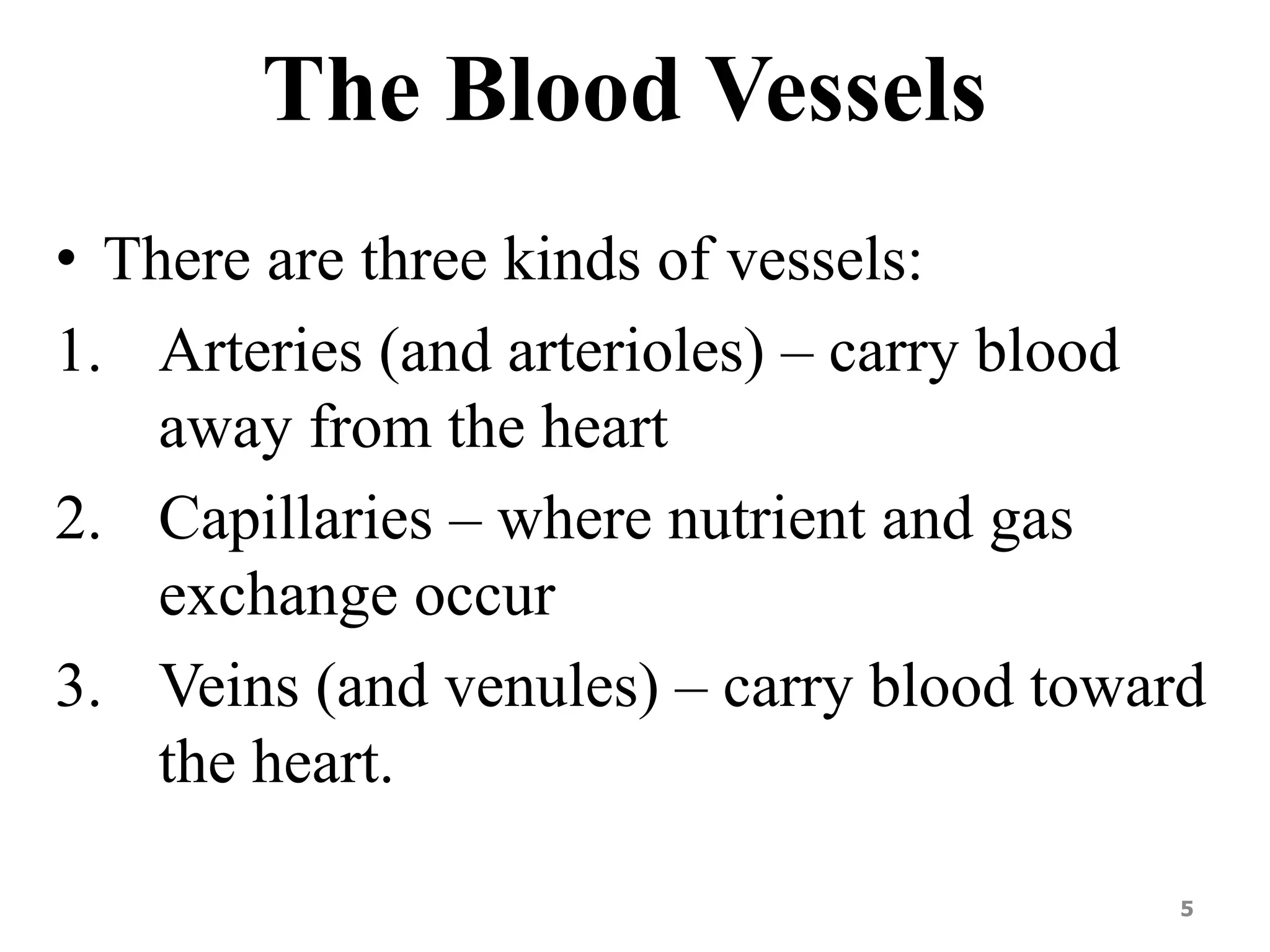
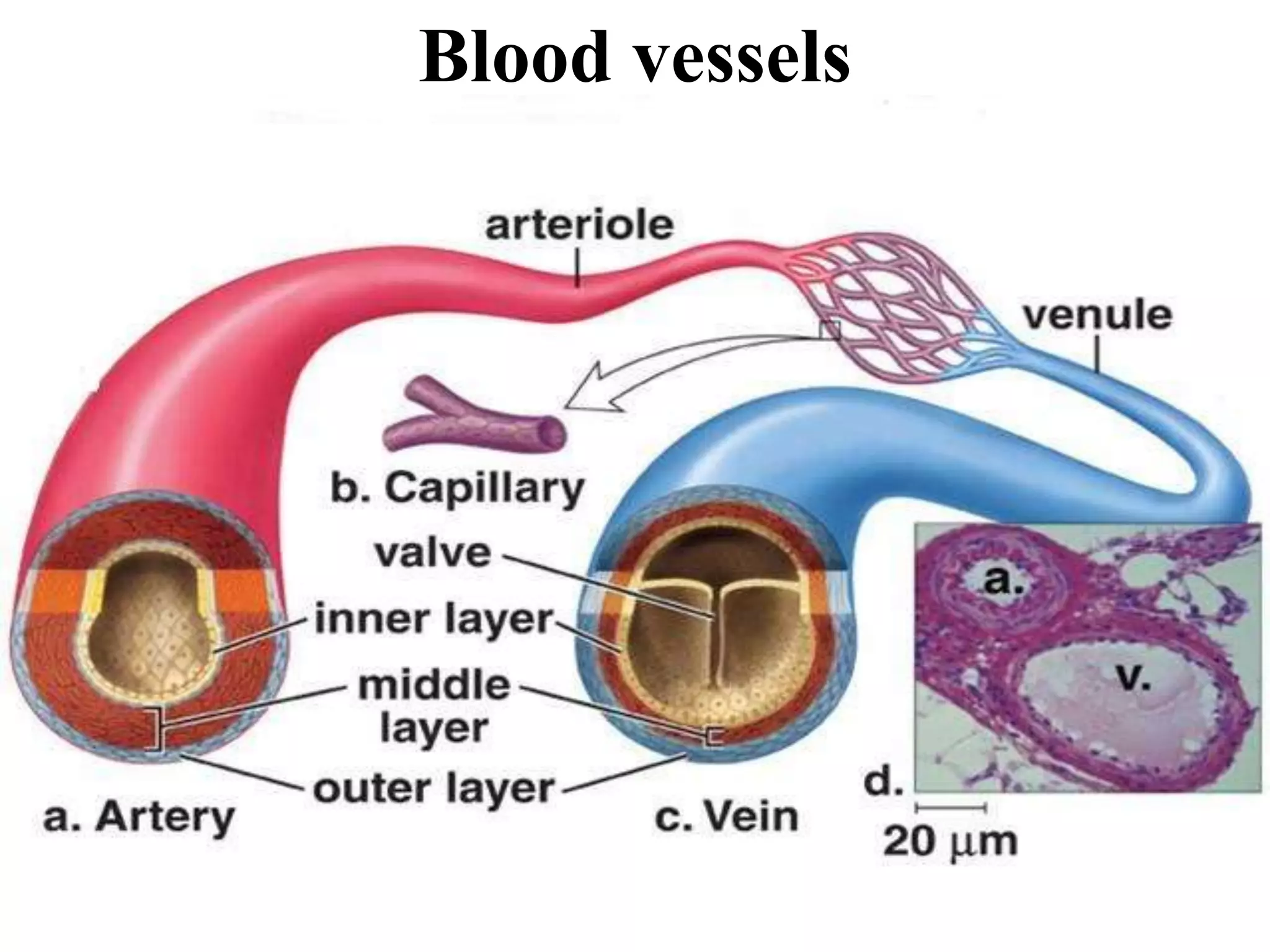
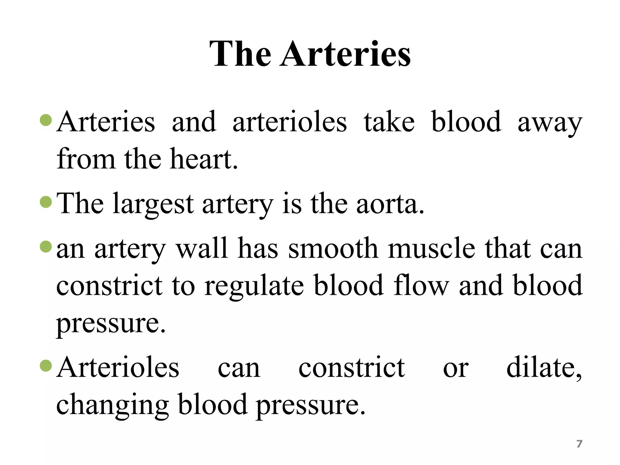
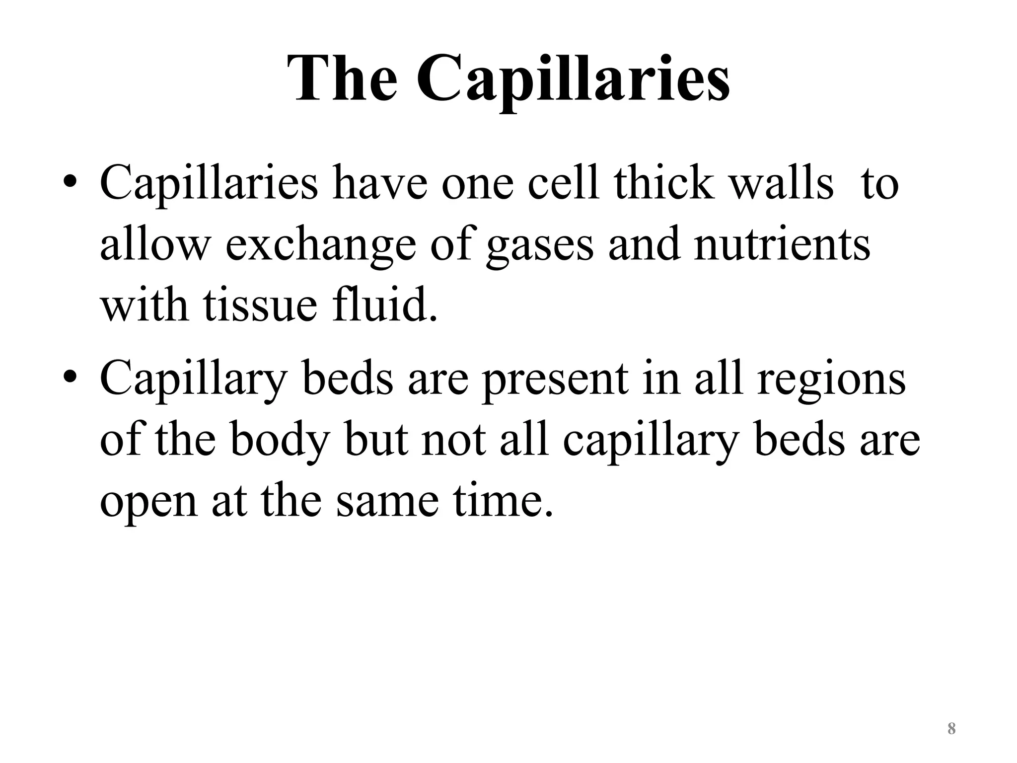
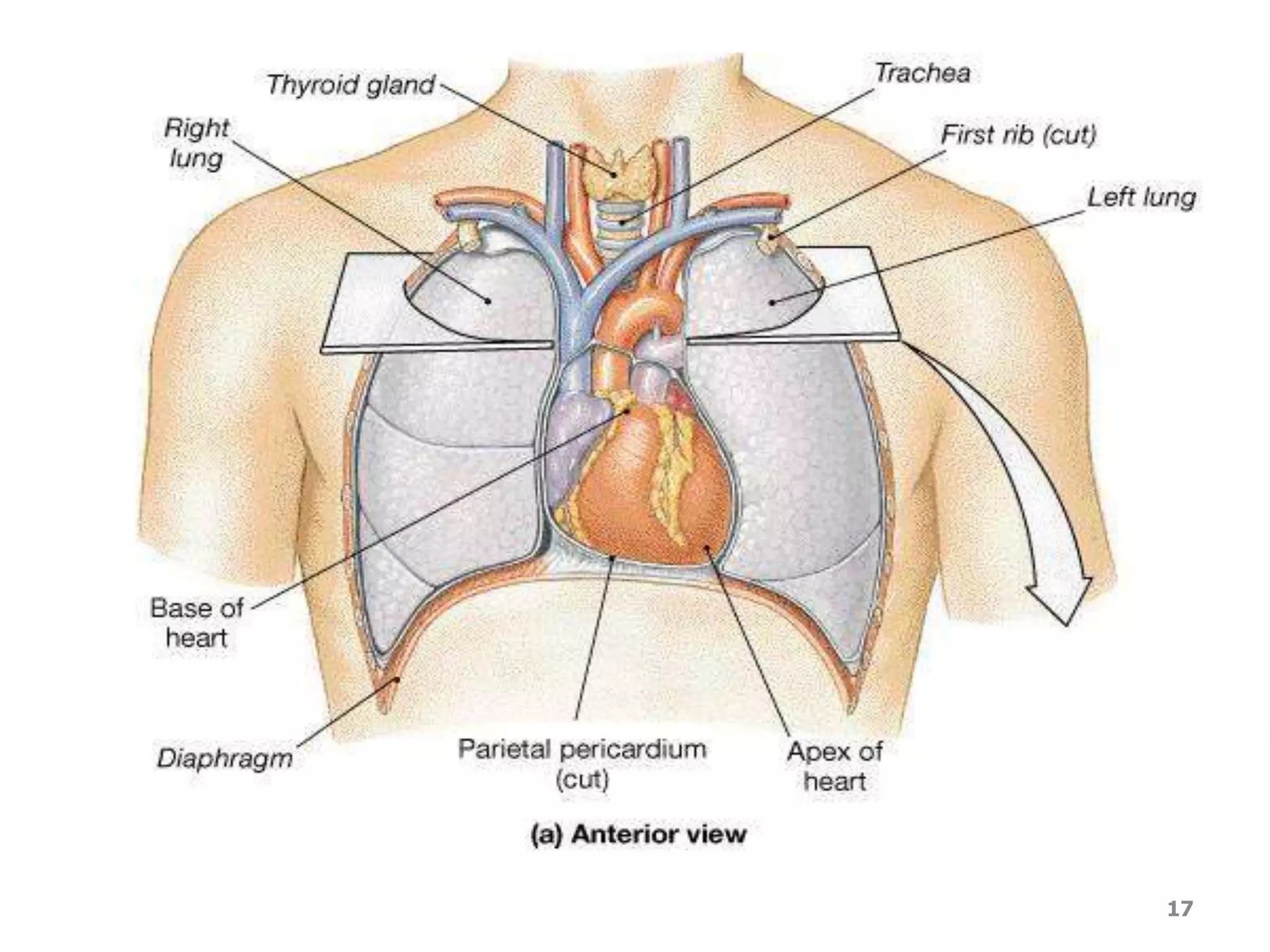
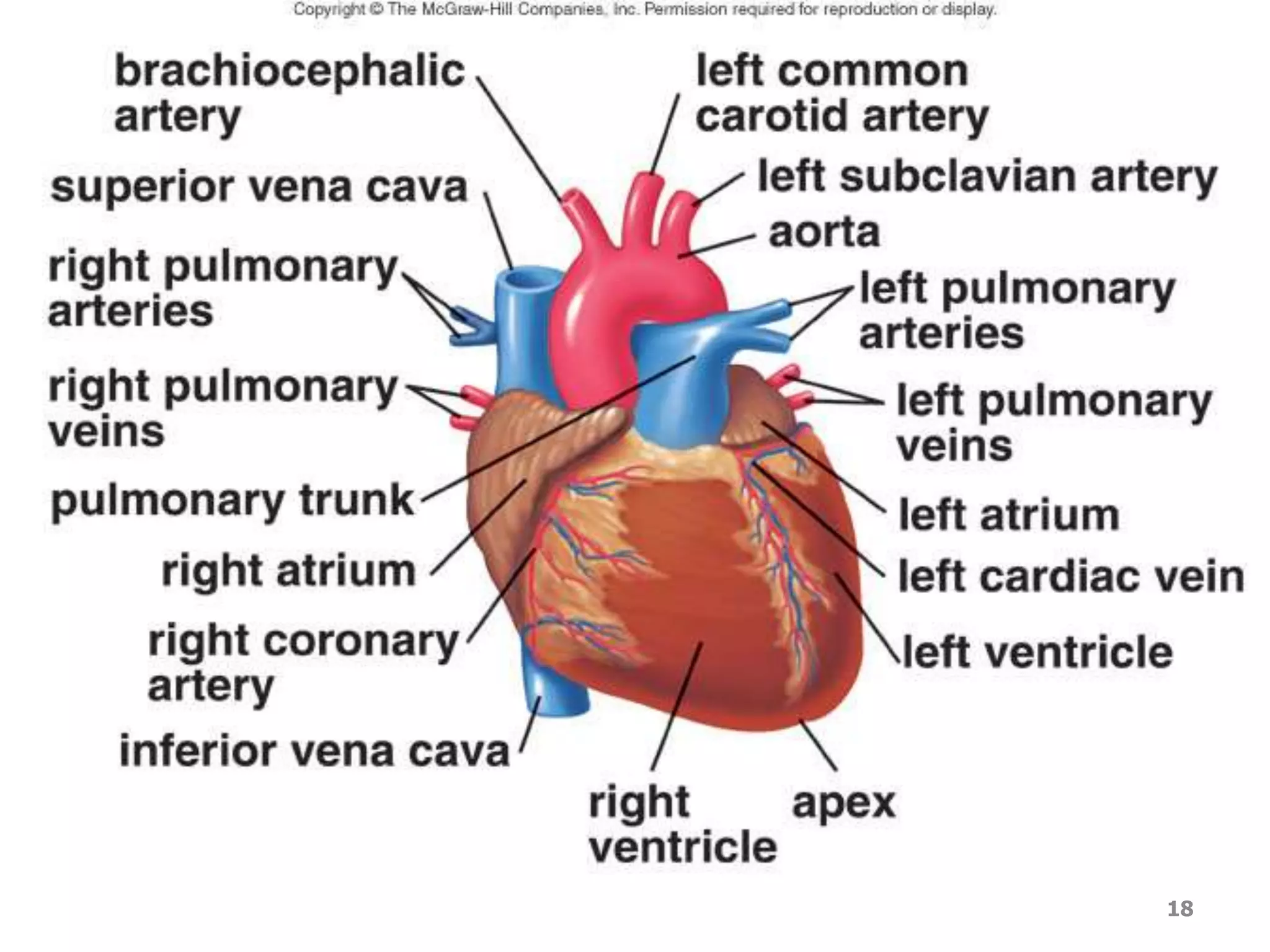
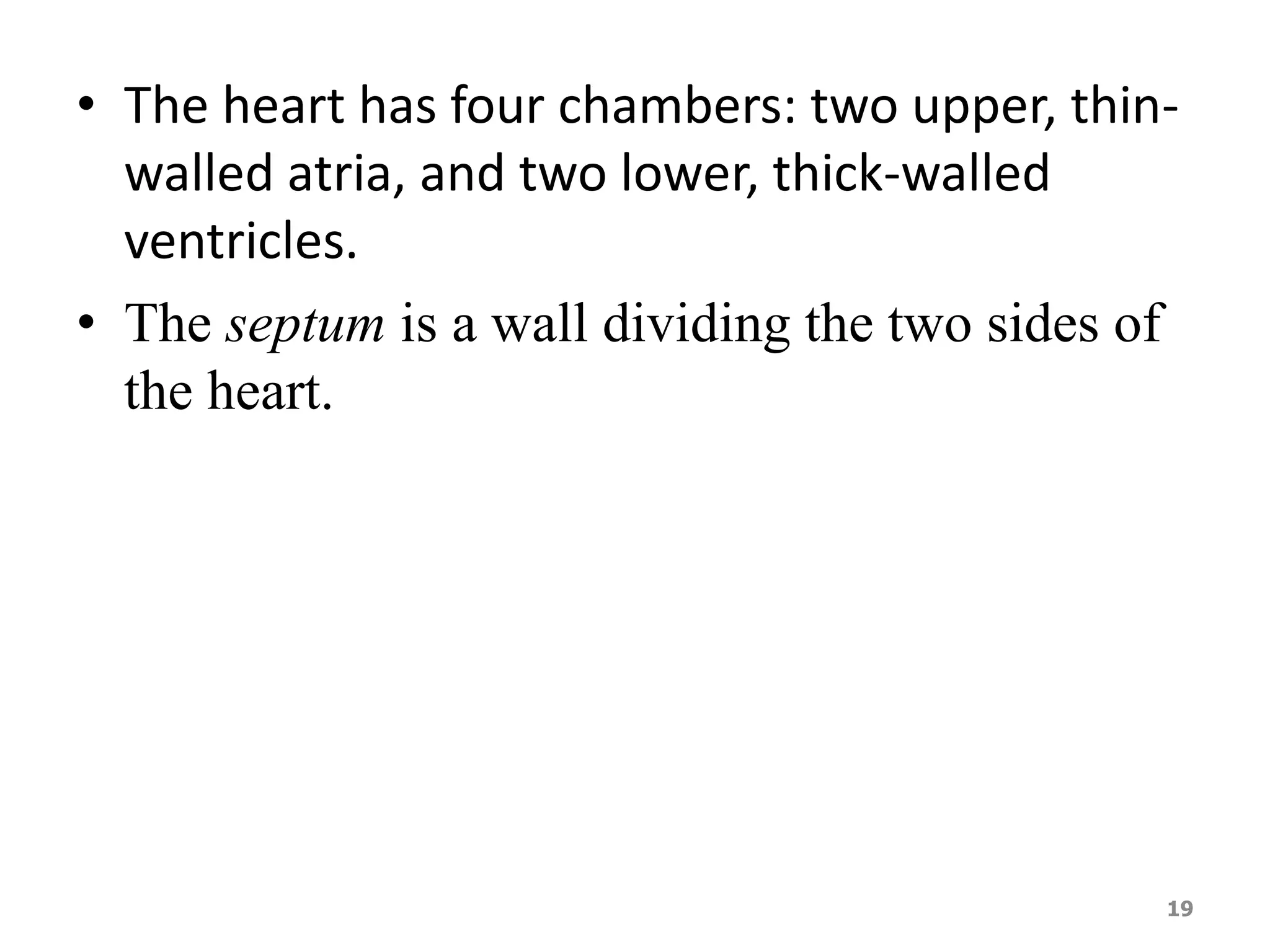
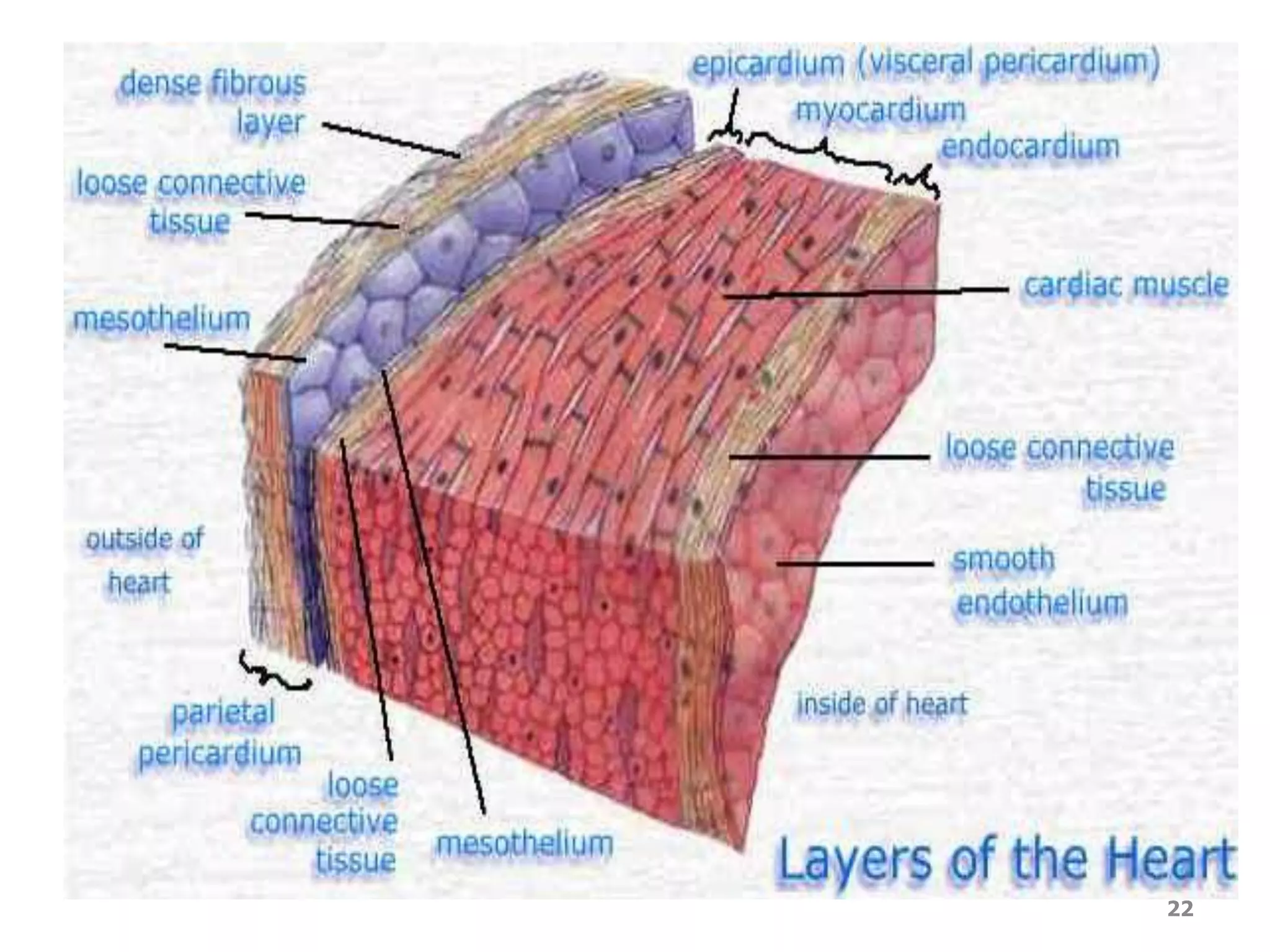
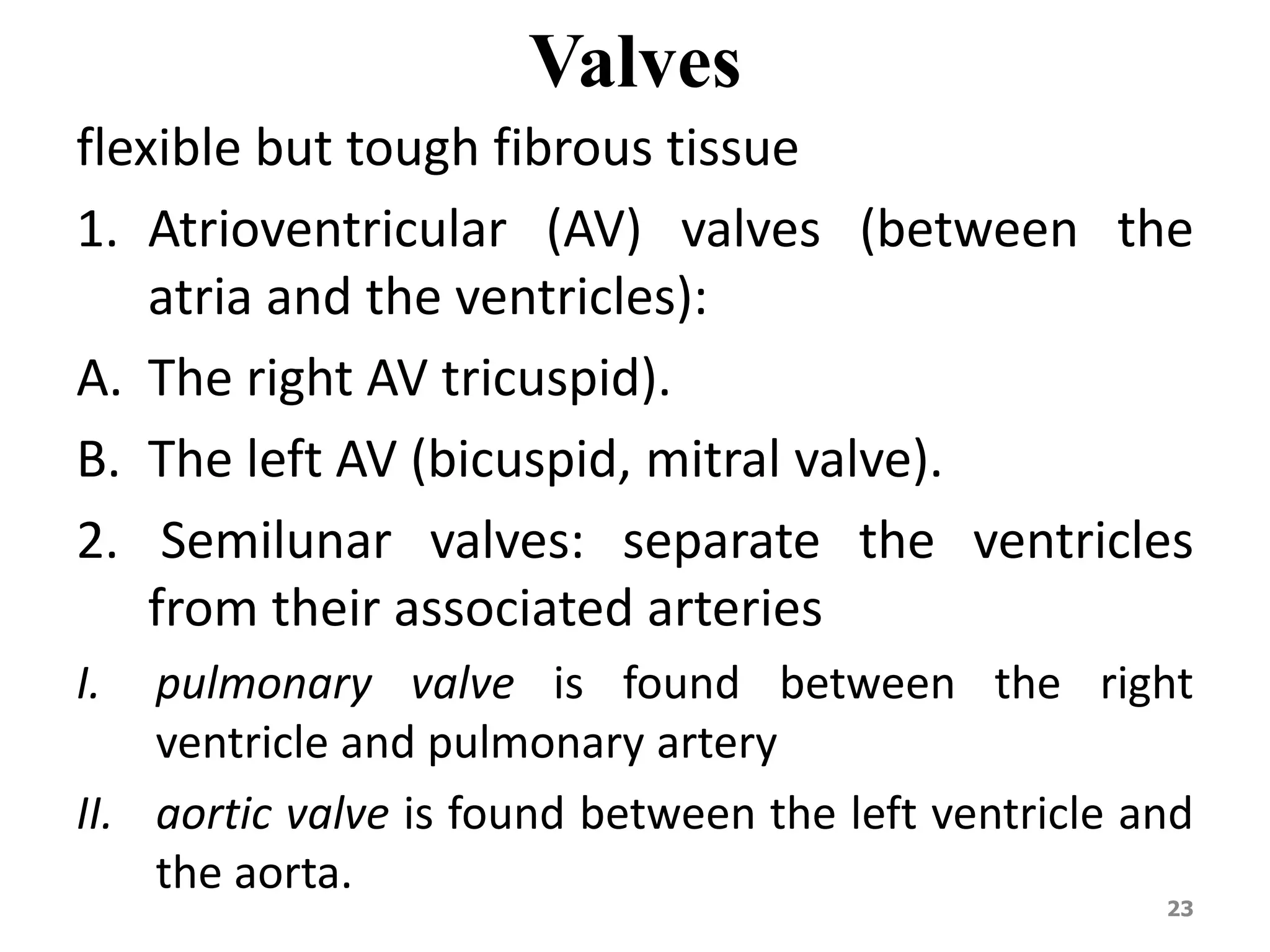
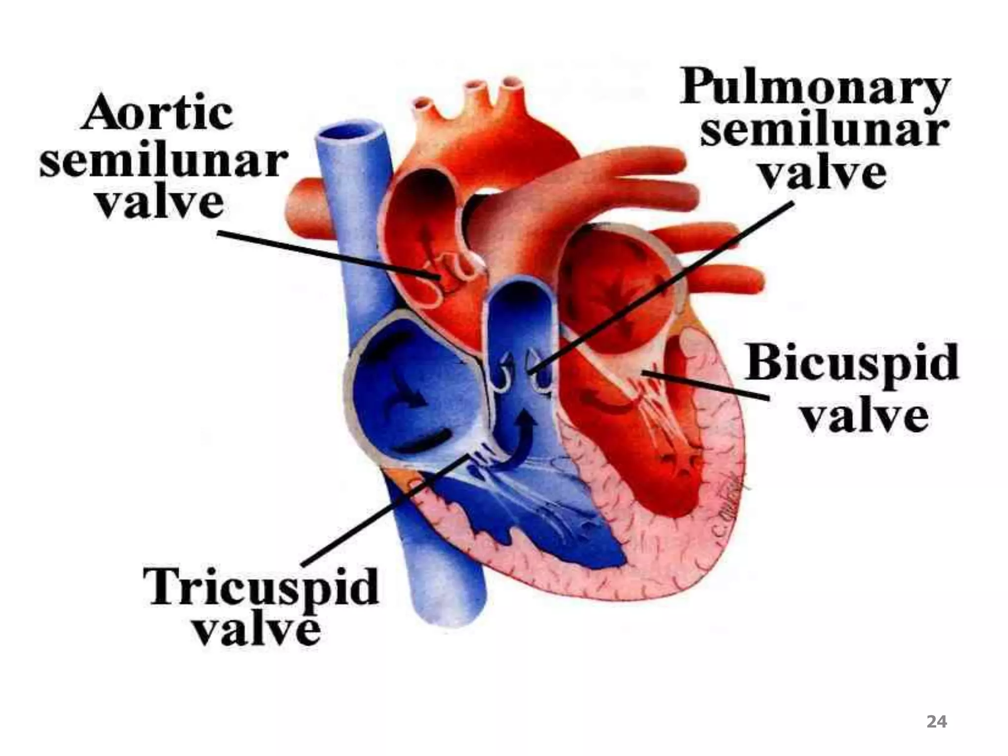
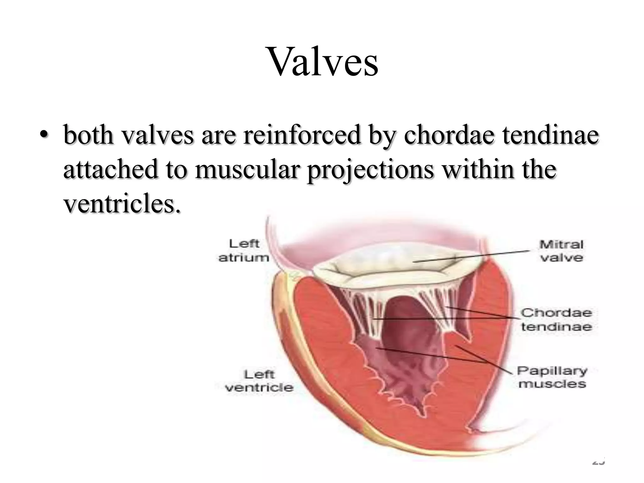
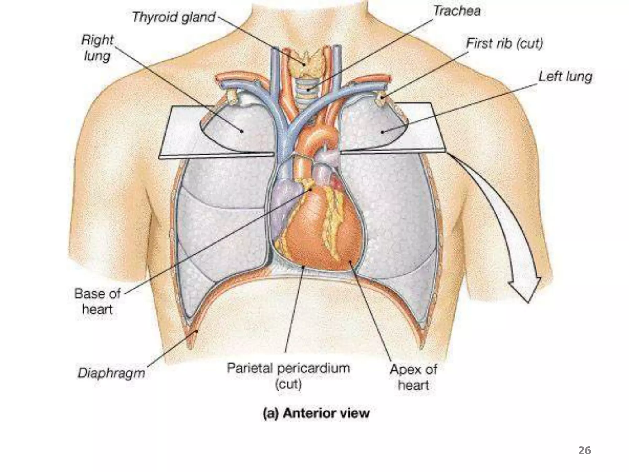
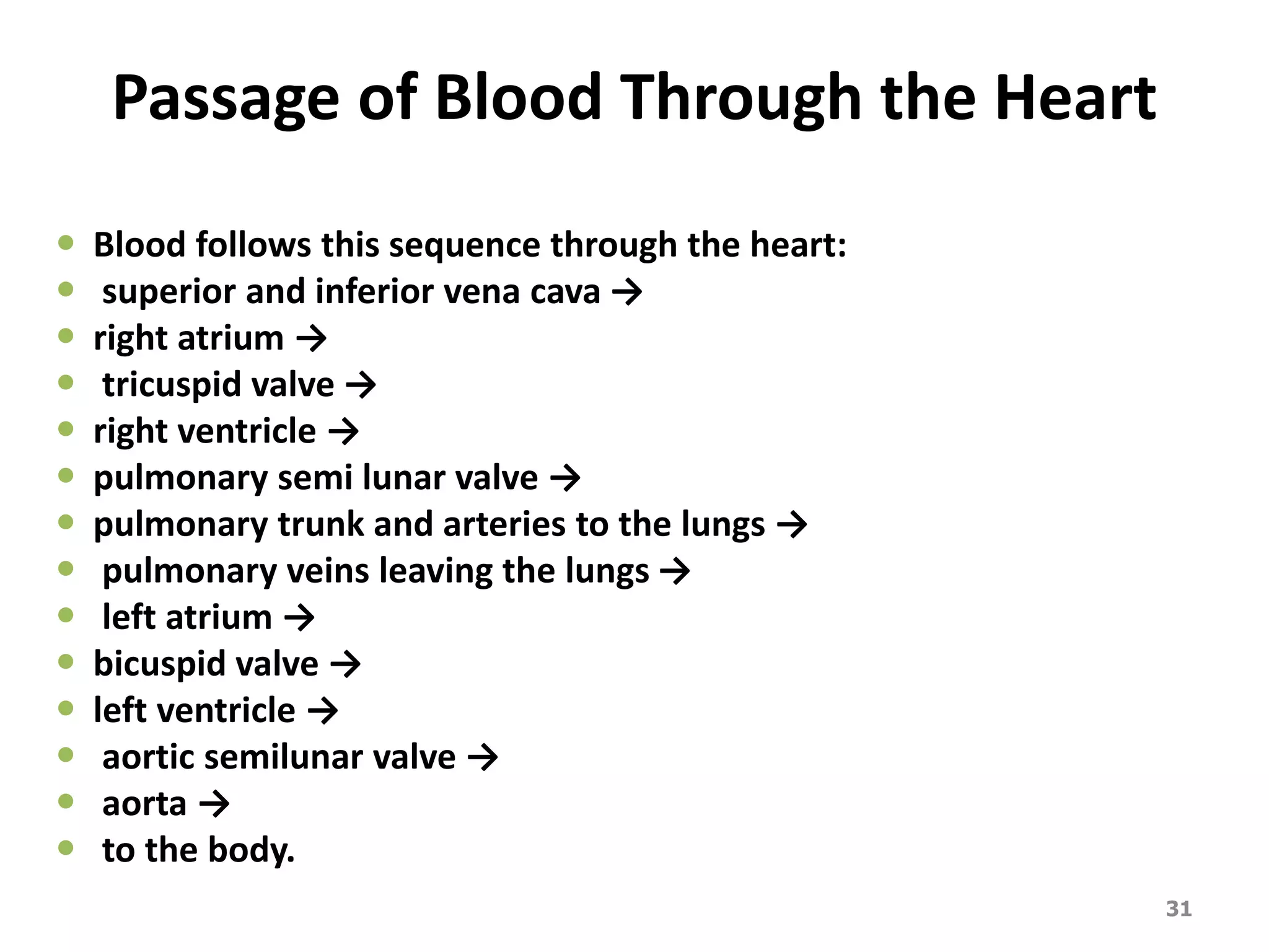
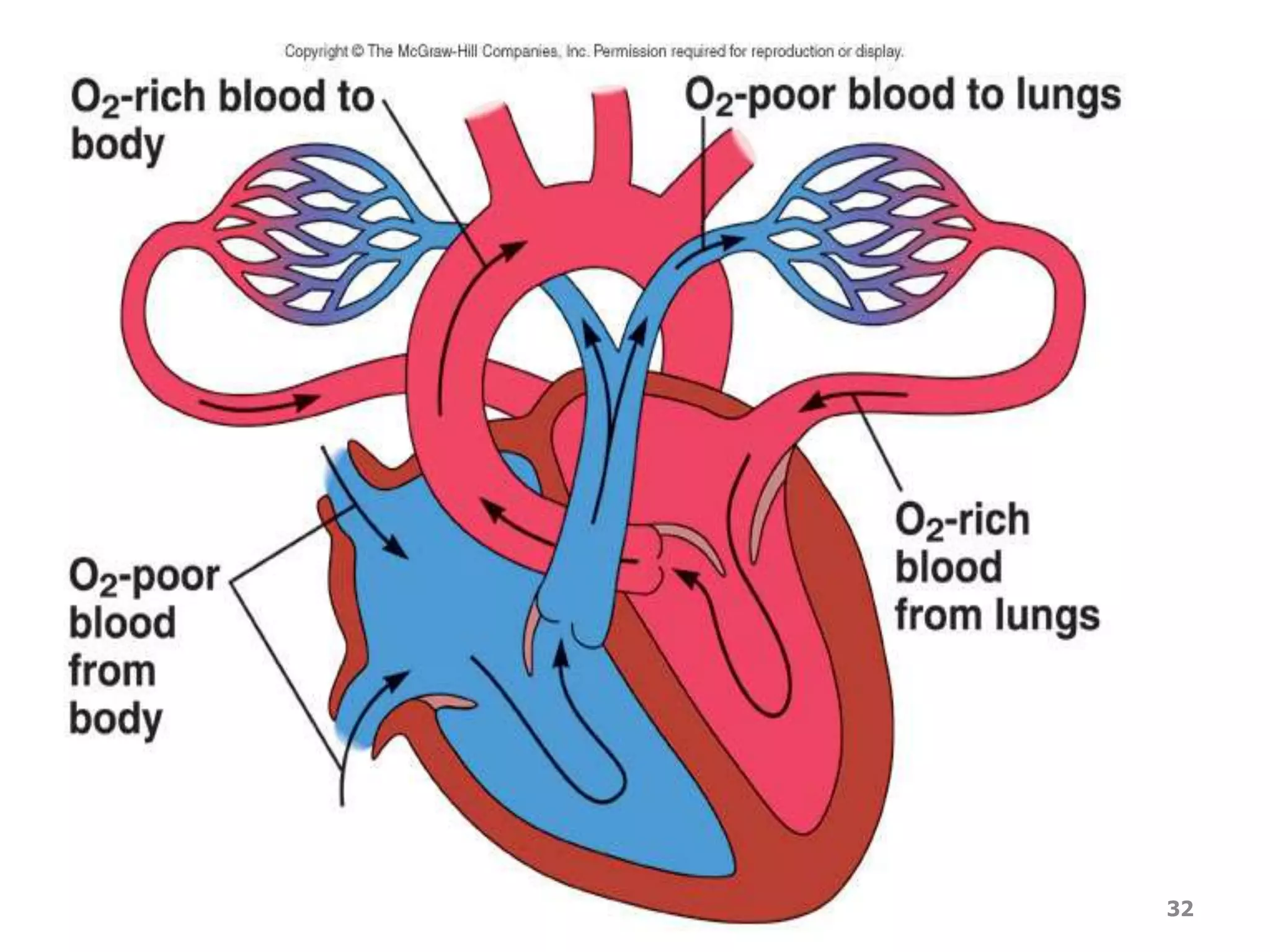
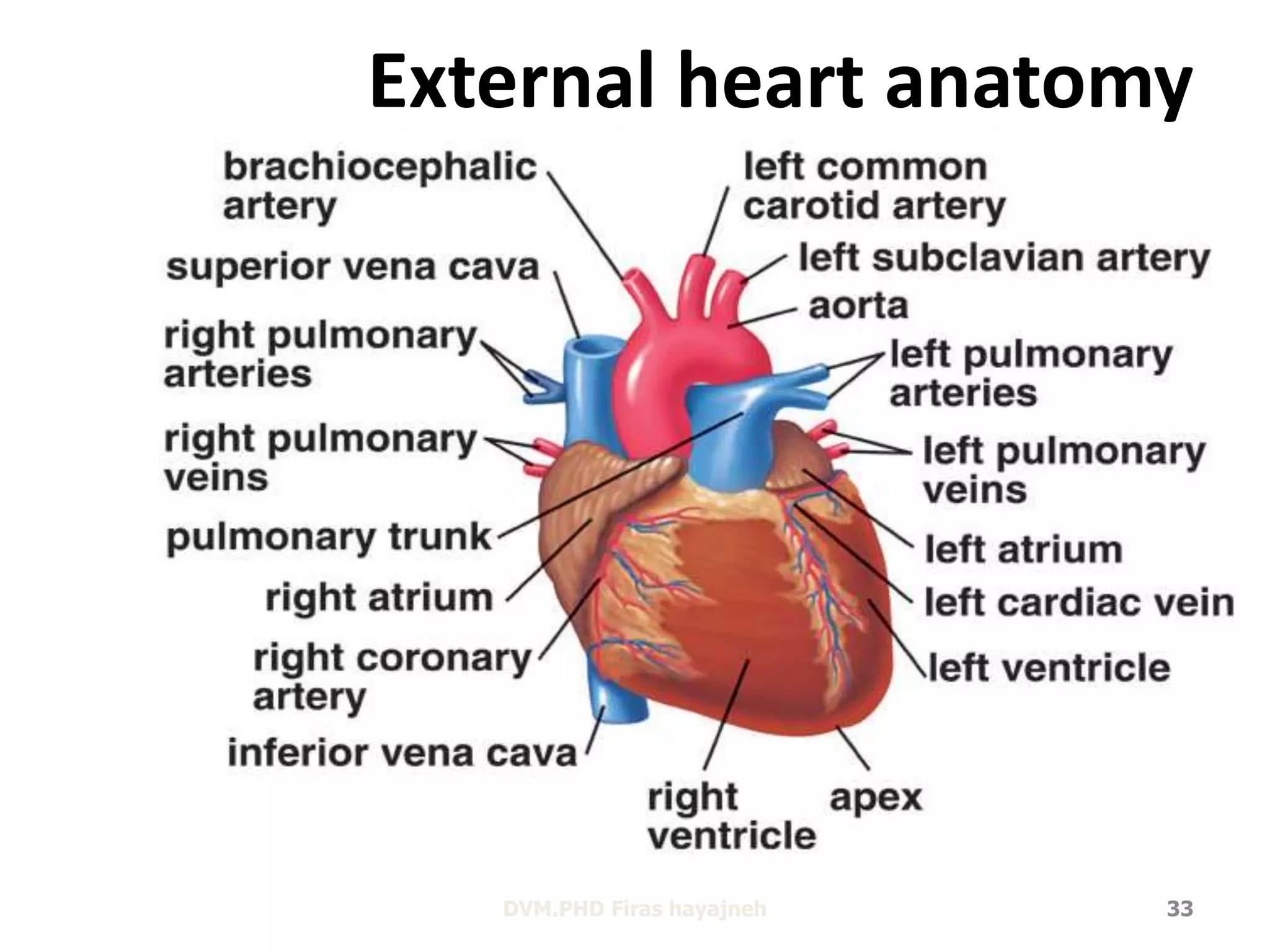
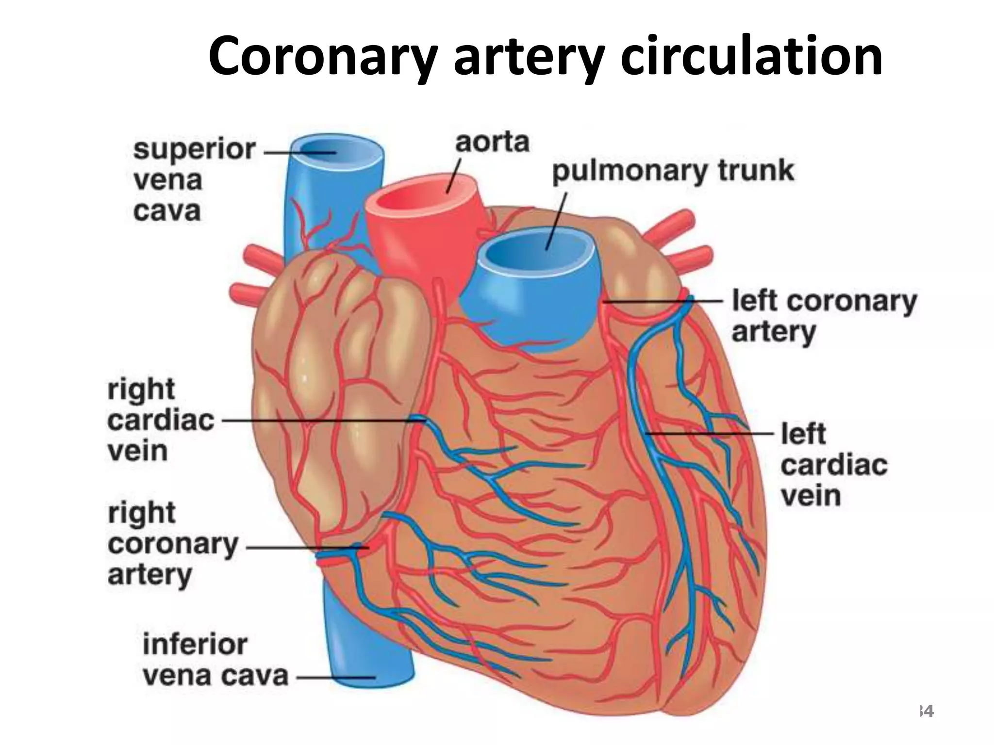
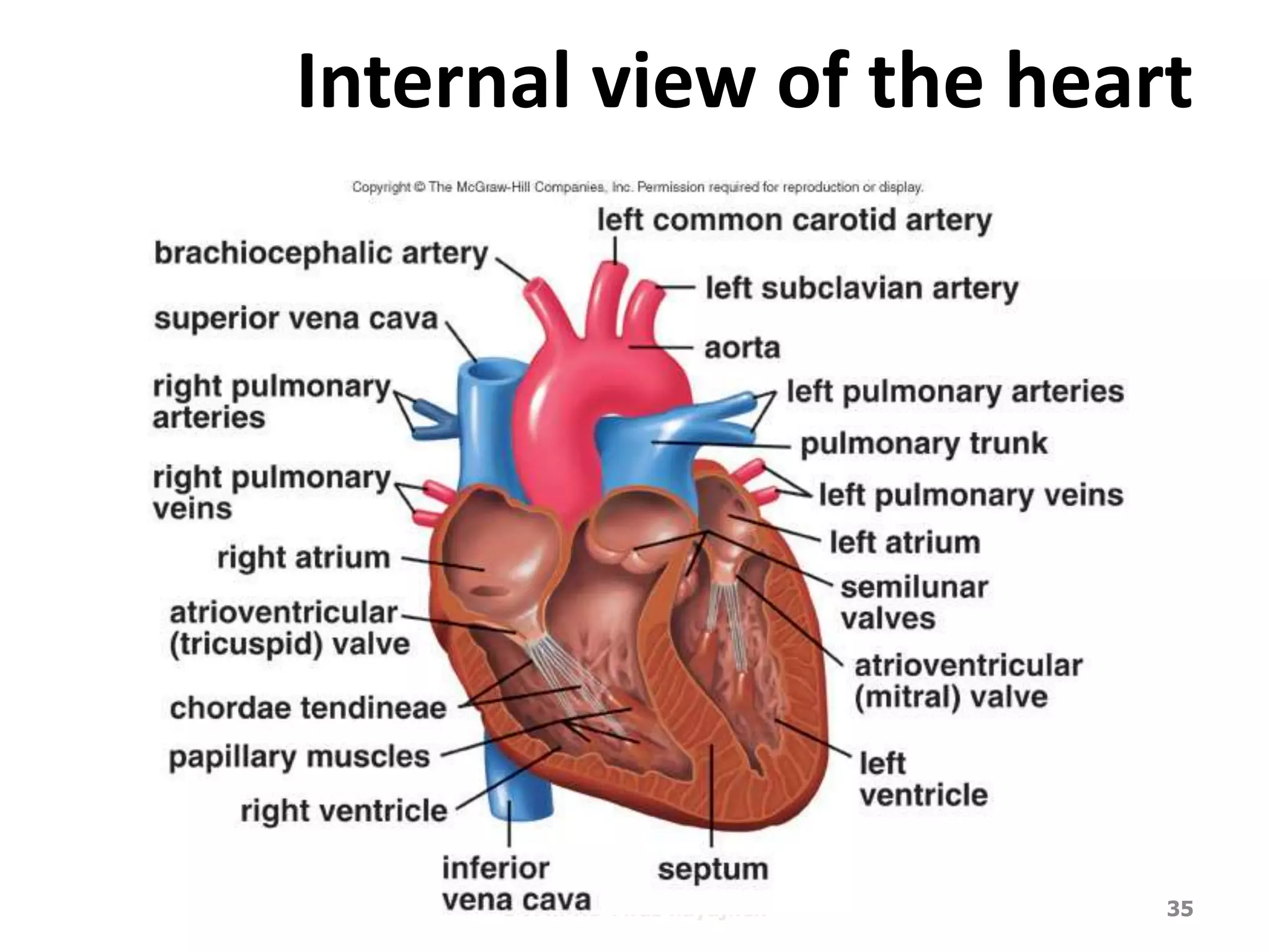
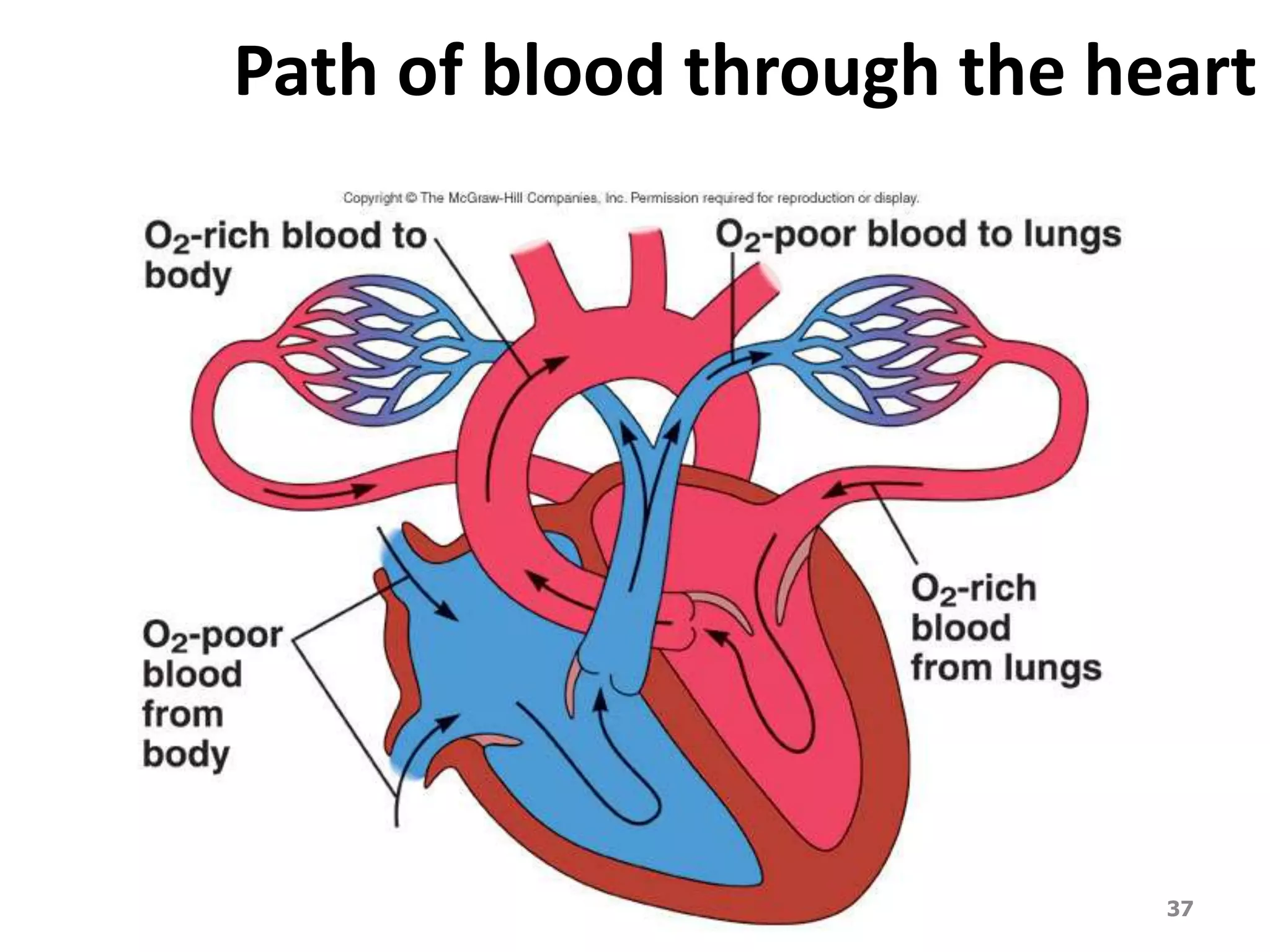
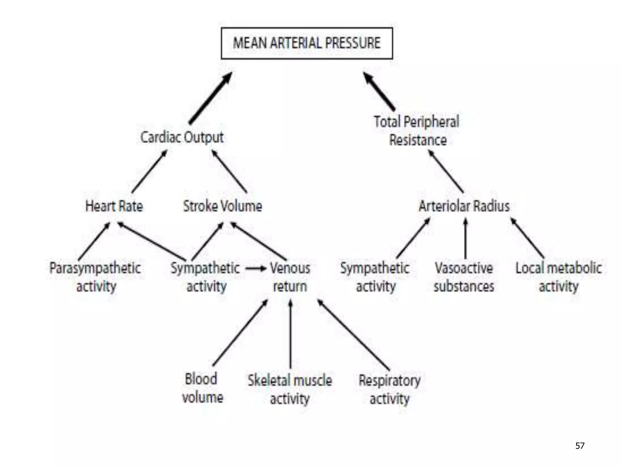
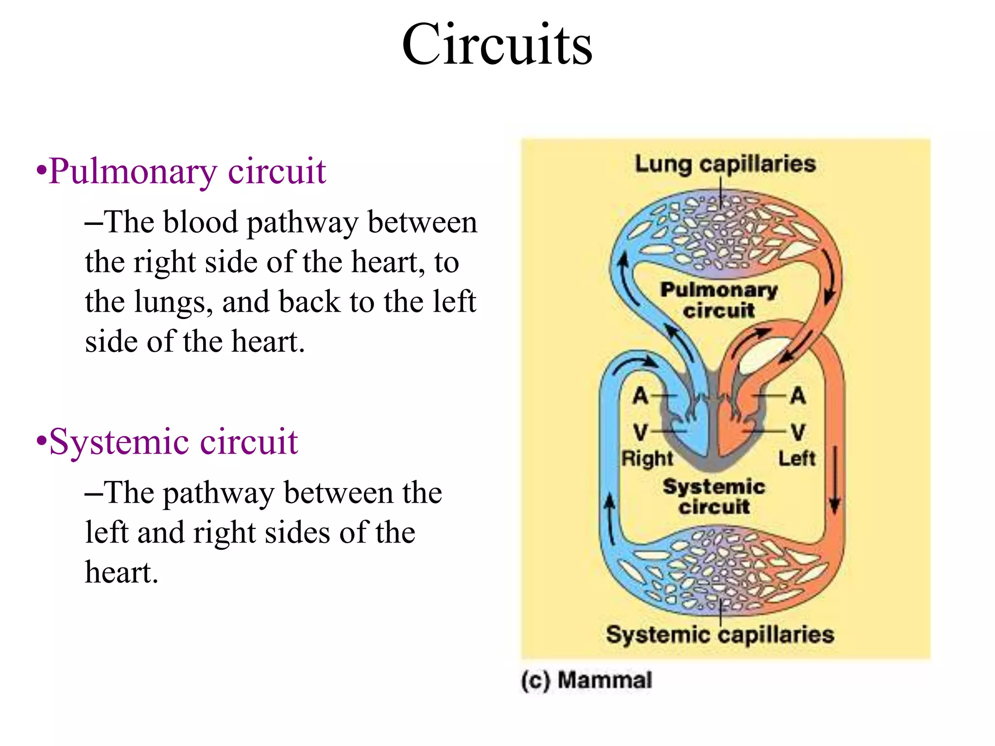
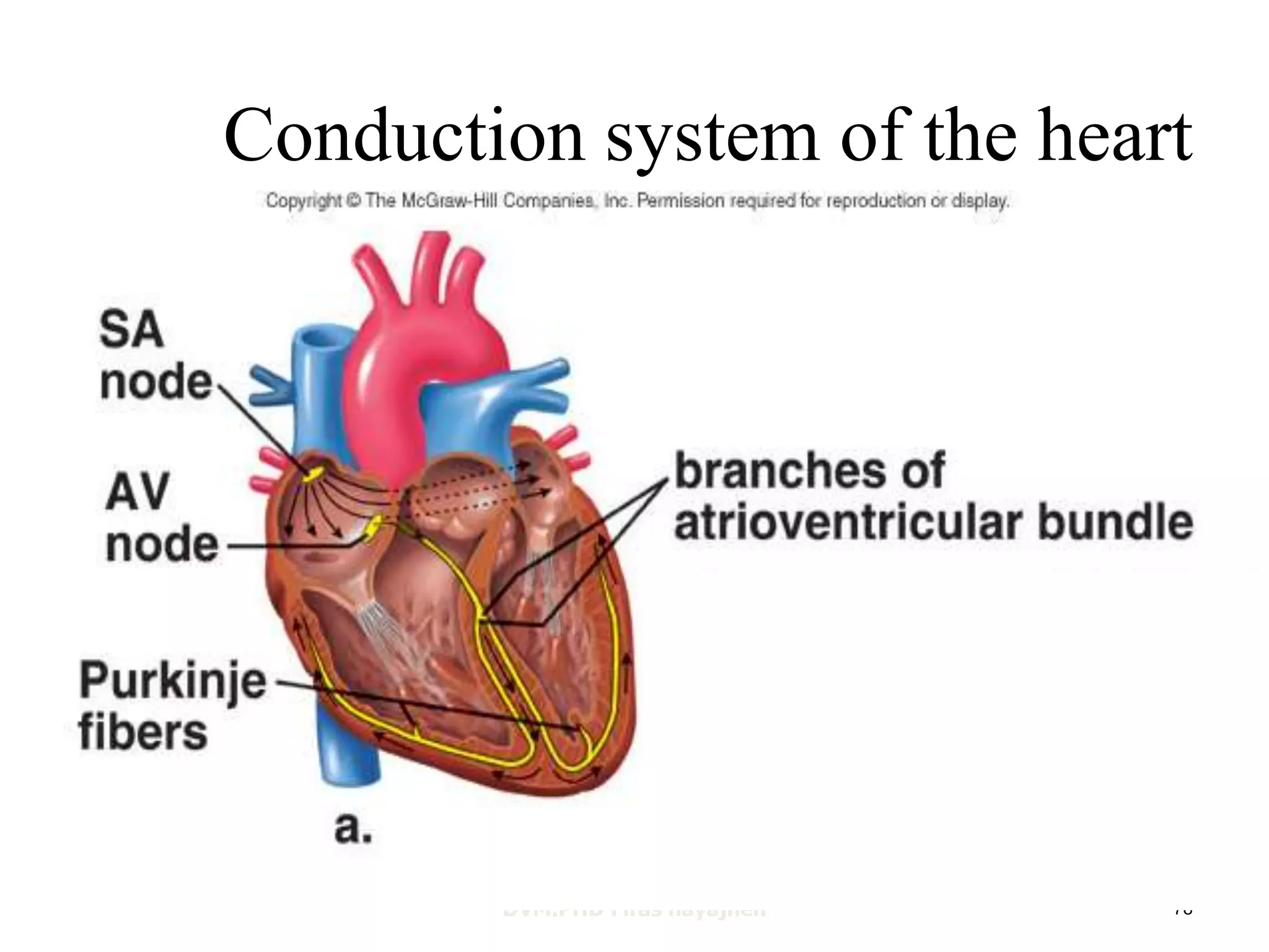
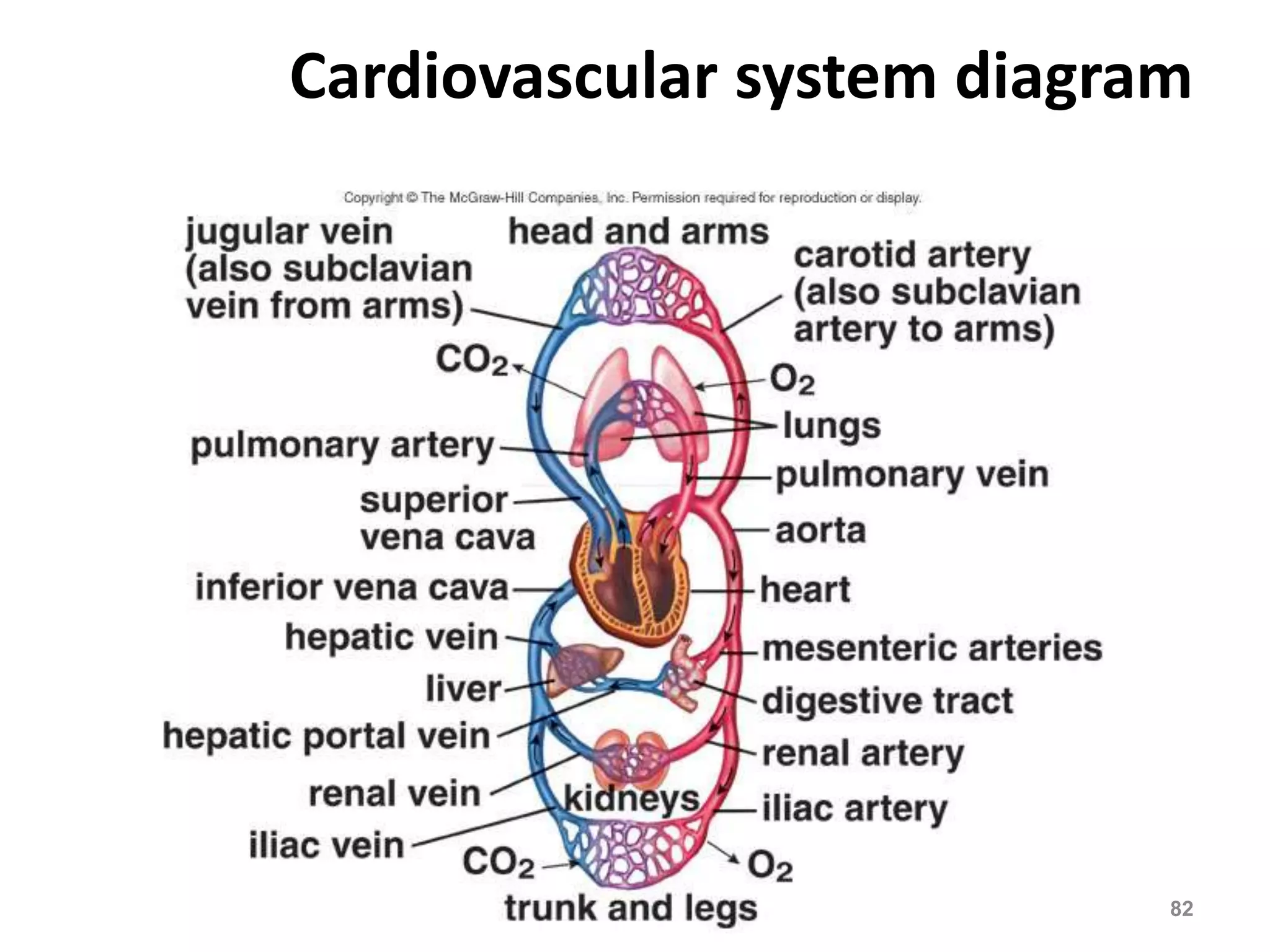
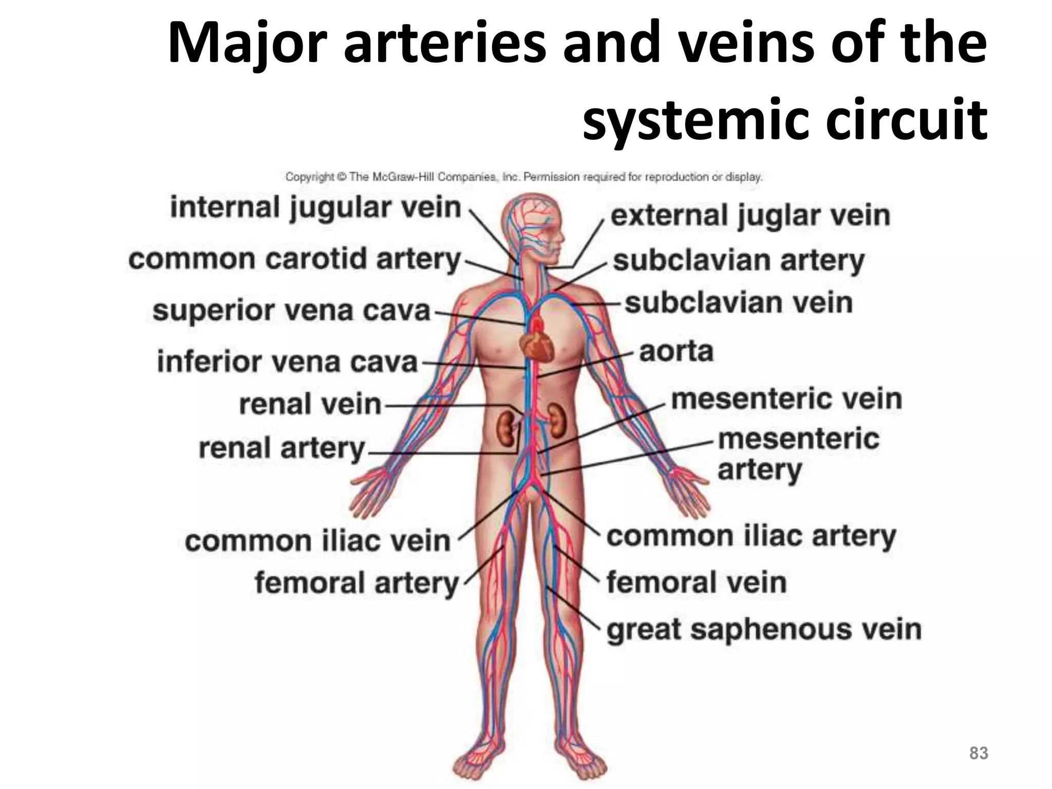
2. FUNCTIONS OF THE
CARDIOVASCULAR SYSTEM
1. Transport of materials (oxygen , nutrients ,
hormones ) throughout the body.
2. it removes metabolic waste products, such as
carbon dioxide, lactic acid, and urea, from
the tissues.
3. contributes to the actions of the immune
system by transporting antibodies and
leukocytes to areas of infection.
2
3. FUNCTIONS OF THE
CARDIOVASCULAR SYSTEM
• The cardiovascular system delivers blood to
tissues in the same time takes blood away to
be reloaded with vital requirements for the
cells.
• Components
1. Pump (heart).
2. Vessels.
3
4. The cardiovascular system
• All tissues are perfused (receive blood),
depending on their function.
• kidneys, only 1% of BW receive 20% (O2),
the GIT receives approximately 27% of the
O2, the skin receives 6 -15% of the blood.
• During exercise blood flow is directed away
from the kidneys and organs of the digestive
system and toward the skeletal and cardiac
muscles.
4
5. The Blood Vessels
• There are three kinds of vessels:
1. Arteries (and arterioles) – carry blood
away from the heart
2. Capillaries – where nutrient and gas
exchange occur
3. Veins (and venules) – carry blood toward
the heart.
5
7. The Arteries
Arteries and arterioles take blood away
from the heart.
The largest artery is the aorta.
an artery wall has smooth muscle that can
constrict to regulate blood flow and blood
pressure.
Arterioles can constrict or dilate,
changing blood pressure.
7
8. The Capillaries
• Capillaries have one cell thick walls to
allow exchange of gases and nutrients
with tissue fluid.
• Capillary beds are present in all regions
of the body but not all capillary beds are
open at the same time.
8
9. • Contraction of a sphincter muscle closes off a
bed and blood can flow through an
arteriovenous shunt that bypasses the capillary
bed.
9
11. The Veins
• Venules drain blood from capillaries,
then join to form veins that take
blood to the heart.
• Veins have much less smooth muscle
and connective tissue than arteries.
11
12. The Veins
• Veins often have valves that prevent
the backward flow of blood when
closed.
• Veins carry about 70% of the body’s
blood and act as a reservoir during
hemorrhage.
• large blood vessels are characterized
by an abundance of collagen fibers
and elastin fibers 12
13. Heart
• Located in the center of the thoracic
cavity.
• The heart is a cone-shaped ( ,)مخروط
muscular organ located between the lungs
behind the sternum.
• The heart muscle forms the myocardium,
with tightly interconnect cells of cardiac
muscle tissue
13
15. Functions of the Heart
• Generating blood pressure
• Routing blood
– Heart separates pulmonary and systemic
circulations
• Ensuring one-way blood flow
– Heart valves ensure one-way flow
• Regulating blood supply
– Changes in contraction rate and force
match blood delivery to changing
metabolic needs
16. Heart
• Located in the center of the thoracic
cavity.
• The pericardium is the outer membranous
sac with lubricating fluid produced by the
membranes of the pericardium.is
composed of:
1) Receiving chambers (atria, single atrium)
2) Delivery chambers (ventricles).
3) Valves (forward flow of blood).
16
19. • The heart has four chambers: two upper, thin-walled
atria, and two lower, thick-walled
ventricles.
• The septum is a wall dividing the two sides of
the heart.
19
20. Pericardium: a double-walled fibrous sac sorrounding
the heart
pericardial fluid produced by the pericardium minimizes
friction of the heart
20
21. The heart
• The wall of the heart has three layers:
1. Epicardium:is the thin membrane on the
external surface of the heart
2. Endocardium: thin delicate layer of cells
lining the chambers of the heart
3. Myocardium: the muscular layer of the heart
21
23. Valves
flexible but tough fibrous tissue
1. Atrioventricular (AV) valves (between the
atria and the ventricles):
A. The right AV tricuspid).
B. The left AV (bicuspid, mitral valve).
2. Semilunar valves: separate the ventricles
from their associated arteries
I. pulmonary valve is found between the right
ventricle and pulmonary artery
II. aortic valve is found between the left ventricle and
the aorta.
23
27. Heart
• The heart is a cone-shaped ( ,)مخروط
muscular organ located between the
lungs behind the sternum.
• The heart muscle forms the myocardium,
with tightly interconnect cells of cardiac
muscle tissue.
27
28. The septum is a wall dividing the two sides of
the heart.
Atrioventricular valves occur between the
atria and ventricles –
1. the tricuspid valve on the right
2. the bicuspid valve on the left.
28
29. • lub-dup heart sounds are due to the closing of
the atrioventricular valves, followed by the
closing of the semilunar valves.
• The pumping of the heart sends out blood
under pressure to the arteries.
• Blood pressure is greatest in the aorta; the
wall of the left ventricle is thicker than
that of the right ventricle and pumps
blood to the entire body.
29
31. Passage of Blood Through the Heart
Blood follows this sequence through the heart:
superior and inferior vena cava →
right atrium →
tricuspid valve →
right ventricle →
pulmonary semi lunar valve →
pulmonary trunk and arteries to the lungs →
pulmonary veins leaving the lungs →
left atrium →
bicuspid valve →
left ventricle →
aortic semilunar valve →
aorta →
to the body.
31
36. • The pumping of the heart sends out
blood under pressure to the arteries.
• Blood pressure is greatest in the aorta;
the wall of the left ventricle is thicker
than that of the right ventricle and
pumps blood to the entire body.
DVM.PHD Firas hayajneh 36
37. Path of blood through the heart
DVM.PHD Firas hayajneh 37
38. The Heart beat
• Each heartbeat is called a cardiac
cycle.
• When the heart beats, the two
atria contract together, then the
two ventricles contract; then the
whole heart relaxes.
• Systole is the contraction of heart
chambers; diastole is their
relaxation.
DVM.PHD Firas hayajneh 38
39. • The heart sounds, lub-dup, are
due to the closing of the
atrioventricular valves, followed
by the closing of the semilunar
valves.
• Blood pressure then decreases as
the cross-sectional area of arteries
and then arterioles increases
DVM.PHD Firas hayajneh 39
40. Blood pressure
• From the left ventricle (main force)……right
atrium.
• Systolic pressure 120mmHg, diastolic pressure
80mmHg.
• Pulse pressure =120-80=40
40
41. Blood flow through vessels
• Is determined by:
1. Pressure gradient.
The difference between the beginning pressure
and the pressure at the end.
1. Vascular resistance . Which depends on
Blood viscosity
Vessel length
Vessel radius
41
42. Mean arterial pressure(MAP)
Is the driving force for blood flow through the
body’s organs and tissues. (very important):
hypotension:
dizziness and fainting
Hypertension:
Increase cardiac workload
Vascular damage (atherosclerosis) and the
rupture of small vesseles, in arteries of the brain
and heart.
42
43. Control of mean arterial pressure
(MAP)
1. Autonomic nervous system (ANS)
2. Vasoactive substances
3. Venous return
4. Local metabolic activity
43
44. Intrinsic Control of Heartbeat
• The SA (sinoatrial) node, or pacemaker,
initiates the heartbeat and causes the atria to
contract on average every 0.85 seconds.
• The AV (atrioventricular) node conveys the
stimulus and initiates contraction of the
ventricles.
• The signal for the ventricles to contract travels
from the AV node through the atrioventricular
bundle to the smaller Purkinje fibers.
44
45. Electrical activity of the heart
The specialized excitation and electrical
conduction system in the heart consists of:
A. Sinoatrial node
B. Interatrial pathway
C. Internodal pathway
D. Atrioventricular node
E. Bundle of His
F. Bundle branches
G. Purkinje fibers
45
46. Intrinsic Control of Heartbeat
A. The SA (sinoatrial) node, or pacemaker, a
group of cells on the wall of the right
atrium, near the entrance of the superior
vena cava , initiates the heartbeat and causes
the atria to contract on average every 0.85
seconds.
• Sympathetic and parasympathetic system
innervates the SA node.
• Sympathetic nerves cause increase in the
heartbeat, parasympathetic cause decrease in
the heart rate. 46
47. Contraction of the heart muscles
• The “resting” membrane potential, or pacemaker
potential is -55mv.
1. depolarization due to influx of (Na, Ca). Untill
threshold is reached (-40mv).
2. Action potential happens.
3. Calcium channels close and potasium channels
open leading to repolarization .
47
48. Nervous system control
• Norepinephrine, which stimulates b-adrenergic
receptors, increases the rate of pacemaker
depolarization by increasing the permeability to Na
and Ca ions.
• Parasympathetic stimulation causes a decrease in
heart rate. Acetylcholine, which stimulates muscarinic
receptors, increases the permeability to potassium.
• From the SA node the heart beat spreads to the atria.
(left atrium)
B. interatrial conduction pathway (from the SA node
to the left atrium
48
49. C. internodal conduction pathway: at the base
of the right atrium near the interventricular
septum, transmits the impulse directly to the
atrioventricular (AV) node
49
51. • atria and ventricles are separated from each
other by fibrous connective tissue
D. the AV node serves as the only pathway
through which the impulse can be
transmitted to the ventricles slight delay (0.1
sec).
• it allows atria to complete their contraction
before ventricular contraction begins. This
timing ensures proper filling of the ventricles
prior to contraction.
51
52. • The AV (atrioventricular) node conveys the
stimulus and initiates contraction of the
ventricles.
E. Bundle of His: impulse spread through the
heart , this bundle penetrates the fibrous
tissue seperating the atria from the ventricles.
F. Purkinje fibers: that extend from the bundle
branches and spread through the
myocardium.
52
54. Extrinsic Control of Heartbeat
• A cardiac control center in the medulla
oblongata speeds up or slows down the
heart rate by way of the autonomic nervous
system branches: parasympathetic system
(slows heart rate) and the sympathetic
system (increases heart rate).
• Hormones epinephrine and norepinephrine
from the adrenal medulla also stimulate
faster heart rate.
55. Autonomic nervous system (ANS)
• parasympathetic system innervates the SA
node and the AV node of the heart by way of
the vagus nerves(decrease HR, which
decreases CO and MAP).
• The sympathetic system innervates most
tissues in the heart including the SA node, AV
node, and ventricular muscle. increase in HR
and increase in the heart contactility.
55
58. Vasomotor center
• Autonomic nervous activity to the
cardiovascular system is regulated by the
vasomotor center, Located in the lower pons
and the medulla of the brainstem.
58
59. 1. Baroreceptors (stretch receptors)
Monitor blood pressure in the aorta and carotid
sinuses, respond to stretch or distension of
blood vessels.
2. Chemoreceptors (carotid bodies, aortic
bodies):
Stimulated by hypoxisa and hypercarpnia and
decrease in arterial pH (acidosis)
3. Low pressure receptors: located in the
walls of the atria and pulmonary arteries.
Stimulated by a change in blood column
59
61. Vasoactive substances
• Substances released from many cells and
tissues in the body, including the endothelium
lining blood vessels, endocrine glands to cause
vasoconstriction or inhibit it to cause
vasodilation.
61
62. • Vasoconstrictors: When blood vessels
constrict, the flow of blood is restricted or
decreased (increased BP)
I. Catecholamines(epinephrine and
norepinephrine) from the adrenal medulla.
• Through α1-adrenergic receptors
• The selective α 1-adrenergic receptor
antagonist, prazosin (hypertension )
62
63. I. Angiotensin II (kidney). Act through AT1
receptors on smooth muscles. ACE inhibitors for
the treatment of hypertenision.
II. Vasopressin (antidiuretic hormone), produced in
the hypothalamus. Secreted from the
adenohypophysis. Increases the reabsorption of
water. Act through V1, V2 receptors.
III. Endothelin: produced by vascular endothelium
IV. Thromboxane A2: produced by vascular
endothelium.
63
64. Pharmacy application
antihypertensive drugs
• consistent elevation in blood pressure such that
systolic/diastolic pressures are ≥140/90 mmHg.
• There are several categories of antihypertensive
agents:
1. Diuretics. Decreases plasma volume ( decreases CO
(cardiac outpu, MAP (mean arterial pressure, VR
(venous return)).
2. Sympatholytics. Centrally acting agents affect the
vasomotor center in the brainstem and inhibit
sympathetic discharge, decrease CO, MAP
64
65. 3. Vasodilators: hydralazine causes direct
relaxation of arteriolar smooth muscle.
4. Ca++ channel blockers.(Verapamil,
nifedipine.
5. Angiotensin-converting enzyme (ACE)
inhibitors.
6. Angiotensin II receptor antagonists..
65
66. Vasodilators
• Vasodilators produced in the human body:
1. Prostacyclin: a metabolite of arachidonic
acid acid
2. Nitric oxide
66
67. Pharmacy application
Angina pectoris ( .(الذبحه الصدريه
• symptom of chronic ischemic heart disease. caused by an
imbalance between the oxygen supply and oxygen
demand of the cardiac
muscle.
• Myocardial oxygen demand increases during exertion,
exercise, and emotional stress.
• treated with nitroglycerin (vasodilation), which might
decrease blood pressure.
• Nitroglycerin is administered sublingual.
67
68. Venous return (VR)
• Is the volume of blood that flows from the
systemic veins into the right atrium per minute.
• In a healthy heart CO is equal to VR.
• VEINS serve as blood reservoirs ( The most
distensible vessels in the circulatory system
are the veins).
• veins help to regulate cardiac output (CO).
• under resting conditions, 64% of the blood
volume is contained within these vessels.
68
70. The Hear tbeat
• Each heartbeat is called a cardiac cycle.
• When the heart beats, the two atria contract
together, then the two ventricles contract;
then the whole heart relaxes.
• Systole is the contraction of heart chambers;
diastole is their relaxation.
DVM.PHD Firas hayajneh 70
71. How does the Heart work?
blood from
the body
blood from
the lungs
The heart beat begins when
the heart muscles relax
and blood flows into the
atria.
STEP ONE
72. How does the Heart work?
STEP TWO
The atria then contract
and the valves open
to allow blood into the
ventricles.
73. How does the Heart work?
The valves close to stop blood
flowing backwards.
The ventricles contract forcing
the blood to leave the heart.
At the same time, the atria are
relaxing and once again filling with
blood.
The cycle then repeats
itself.
STEP THREE
74. Circuits
•Pulmonary circuit
–The blood pathway between
the right side of the heart, to
the lungs, and back to the left
side of the heart.
•Systemic circuit
–The pathway between the
left and right sides of the
heart.
75. The Pulmonary Circuit
• The pulmonary circuit begins with the
pulmonary trunk from the right ventricle
which branches into two pulmonary
arteries that take oxygen-poor blood to the
lungs.
• In the lungs, oxygen diffuses into the blood,
and carbon dioxide diffuses out of the
blood to be expelled by the lungs.
• Four pulmonary veins return oxygen-rich
blood to the left atrium.
75
76. The Systemic Circuit
• The systemic circuit starts with the aorta
carrying O2-rich blood from the left
ventricle.
• The aorta branches with an artery going to
each specific organ.
• Generally, an artery divides into arterioles
and capillaries which then lead to venules.
DVM.PHD Firas hayajneh 76
77. • The vein that takes blood to the vena cava
often has the same name as the artery that
delivered blood to the organ.
• In the adult systemic circuit, arteries carry
blood that is relatively high in oxygen and
relatively low in carbon dioxide, and veins
carry blood that is relatively low in oxygen
and relatively high in carbon dioxide.
• This is the reverse of the pulmonary circuit.
DVM.PHD Firas hayajneh 77
79. Extrinsic Control of Heartbeat
• A cardiac control center in the medulla
oblongata :
*parasympathetic system (slows heart rate) .
* sympathetic system (increases heart rate)..
DVM.PHD Firas hayajneh 79
80. Hormones epinephrine and
norepinephrine from the adrenal
medulla also stimulate faster heart rate
DVM.PHD Firas hayajneh 80
83. Major arteries and veins of the
systemic circuit
DVM.PHD Firas hayajneh 83
84. • The coronary arteries serve the heart
muscle itself; they are the first branch off
the aorta.
• Since the coronary arteries are so small,
they are easily clogged, leading to heart
disease.
• The hepatic portal system carries blood
rich in nutrients from digestion in the
small intestine to the liver, the organ that
monitors the composition of the blood.
DVM.PHD Firas hayajneh 84
85. Blood Flow in Arteries
• Blood pressure due to the pumping of the
heart accounts for the flow of blood in the
arteries.
• Systolic pressure is high when the heart
expels the blood.
• Diastolic pressure occurs when the heart
ventricles are relaxing.
• Both pressures decrease with distance from
the left ventricle because blood enters more
and more arterioles and arteries.
DVM.PHD Firas hayajneh 85
86. Blood Flow in Capillaries
Blood moves slowly in capillaries because
there are more capillaries than arterioles.
This allows time for substances to be
exchanged between the blood and
tissues.
DVM.PHD Firas hayajneh 86
87. Blood Flow in Veins
• Venous blood flow is dependent upon:
1) skeletal muscle contraction,
2) presence of valves in veins, and
3) respiratory movements.
• Compression of veins causes blood to
move forward past a valve that then
prevents it from returning backward.
DVM.PHD Firas hayajneh 87
88. • Changes in thoracic and abdominal pressure
that occur with breathing also assist in the
return of blood.
• Varicose veins develop when the valves of
veins become weak.
• Hemorrhoids (piles) are due to varicose veins
in the rectum.
• Phlebitis is inflammation of a vein and can
lead to a blood clot and possible death if the
clot is dislodged and is carried to a
pulmonary vessel.
DVM.PHD Firas hayajneh 88
89. Capillary Exchange
• At the arteriole end of a capillary, water
moves out of the blood due to the force
of blood pressure.
• At the venule end, water moves into the
blood due to osmotic pressure of the
blood.
• Substances that leave the blood
contribute to tissue fluid, the fluid
between the body’s cells.
DVM.PHD Firas hayajneh 89
90. • In the midsection of the capillary,
nutrients diffuse out and wastes
diffuse into the blood.
• Since plasma proteins are too large to
readily pass out of the capillary, tissue
fluid tends to contain all components
of plasma except it has lesser amounts
of protein.
• Excess tissue fluid is returned to the
blood stream as lymph in lymphatic
vessels.
DVM.PHD Firas hayajneh 90
91. The Electrocardiogram
• An electrocardiogram (ECG) is a
recording of the electrical changes that
occur in the myocardium during a cardiac
cycle.
• Atrial depolarization creates the P wave,
ventricle depolarization creates the QRS
wave, and repolarization of the ventricles
produces the T wave.
DVM.PHD Firas hayajneh 91
93. The ECG provides information
concerning:
1. Relative size of heart chambers
2. Various disturbances of rhythm and
electrical conduction
3. Extent and location of ischemic damage to
the myocardium
4. Effects of altered electrolyte concentrations
5. Influence of certain drugs (e.g., digitalis and
antiarrhythmic drugs)
DVM.PHD Firas hayajneh 93
94. Pharmacy applications
antiarrhythmic drugs
• Arrhythmia: Any abnormality of the initiation
or propagation of the impulse.
1. Verapamil (Class IV antiarrhythmic drug) is an
effective agent for atrial or supraventricular
tachycardia. A Ca++ channel blocker.
2. Procainamide (Class IA antiarrhythmic drug),
blockade of the fast Na+ channels
94
95. Cardiac cycle
• the period of time from beginning of one
heart beat to beginning of the next.
• Systole, in which the chambers contract and
eject the blood
• Diastole, in which the chambers relax allowing
blood to fill them
95
96. • capillary blood flow is not interrupted by this
cycle because blood flow to the tissues is
continuous, due to the elastic properties of
the arterial walls.
• large blood vessels are characterized by an
abundance of collagen fibers and elastin fibers
96
97. Cardiac output
• cardiac output (CO):the volume of blood
pumped into the aorta per minute.
• Cardiac output (CO) = heart rate (HR)*stroke
volume (SV).
• At rest (adult)
• CO = 70 beats/min*70 ml/beat = 4900 ml/min
=5 l/min
97
99. Control of heart rate
• the typical average heart rate is about 70
beats per minute ( much greater in children).
• Heart rate depends on:
1. Autonomic nervous system influence
2. Catecholamines
3. Body temperature
99
100. 1. Autonomic nervous system
influence
• sympathetic and parasympathetic systems
have antagonistic effects on the heart.
100
SA node
AV node
sympathetic and
sympathetic
parasympathetic
systems.
101. Sympathetic stimulation:
norepinephren binds to beta
adrenergic receptorrs:
1. Increased rate of discharge of the SA node
2. Increased rate of conduction through the AV
node
3. Increased rate of conduction through the
bundle of His and the Purkinje fibers
DVM.PHD Firas hayajneh 101
102. enhanced depolarization
• potassium permeability sodium, calcium
permeability. the inside of the cell becomes
less (threshold more rapidly). In this way,
action potentials are generated faster and
travel through the conduction pathway more
quickly so that the heart can generate more
heartbeats per minute
DVM.PHD Firas hayajneh 102
103. Parasympathetic stimulation
• heart rate. Acetylcholine (vagus nerve) binds
to muscarinic receptors and causes the
following effects:
1. Decreased rate of discharge of the SA node
2. Decreased rate of conduction through the
AV node.
DVM.PHD Firas hayajneh 103
104. Parasympathetic stimulation
permeability to potassium……SA node (cells
are hyperpolarized…a far threshold…greater
depolarization is needed.
• Rate of depolarization is reduced.
• at rest parasymathetic nervous system
predominates.
DVM.PHD Firas hayajneh 104
105. 2. Catecholamines ( epinephrine and norrepinephrine
Released from From adrenal medulla Increase the heart
rate.
3. Body temperature affects heart rate by altering the
rate of discharge of the SA node.
An increase of 1 F in body temperature results in an
increase in heart rate of about 10 beats per minute.
DVM.PHD Firas hayajneh 105
106. stroke volume
• The volume of blood pumped from one
ventricle of the heart with each beat depends
on:
1. Length of diastole
2. Venous return (preload)
3. Contractility of the myocardium
4. Afterload
5. Heart rate
DVM.PHD Firas hayajneh 106
107. • Diastole: is the period in the cardiac cycle in
which relaxation of the myocardium and
ventricular filling take place.
• Venous return: the volume of blood returned
to the right atrium per minute.
DVM.PHD Firas hayajneh 107
108. Cardiovascular Disorders
• Cardiovascular disease (CVD) is the leading
cause of death in Western countries.
• Modern research efforts have improved
diagnosis, treatment, and prevention.
• Major cardiovascular disorders include
atherosclerosis, stroke, heart attack,
aneurysm, and hypertension.
DVM.PHD Firas hayajneh 108
109. Atherosclerosis
• Atherosclerosis:arteriosclerotic vascular disease
or ASVD) is a condition in which an artery wall
thickens as a result of the accumulation of fatty
materials such as cholestrol under the inner lining
of arteries.
• The plaque can cause a thrombus (blood clot) to
form.
• The thrombus can dislodge as an embolus and
lead to thromboembolism.
DVM.PHD Firas hayajneh 109
110. Stroke, Heart Attack, and
Aneurysm
• A cerebrovascular accident, or stroke, results
when an embolus lodges in a cerebral blood
vessel or a cerebral blood vessel bursts; a
portion of the brain dies due to lack of oxygen.
• A myocardial infarction, or heart attack,
occurs when a portion of heart muscle dies
due to lack of oxygen.
DVM.PHD Firas hayajneh 110
111. • Partial blockage of a coronary artery causes
angina pectoris, or chest pain.
• An aneurysm is a ballooning of a blood vessel,
usually in the abdominal aorta or arteries
leading to the brain.
• Death results if the aneurysm is in a large
vessel and the vessel bursts.
• Atherosclerosis and hypertension weaken
blood vessels over time, increasing the risk of
aneurysm.
DVM.PHD Firas hayajneh 111
112. Coronary Bypass Operations
• A coronary bypass operation involves
removing a segment of another blood
vessel and replacing a clogged coronary
artery.
• It may be possible to replace this surgery
with gene therapy that stimulates new
blood vessels to grow where the heart
needs more blood flow.
DVM.PHD Firas hayajneh 112
114. Clearing Clogged Arteries
• Angioplasty uses a long tube threaded
through an arm or leg vessel to the point
where the coronary artery is blocked;
inflating the tube forces the vessel open.
• Small metal stents are expanded inside the
artery to keep it open.
• Stents are coated with heparin to prevent
blood clotting and with chemicals to
prevent arterial closing.
DVM.PHD Firas hayajneh 114
116. Dissolving Blood Clots
• Medical treatments for dissolving blood clots
include use of t-PA (tissue plasminogen
activator) that converts plasminogen into
plasmin, an enzyme that dissolves blood clots,
but can cause brain bleeding.
• Aspirin reduces the stickiness of platelets and
reduces clot formation and lowers the risk of
heart attack.
DVM.PHD Firas hayajneh 116
117. Heart Transplants and Artificial Hearts
• Heart transplants are routinely performed
but immunosuppressive drugs must be
taken thereafter.
• There is a shortage of human organ donors.
• Work is currently underway to improve self-contained
artificial hearts, and muscle cell
transplants may someday be useful.
DVM.PHD Firas hayajneh 117
118. Hypertension
• About 20% of Americans suffer from hypertension
(high blood pressure).
• Hypertension is present when systolic pressure is
140 or greater or diastolic pressure is 100 or
greater; diastolic pressure is emphasized when
medical treatment is considered.
• A genetic predisposition for hypertension occurs
in those who have a gene that codes for
angiotensinogen, a powerful vasoconstrictor.
DVM.PHD Firas hayajneh 118