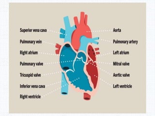
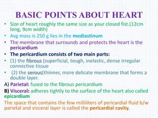
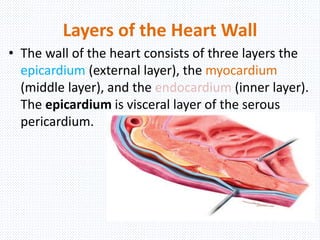
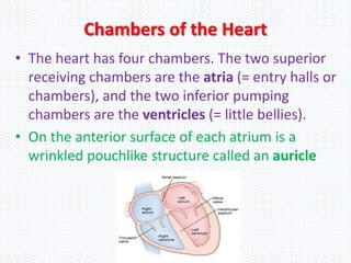
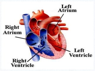
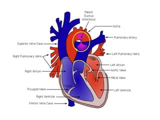
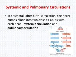
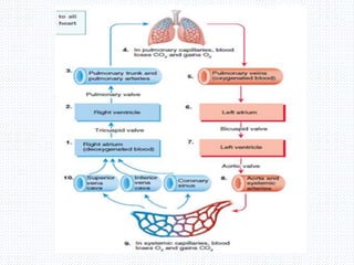
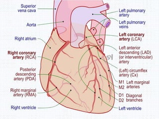
CARDIOVASCULAR SYSTEM
THE HEART
Dr. S P Srinivas Nayak,
PharmD, RPh, (MSc), (PGDND) (PhD)
Assistant Professor, Dept. of Pharmacy Practice, PU.
BASIC POINTS ABOUT HEART
โ€ข Size of heart roughly the same size as your closed fist.(12cm
long, 9cm width)
โ€ข Avg mass is 250 g lies in the mediastinum
โ€ข The membrane that surrounds and protects the heart is the
pericardium
โ€ข The pericardium consists of two main parts:
โ€ข (1) the fibrous (superficial, tough, inelastic, dense irregular
connective tissue
โ€ข (2) the serous(thinner, more delicate membrane that forms a
double layer.
A) Parietal: fused to the fibrous pericardium
B) Visceral: adheres tightly to the surface of the heart also called
epicardium
The space that contains the few milliliters of pericardial fluid b/w
parietal and visceral layer is called the pericardial cavity.
Layers of the Heart Wall
โ€ข The wall of the heart consists of three layers the
epicardium (external layer), the myocardium
(middle layer), and the endocardium (inner layer).
The epicardium is visceral layer of the serous
pericardium.
Chambers of the Heart
โ€ข The heart has four chambers. The two superior
receiving chambers are the atria (= entry halls or
chambers), and the two inferior pumping
chambers are the ventricles (= little bellies).
โ€ข On the anterior surface of each atrium is a
wrinkled pouchlike structure called an auricle
Right Atrium
โ€ข The right atrium forms the right surface of the
heart and receives blood from three veins: the
superior vena cava, inferior vena cava, and
coronary sinus
โ€ข A prominent feature of this septum is an oval
depression called the fossa ovalis, the remnant of
the foramen ovale, an opening in the interatrial
septum of the fetal heart that normally closes
soon after birth.
โ€ข Blood passes from the right atrium into the right
ventricle through a valve that is called the
tricuspidvalve
RIGHT VENTRICLE
โ€ข The right ventricle is about 4โ€“5 mm (0.16โ€“0.2 in.) in average
thickness and forms most of the anterior surface of the heart.
The inside of the right ventricle contains a series of ridges
formed by raised bundles of cardiac muscle fibers called
trabeculae carneae.
โ€ข Internally, the right ventricle is separated from the left
ventricle by a partition called the interventricular septum.
โ€ข Blood passes from the right ventricle through the pulmonary
valve (pulmonary semilunar valve) into a large artery called
the pulmonary trunk, which divides into right and left
pulmonary arteries and carries blood to the lungs.
โ€ข Arteries always take blood away from the heart (a mnemonic
to help you: artery = away).
LEFT ATRIUM
โ€ข The left atrium is about the same thickness as
the right atrium and forms most of the base of
the heart It receives blood from the lungs
through four pulmonary veins.
โ€ข Blood passes from the left atrium into the left
ventricle through the bicuspid (mitral) valve
LEFT VENTRICLE
โ€ข The left ventricle is the thickest chamber of the heart,
averaging 10โ€“15 mm (0.4โ€“0.6 in.), and forms the apex of
the heart.
โ€ข Blood passes from the left ventricle through the aortic
valve
โ€ข During fetal life, a temporary blood vessel, called the
ductus arteriosus, shunts blood from the pulmonary trunk
into the aorta.
โ€ข Hence, only a small amount of blood enters the
nonfunctioning fetal lungs.
โ€ข The ductus arteriosus normally closes shortly after birth,
leaving a remnant known as the ligamentum arteriosum
which connects the arch of the aorta and pulmonary trunk
HEART VALVES AND
CIRCULATION OF BLOOD
โ€ข Because they are located between an atrium
and a ventricle, the tricuspid and bicuspid
valves are termed atrioventricular (AV) valves
โ€ข The aortic and pulmonary valves are known as
the semilunar (SL) valves because they are
made up of three crescent moonโ€“shaped
cusps . Each cusp attaches to the arterial wall
by its convex outer margin
Systemic and Pulmonary Circulations
โ€ข In postnatal (after birth) circulation, the heart
pumps blood into two closed circuits with
each beatโ€”systemic circulation and
pulmonary circulation
The circulation
โ€ข The right side of the heart is the pump for
pulmonary circulation; it receives all of the dark-
red deoxygenated blood returning from the
systemic circulation.
โ€ข Blood ejected from the right ventricle flows into
the pulmonary trunk, which branches into
pulmonary arteries that carry blood to the right
and left lungs. In pulmonary capillaries, blood
unloads CO2, which is exhaled, and picks up O2
from inhaled air.
โ€ข The freshly oxygenated blood then flows into
pulmonary veins and returns to the left atrium.
CORONARY CIRCULATION
โ€ข Nutrients are not able to diffuse quickly enough
from blood in the chambers of the heart to
supply all layers of cells that make up the heart
wall.
โ€ข For this reason, the myocardium has its own
network of blood vessels, the coronary
circulation
โ€ข the left and right coronary arteries, branch
from the ascending aorta
LEFT CORONARY ARTERY
The left coronary artery
passes inferior to the left
auricle and divides into the
anterior interventricular and
circumflex branches.
The anterior interventricular
branch or left anterior
descending (LAD) artery is in
the anterior interventricular
sulcus and supplies
oxygenated blood to the walls
of both ventricles
The circumflex branch (SER-
kum-fleks) lies in the coronary
sulcus and distributes
oxygenated blood to the walls
of the left ventricle and left
atrium
THE RIGHT CORONARY ARTERY(RCA)
RCA supplies small branches
(atrial branches) to the right
atrium. It continues inferior to
the right auricle and ultimately
divides into the posterior
interventricular
and marginal branches.
The posterior interventricular
artery divides into PDA and
circulates walls of posterior
ventricles
The marginal branch runs along
the right margin of the heart and
transports oxygenated blood to
the wall of the right ventricle.
CORONARY VEINS
After blood passes through the arteries of the
coronary circulation, it flows into capillaries,
where it delivers oxygen and nutrients to the
heart muscle and collects carbon dioxide and
waste, and then moves into coronary veins.
Most of the deoxygenated blood from the
myocardium drains into a large vascular sinus in
the coronary sulcus on the posterior surface of
the heart, called the coronary sinus
Great cardiac vein in the anterior
interventricular sulcus, which drains
the areas of the heart supplied by
the left coronary artery (left and
right ventricles and left atrium)
โ€ข Middle cardiac vein in the
posterior interventricular sulcus,
which drains the areas supplied by
the posterior interventricular
branch of the right coronary artery
(left and right ventricles)
โ€ข Small cardiac vein in the coronary
sulcus, which drains the right
atrium and right ventricle
โ€ข Anterior cardiac veins, which
drain the right ventricle and open
directly into the right atrium
AUTORHYTHMIC FIBERS
THE CONDUCTION SYSTEM
โ€ข An inherent and rhythmical electrical activity is the reason
for the heartโ€™s lifelong beat. The source of this electrical
activity is a network of specialized cardiac muscle fibers
called autorhythmic fibers because they are self-excitable.
โ€ข Autorhythmic fibers repeatedly generate action potentials
that trigger heart contractions.
โ€ข During embryonic development, only about 1% of the
cardiac muscle fibers become autorhythmic fibers; these
relatively rare fibers have two important functions:
1. They act as a pacemaker, setting the rhythm of electrical
excitation that causes contraction of the heart.
2. They form the cardiac conduction system, a network of
specialized cardiac muscle fibers that provide a path for each
cycle of cardiac excitation to progress through the heart
THE CONDUCTION SYSTEM
โ€ข Cardiac excitation normally begins in the sinoatrial
(SA) node, located in the right atrial wall just
inferior and lateral to the opening of the superior
vena cava. SA node cells do not have a stable resting
potential. Rather, they repeatedly depolarize to
threshold spontaneously. The spontaneous
depolarization is a pacemaker potential. When the
pacemaker potential reaches
โ€ข threshold, it triggers an action potential. Each action
potential from the SA node propagates throughout
both atria via gap junctions in the intercalated discs
of atrial muscle fibers. Following the action
potential, the two atria contract at the same time.
Next class we will study aboutโ€ฆ
ECG, Heart Sounds and Cardiac Disorders
THYANK YOU

CARDIO VASCULAR SYSTEM THE HEART

  • 1.
    CARDIOVASCULAR SYSTEM THE HEART Dr.S P Srinivas Nayak, PharmD, RPh, (MSc), (PGDND) (PhD) Assistant Professor, Dept. of Pharmacy Practice, PU.
  • 3.
    BASIC POINTS ABOUTHEART โ€ข Size of heart roughly the same size as your closed fist.(12cm long, 9cm width) โ€ข Avg mass is 250 g lies in the mediastinum โ€ข The membrane that surrounds and protects the heart is the pericardium โ€ข The pericardium consists of two main parts: โ€ข (1) the fibrous (superficial, tough, inelastic, dense irregular connective tissue โ€ข (2) the serous(thinner, more delicate membrane that forms a double layer. A) Parietal: fused to the fibrous pericardium B) Visceral: adheres tightly to the surface of the heart also called epicardium The space that contains the few milliliters of pericardial fluid b/w parietal and visceral layer is called the pericardial cavity.
  • 5.
    Layers of theHeart Wall โ€ข The wall of the heart consists of three layers the epicardium (external layer), the myocardium (middle layer), and the endocardium (inner layer). The epicardium is visceral layer of the serous pericardium.
  • 6.
    Chambers of theHeart โ€ข The heart has four chambers. The two superior receiving chambers are the atria (= entry halls or chambers), and the two inferior pumping chambers are the ventricles (= little bellies). โ€ข On the anterior surface of each atrium is a wrinkled pouchlike structure called an auricle
  • 8.
    Right Atrium โ€ข Theright atrium forms the right surface of the heart and receives blood from three veins: the superior vena cava, inferior vena cava, and coronary sinus โ€ข A prominent feature of this septum is an oval depression called the fossa ovalis, the remnant of the foramen ovale, an opening in the interatrial septum of the fetal heart that normally closes soon after birth. โ€ข Blood passes from the right atrium into the right ventricle through a valve that is called the tricuspidvalve
  • 11.
    RIGHT VENTRICLE โ€ข Theright ventricle is about 4โ€“5 mm (0.16โ€“0.2 in.) in average thickness and forms most of the anterior surface of the heart. The inside of the right ventricle contains a series of ridges formed by raised bundles of cardiac muscle fibers called trabeculae carneae. โ€ข Internally, the right ventricle is separated from the left ventricle by a partition called the interventricular septum. โ€ข Blood passes from the right ventricle through the pulmonary valve (pulmonary semilunar valve) into a large artery called the pulmonary trunk, which divides into right and left pulmonary arteries and carries blood to the lungs. โ€ข Arteries always take blood away from the heart (a mnemonic to help you: artery = away).
  • 13.
    LEFT ATRIUM โ€ข Theleft atrium is about the same thickness as the right atrium and forms most of the base of the heart It receives blood from the lungs through four pulmonary veins. โ€ข Blood passes from the left atrium into the left ventricle through the bicuspid (mitral) valve
  • 15.
    LEFT VENTRICLE โ€ข Theleft ventricle is the thickest chamber of the heart, averaging 10โ€“15 mm (0.4โ€“0.6 in.), and forms the apex of the heart. โ€ข Blood passes from the left ventricle through the aortic valve โ€ข During fetal life, a temporary blood vessel, called the ductus arteriosus, shunts blood from the pulmonary trunk into the aorta. โ€ข Hence, only a small amount of blood enters the nonfunctioning fetal lungs. โ€ข The ductus arteriosus normally closes shortly after birth, leaving a remnant known as the ligamentum arteriosum which connects the arch of the aorta and pulmonary trunk
  • 17.
    HEART VALVES AND CIRCULATIONOF BLOOD โ€ข Because they are located between an atrium and a ventricle, the tricuspid and bicuspid valves are termed atrioventricular (AV) valves โ€ข The aortic and pulmonary valves are known as the semilunar (SL) valves because they are made up of three crescent moonโ€“shaped cusps . Each cusp attaches to the arterial wall by its convex outer margin
  • 19.
    Systemic and PulmonaryCirculations โ€ข In postnatal (after birth) circulation, the heart pumps blood into two closed circuits with each beatโ€”systemic circulation and pulmonary circulation
  • 21.
    The circulation โ€ข Theright side of the heart is the pump for pulmonary circulation; it receives all of the dark- red deoxygenated blood returning from the systemic circulation. โ€ข Blood ejected from the right ventricle flows into the pulmonary trunk, which branches into pulmonary arteries that carry blood to the right and left lungs. In pulmonary capillaries, blood unloads CO2, which is exhaled, and picks up O2 from inhaled air. โ€ข The freshly oxygenated blood then flows into pulmonary veins and returns to the left atrium.
  • 22.
    CORONARY CIRCULATION โ€ข Nutrientsare not able to diffuse quickly enough from blood in the chambers of the heart to supply all layers of cells that make up the heart wall. โ€ข For this reason, the myocardium has its own network of blood vessels, the coronary circulation โ€ข the left and right coronary arteries, branch from the ascending aorta
  • 24.
    LEFT CORONARY ARTERY Theleft coronary artery passes inferior to the left auricle and divides into the anterior interventricular and circumflex branches. The anterior interventricular branch or left anterior descending (LAD) artery is in the anterior interventricular sulcus and supplies oxygenated blood to the walls of both ventricles The circumflex branch (SER- kum-fleks) lies in the coronary sulcus and distributes oxygenated blood to the walls of the left ventricle and left atrium
  • 25.
    THE RIGHT CORONARYARTERY(RCA) RCA supplies small branches (atrial branches) to the right atrium. It continues inferior to the right auricle and ultimately divides into the posterior interventricular and marginal branches. The posterior interventricular artery divides into PDA and circulates walls of posterior ventricles The marginal branch runs along the right margin of the heart and transports oxygenated blood to the wall of the right ventricle.
  • 26.
    CORONARY VEINS After bloodpasses through the arteries of the coronary circulation, it flows into capillaries, where it delivers oxygen and nutrients to the heart muscle and collects carbon dioxide and waste, and then moves into coronary veins. Most of the deoxygenated blood from the myocardium drains into a large vascular sinus in the coronary sulcus on the posterior surface of the heart, called the coronary sinus
  • 28.
    Great cardiac veinin the anterior interventricular sulcus, which drains the areas of the heart supplied by the left coronary artery (left and right ventricles and left atrium) โ€ข Middle cardiac vein in the posterior interventricular sulcus, which drains the areas supplied by the posterior interventricular branch of the right coronary artery (left and right ventricles) โ€ข Small cardiac vein in the coronary sulcus, which drains the right atrium and right ventricle โ€ข Anterior cardiac veins, which drain the right ventricle and open directly into the right atrium
  • 29.
    AUTORHYTHMIC FIBERS THE CONDUCTIONSYSTEM โ€ข An inherent and rhythmical electrical activity is the reason for the heartโ€™s lifelong beat. The source of this electrical activity is a network of specialized cardiac muscle fibers called autorhythmic fibers because they are self-excitable. โ€ข Autorhythmic fibers repeatedly generate action potentials that trigger heart contractions. โ€ข During embryonic development, only about 1% of the cardiac muscle fibers become autorhythmic fibers; these relatively rare fibers have two important functions: 1. They act as a pacemaker, setting the rhythm of electrical excitation that causes contraction of the heart. 2. They form the cardiac conduction system, a network of specialized cardiac muscle fibers that provide a path for each cycle of cardiac excitation to progress through the heart
  • 30.
    THE CONDUCTION SYSTEM โ€ขCardiac excitation normally begins in the sinoatrial (SA) node, located in the right atrial wall just inferior and lateral to the opening of the superior vena cava. SA node cells do not have a stable resting potential. Rather, they repeatedly depolarize to threshold spontaneously. The spontaneous depolarization is a pacemaker potential. When the pacemaker potential reaches โ€ข threshold, it triggers an action potential. Each action potential from the SA node propagates throughout both atria via gap junctions in the intercalated discs of atrial muscle fibers. Following the action potential, the two atria contract at the same time.
  • 31.
    Next class wewill study aboutโ€ฆ ECG, Heart Sounds and Cardiac Disorders THYANK YOU