• Students atthe end of this course should acquire knowledge about the
following;
Congenital heart diseases
Cardiomyopathies
Valvular heart diseases
Ischemic/coronary heart diseases
Hypertensive heart disease
Heart failure
Pericardial diseases
3.
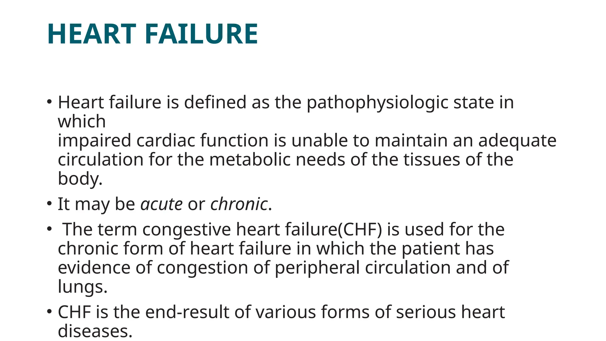
HEART FAILURE
• Heartfailure is defined as the pathophysiologic state in
which
impaired cardiac function is unable to maintain an adequate
circulation for the metabolic needs of the tissues of the
body.
• It may be acute or chronic.
• The term congestive heart failure(CHF) is used for the
chronic form of heart failure in which the patient has
evidence of congestion of peripheral circulation and of
lungs.
• CHF is the end-result of various forms of serious heart
diseases.
4.
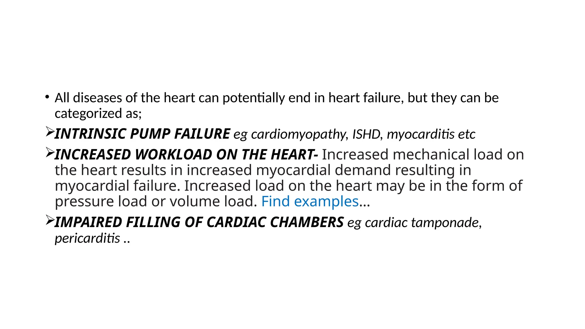
• All diseasesof the heart can potentially end in heart failure, but they can be
categorized as;
INTRINSIC PUMP FAILURE eg cardiomyopathy, ISHD, myocarditis etc
INCREASED WORKLOAD ON THE HEART- Increased mechanical load on
the heart results in increased myocardial demand resulting in
myocardial failure. Increased load on the heart may be in the form of
pressure load or volume load. Find examples…
IMPAIRED FILLING OF CARDIAC CHAMBERS eg cardiac tamponade,
pericarditis ..
5.
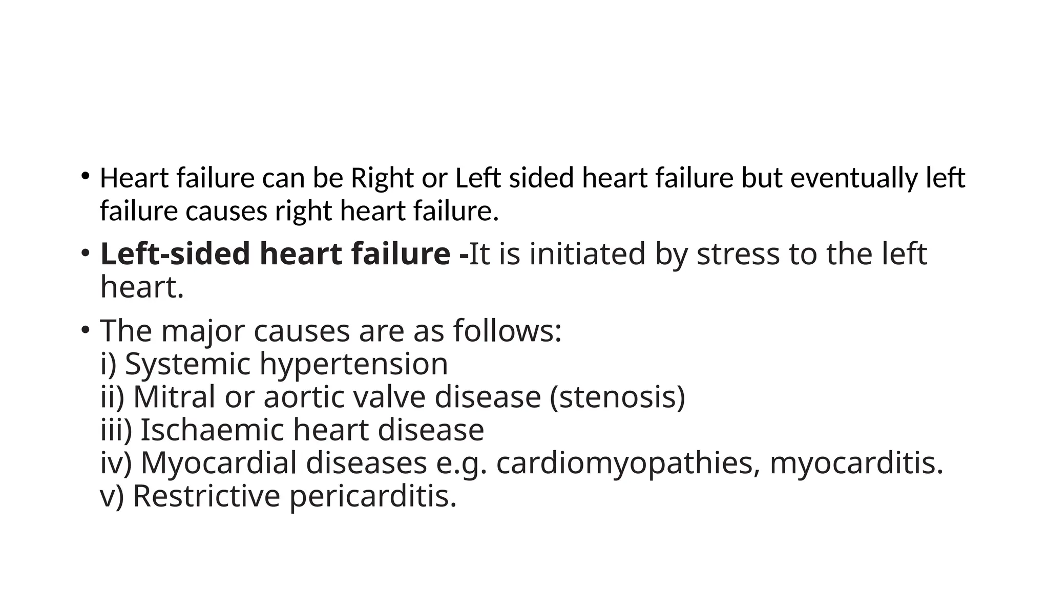
• Heart failurecan be Right or Left sided heart failure but eventually left
failure causes right heart failure.
• Left-sided heart failure -It is initiated by stress to the left
heart.
• The major causes are as follows:
i) Systemic hypertension
ii) Mitral or aortic valve disease (stenosis)
iii) Ischaemic heart disease
iv) Myocardial diseases e.g. cardiomyopathies, myocarditis.
v) Restrictive pericarditis.
6.
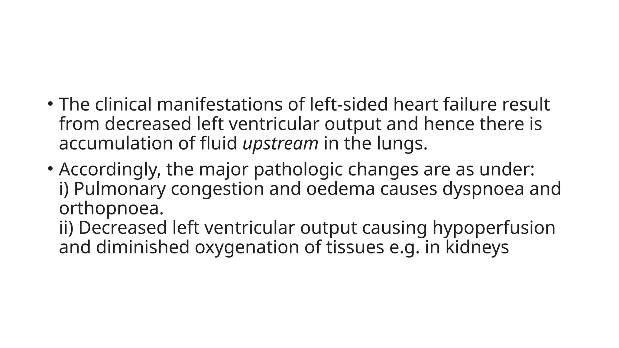
• The clinicalmanifestations of left-sided heart failure result
from decreased left ventricular output and hence there is
accumulation of fluid upstream in the lungs.
• Accordingly, the major pathologic changes are as under:
i) Pulmonary congestion and oedema causes dyspnoea and
orthopnoea.
ii) Decreased left ventricular output causing hypoperfusion
and diminished oxygenation of tissues e.g. in kidneys
7.
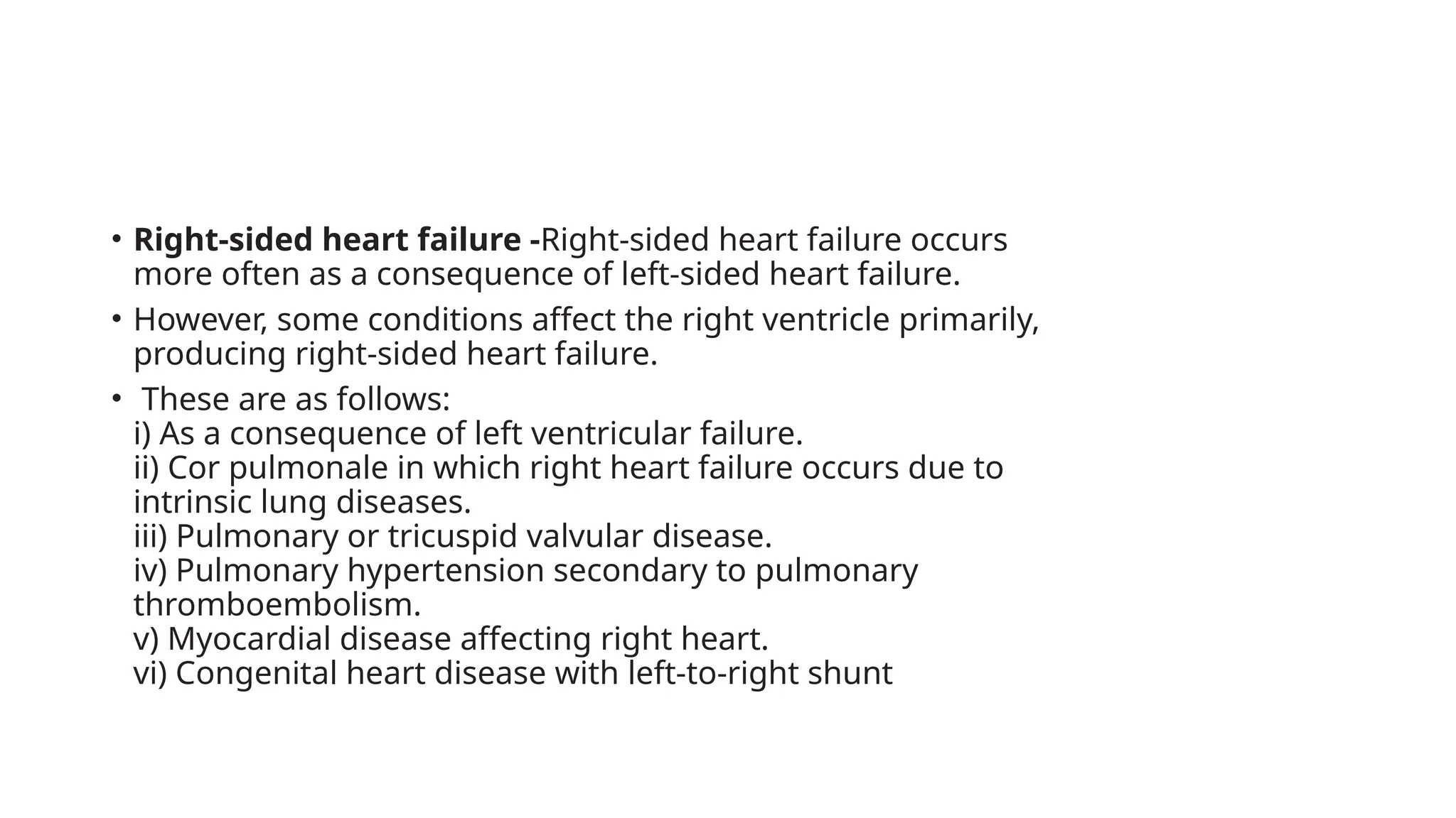
• Right-sided heartfailure -Right-sided heart failure occurs
more often as a consequence of left-sided heart failure.
• However, some conditions affect the right ventricle primarily,
producing right-sided heart failure.
• These are as follows:
i) As a consequence of left ventricular failure.
ii) Cor pulmonale in which right heart failure occurs due to
intrinsic lung diseases.
iii) Pulmonary or tricuspid valvular disease.
iv) Pulmonary hypertension secondary to pulmonary
thromboembolism.
v) Myocardial disease affecting right heart.
vi) Congenital heart disease with left-to-right shunt
8.
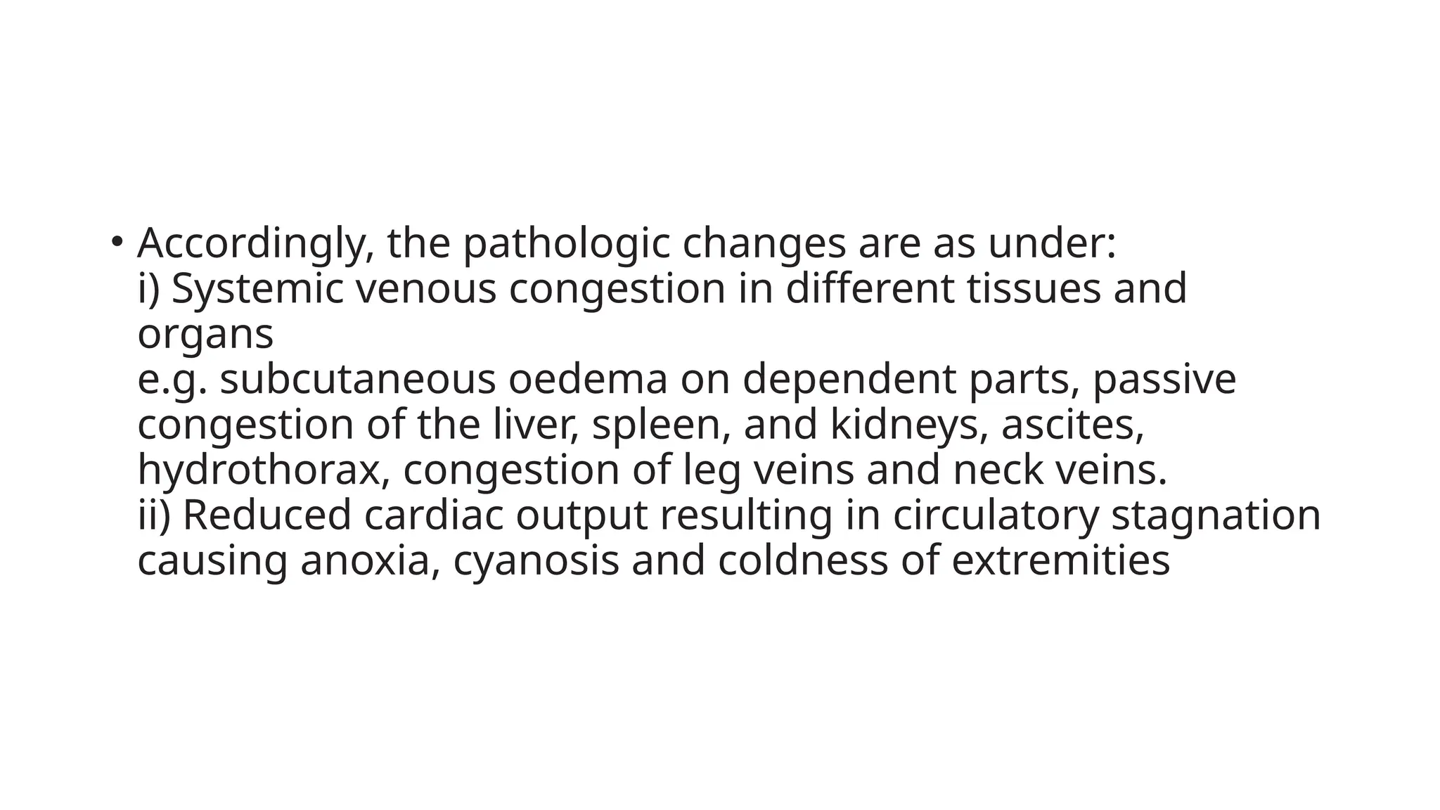
• Accordingly, thepathologic changes are as under:
i) Systemic venous congestion in different tissues and
organs
e.g. subcutaneous oedema on dependent parts, passive
congestion of the liver, spleen, and kidneys, ascites,
hydrothorax, congestion of leg veins and neck veins.
ii) Reduced cardiac output resulting in circulatory stagnation
causing anoxia, cyanosis and coldness of extremities
9.
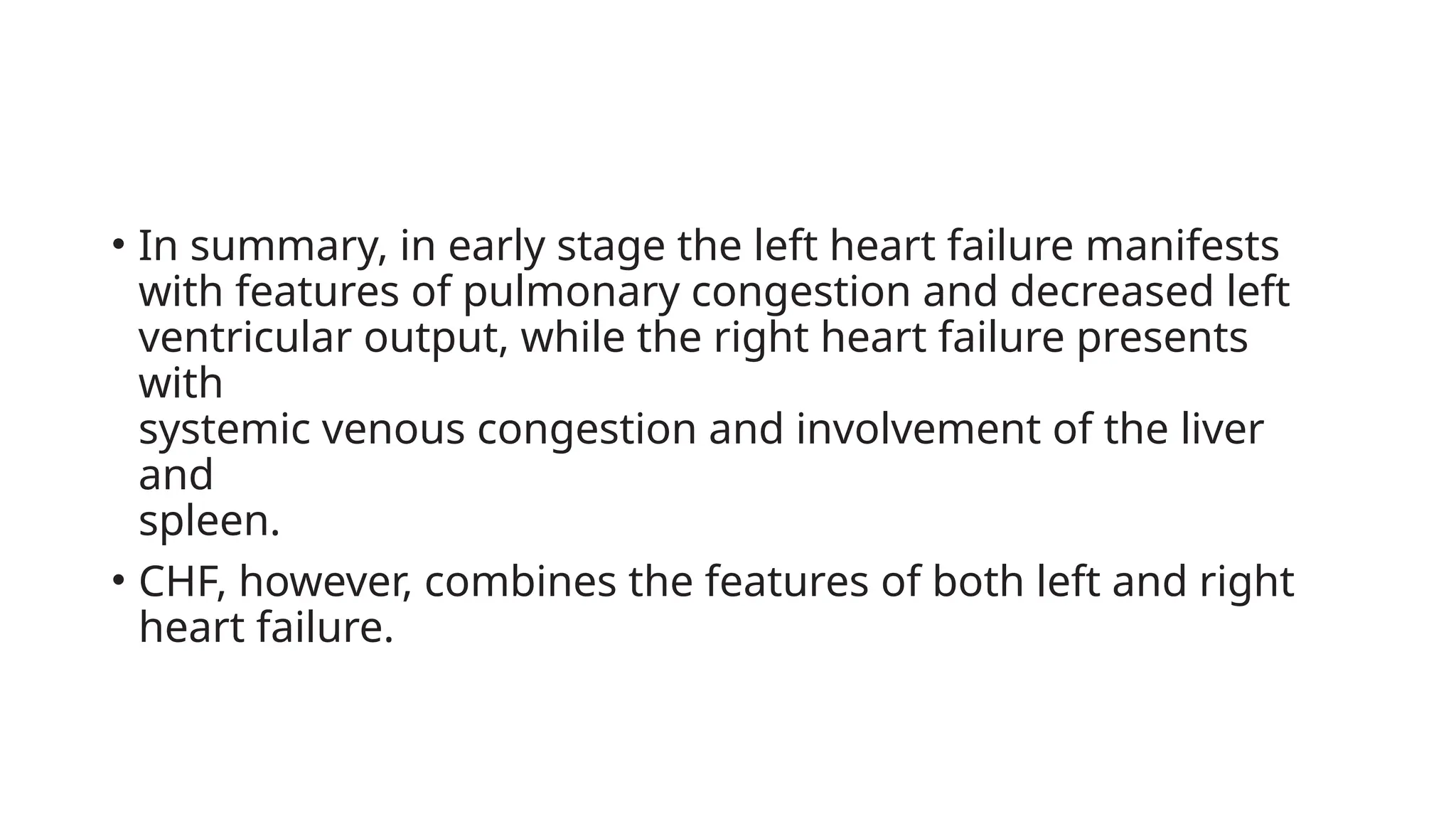
• In summary,in early stage the left heart failure manifests
with features of pulmonary congestion and decreased left
ventricular output, while the right heart failure presents
with
systemic venous congestion and involvement of the liver
and
spleen.
• CHF, however, combines the features of both left and right
heart failure.
10.
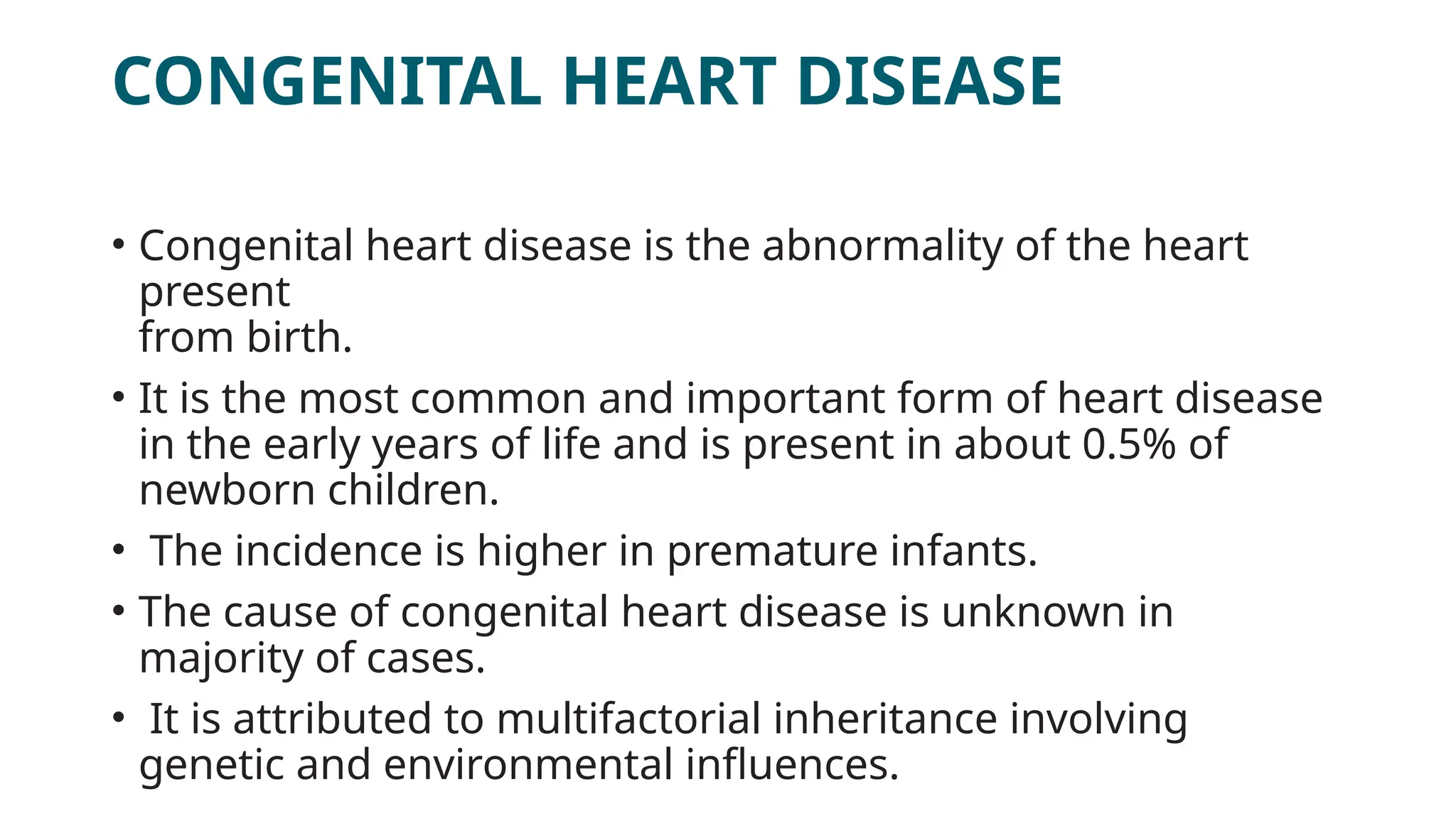
CONGENITAL HEART DISEASE
•Congenital heart disease is the abnormality of the heart
present
from birth.
• It is the most common and important form of heart disease
in the early years of life and is present in about 0.5% of
newborn children.
• The incidence is higher in premature infants.
• The cause of congenital heart disease is unknown in
majority of cases.
• It is attributed to multifactorial inheritance involving
genetic and environmental influences.
12.
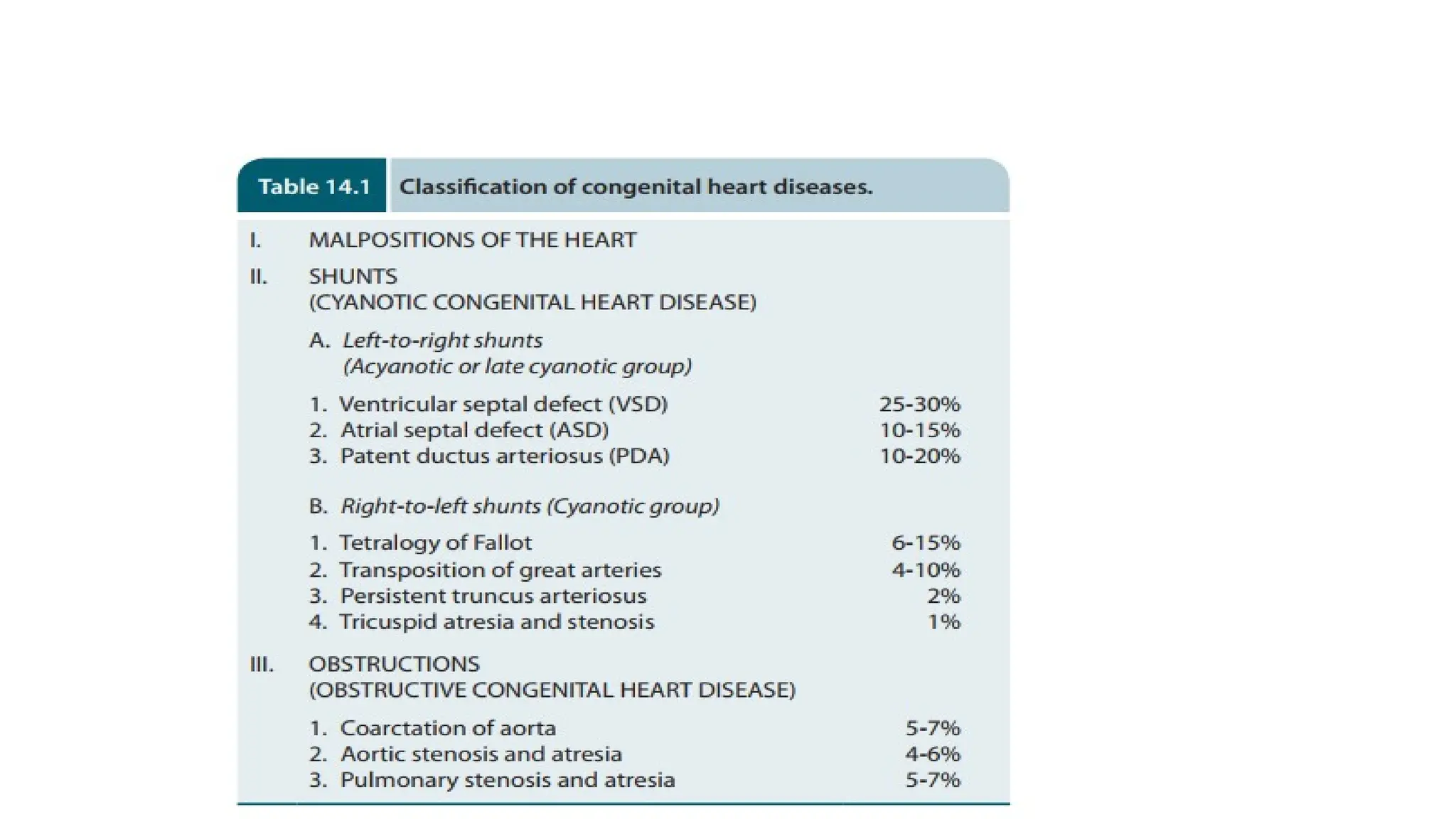
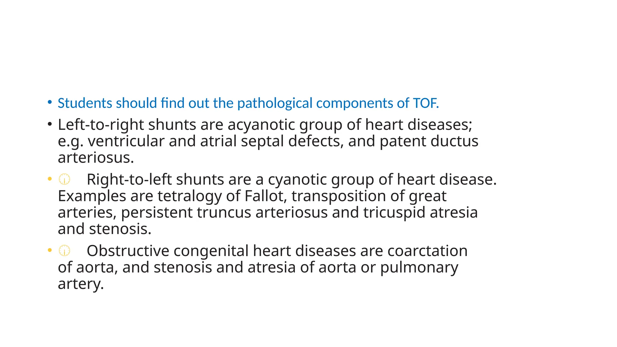
• Students shouldfind out the pathological components of TOF.
• Left-to-right shunts are acyanotic group of heart diseases;
e.g. ventricular and atrial septal defects, and patent ductus
arteriosus.
• Œ Right-to-left shunts are a cyanotic group of heart disease.
Examples are tetralogy of Fallot, transposition of great
arteries, persistent truncus arteriosus and tricuspid atresia
and stenosis.
• Œ Obstructive congenital heart diseases are coarctation
of aorta, and stenosis and atresia of aorta or pulmonary
artery.
13.
ISCHAEMIC HEART DISEASE
•Ischaemic heart disease (IHD) is defined as acute or chronic
form of cardiac disability arising from imbalance between the
myocardial supply and demand for oxygenated blood.
• Since narrowing or obstruction of the coronary arterial system is
the most common cause of myocardial anoxia, the alternate
term ‘coronary artery disease (CAD)’ is used synonymously
with IHD.
• Men develop IHD earlier than women and death rates are also
slightly higher for men than for women.
14.
• IHD isinvariably caused by disease affecting the coronary
arteries, the most prevalent being atherosclerosis accounting
for more than 90% cases, while other causes are responsible
for less than 10% cases of IHD.
• Coronary atherosclerosis resulting in ‘fixed’ obstruction is
the major cause of IHD in more than 90% cases.
• The highest incidence being in the anterior descending branch of the left
coronary (LAD), followed in decreasing frequency, by the right coronary
artery (RCA) and still less in circumflex branch of the left coronary (CXA).
15.
• Significant stenoticlesions that may produce chronic
myocardial ischaemia show more than 75% (three-fourth)
reduction in the cross-sectional area of a coronary artery or
its branch.
• The general features of atheromas of coronary arteries are
similar
to those affecting elsewhere in the body and may develop
similar complications like calcification, coronary thrombosis,
ulceration, haemorrhage, rupture and aneurysm formation.
16.
• SUPERADDED CHANGESIN CORONARY
ATHEROSCLEROSIS
• The attacks of acute coronary syndromes, which include acute
myocardial infarction, unstable angina and sudden
ischaemic
death, are precipitated by certain changes superimposed on
a pre-existing fixed coronary atheromatous plaque. Eg
thrombosis, ulceration etc..
17.
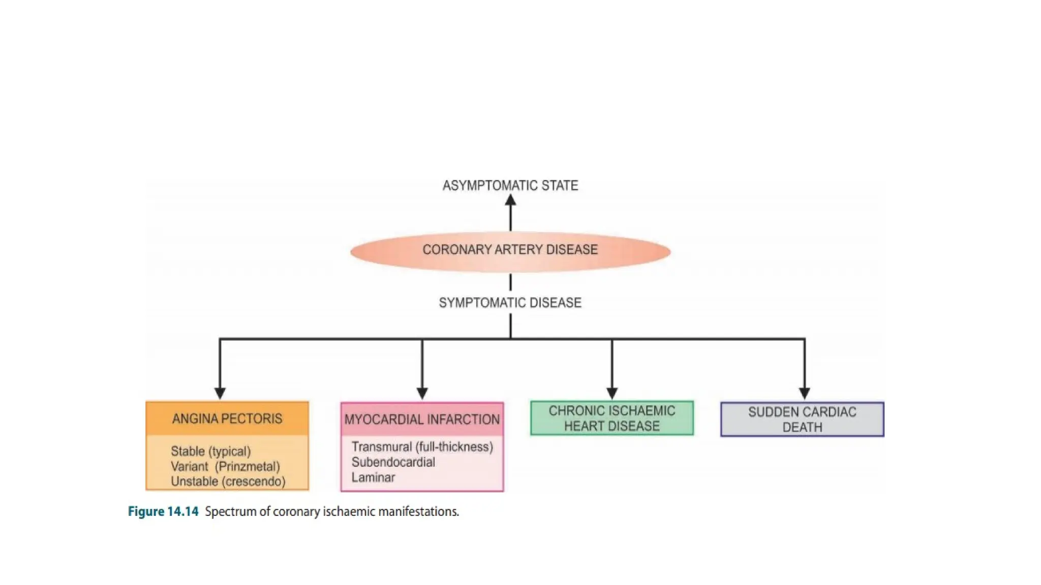
EFFECTS OF MYOCARDIALISCHAEMIA
• Development of lesions in the coronaries is not always
accompanied by cardiac disease.
• Depending upon the suddenness of onset, duration,
degree, location and extent of the area affected by
myocardial ischaemia, the range of changes and clinical
features may range from an asymptomatic state at one
extreme to immediate mortality at another.
18.
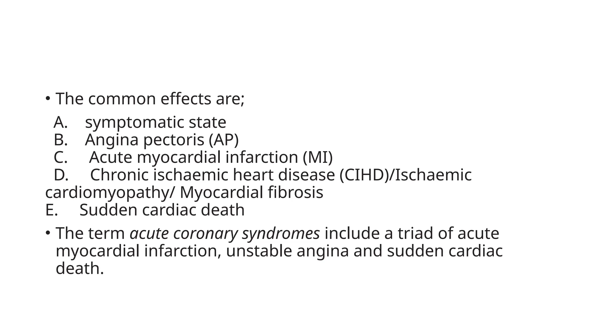
• The commoneffects are;
A. symptomatic state
B. Angina pectoris (AP)
C. Acute myocardial infarction (MI)
D. Chronic ischaemic heart disease (CIHD)/Ischaemic
cardiomyopathy/ Myocardial fibrosis
E. Sudden cardiac death
• The term acute coronary syndromes include a triad of acute
myocardial infarction, unstable angina and sudden cardiac
death.
19.
• ANGINA PECTORIS;
•Angina pectoris is a clinical syndrome of IHD resulting
from transient myocardial ischaemia.
• It is characterised by paroxysmal pain in the substernal or
precordial region of the chest which is aggravated by an
increase in the demand of the heart and relieved by a
decrease in the work of the heart .
21.
HYPERTENSIVE HEART DISEASE
•Hypertensive heart disease or hypertensive cardiomyopathy is
the disease of the heart resulting from systemic hypertension
of prolonged duration and manifesting by left ventricular
hypertrophy.
• It is the second most common form of heart disease after IHD.
• Amongst the causes of death in hypertensive patients,
cardiac decompensation leading to CHF accounts for about
one-third of the patients; other causes of death are IHD,
cerebrovascular stroke, renal failure following arteriolar
nephrosclerosis, dissecting aneurysm of the aorta and sudden
cardiac death.
22.
• Grossly, themost significant finding is marked hypertrophy
of the heart, chiefly of the left ventricle.
• The weight of the heart increases to 500 gm or more
(normal weight about 300gm).
• The thickness of the left ventricular wall increases from its
normal 13 to 15 mm up to 20 mm or more.
23.
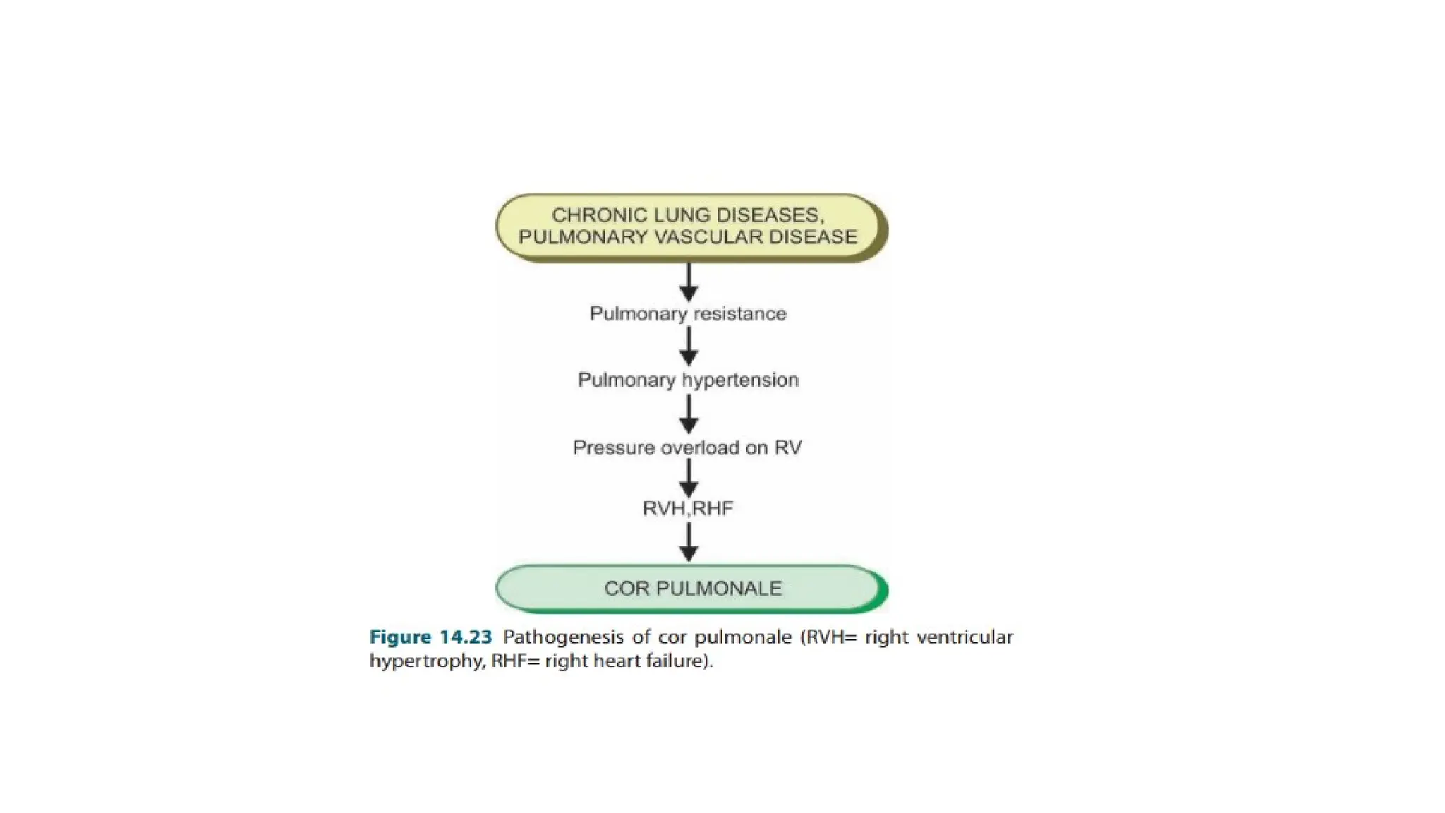
COR PULMONALE
• Corpulmonale (cor = heart; pulmonale = lung) or pulmonary
heart disease is the disease of right side of the heart
resulting from disorders of the lungs.
• It is characterised by right ventricular dilatation or
hypertrophy, or both.
• Thus, cor pulmonale is the right-sided counterpart of the
hypertensive heart disease.
24.
• Chronic lungdiseases as well as diseases of the pulmonary
vessels cause increased pulmonary vascular resistance.
• Pulmonary hypertension causes pressure overload on the
right ventricle and hence right ventricular enlargement.
• Initially, there is right ventricular hypertrophy, but as
cardiac decompensation sets in and right heart failure
ensues, dilatation of right ventricle occurs.
26.
RHEUMATIC FEVER AND
RHEUMATICHEART DISEASE
• Rheumatic fever (RF) is a systemic, post-streptococcal, nonsuppurative
inflammatory disease, principally affecting the heart, joints, central
nervous system, skin and subcutaneous tissues.
• The chronic stage of RF involves all the layers of the heart (pancarditis)
causing major cardiac sequelae referred to as rheumatic heart disease
(RHD).
• In spite of its name suggesting an acute arthritis migrating from joint to
joint, it is well known that it is the heart rather than the joints which is
first and major organ affected.
• Decades ago, William Boyd gave the dictum ‘rheumatism licks the joint, but
bites the whole heart .
27.
• The diseaseappears most commonly in children between
the age of 5 to 15 years when the streptococcal infection
is most frequent and intense.
• Both the sexes are affected equally.
• The geographic distribution, incidence and severity of RF
and RHD are generally related to the frequency and severity of
streptococcal pharyngeal infection.
• The disease is seen more commonly in poor socioeconomic strata of
the society living in damp and overcrowded places which promote
interpersonal spread of the streptococcal infection.
29.
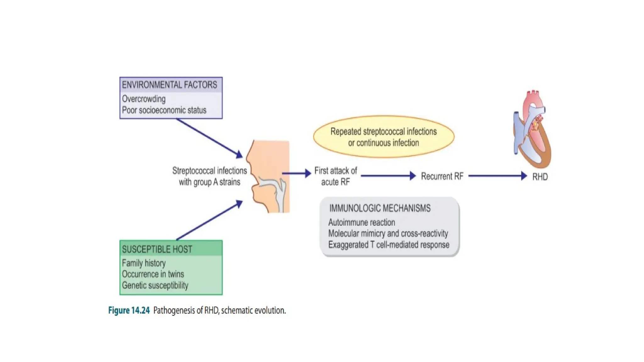
• There are3 types of factors in the etiology and pathogenesis of
RF and RHD: environmental factors, host susceptibility and
immunologic evidences.
• There is often a history of infection of the pharynx, upper
respiratory tract with this microorganism about 2 to 3 weeks
prior to the attack of RF.
• This period is usually the latent period required for sensitisation
to the bacteria.
30.
• Patients withRF have elevated titres of antibodies to the
antigens of b-haemolytic streptococci of group A such as
anti-streptolysin O (ASO) and S, anti-streptokinase,
antistreptohyaluronidase and anti-DNAase B
• Molecular mimicry and cross-reactivity between streptococcal
M protein in particular and the human molecules forms
the basis of autoimmune damage to human target tissues
in
RHD i.e. cardiac muscle, valves, joints, skin, neurons etc.
31.
• The cardiaclesions of RF in the form of pancarditis,
particularly the valvular lesions, are its major manifestations.
• However, supportive connective tissues at other sites like the
synovial membrane, periarticular tissue, skin and subcutaneous tissue,
arterial wall, lungs, pleura and the CNS are all
affected (extracardiac lesions).
• The cardiac manifestations of RF are in the form of focal
inflammatory involvement of the interstitial tissue of all
the three layers of the heart, the so-called pancarditis.
• The pathognomonic feature of pancarditis in RF is the presence
of distinctive Aschoff nodules or Aschoff bodies.
32.
• RHEUMATIC ENDOCARDITIS
•Endocardial lesions of RF may involve the valvular and mural
endocardium, causing rheumatic valvulitis and mural
endocarditis,
respectively.
• Rheumatic valvulitis is chiefly responsible for the major
cardiac manifestations in chronic RHD.
•
33.
• Grossly, thevalves in acute RF show thickening and loss of
translucency of the valve leaflets or cusps.
• This is followed by the formation of characteristic, small (1 to
3 mm in diameter), multiple, warty vegetations or verrucae,
chiefly along the line of closure of the leaflets and cusps.
34.
• The vegetationsin RF appear grey-brown, translucent and
are firmly attached so that they are not likely to get detached
to form emboli, unlike the friable vegetations of infective
endocarditis.
• Though all the four heart valves are affected, their
frequency and severity of involvement varies: mitral valve
alone being the most common site, followed in decreasing
order of frequency, by combined mitral and aortic valve.
• The tricuspid and pulmonary valves usually show infrequent and
slight involvement.
35.
• The chronicstage of RHD is characterised by permanent
deformity of one or more valves, especially the mitral (in
98% cases alone or along with other valves) and aortic.
• Mitral valve is almost always involved in RHD.
• Gross appearance of chronic healed mitral valve in RHD
is characteristically ‘fish mouth’ or ‘button hole’ stenosis.
• Mitral stenosis and insufficiency are commonly combined
in chronic RHD.
• Calcific aortic stenosis may also be found.
• These healed chronic valvular lesions in RHD occur due to
diffuse fibrocollagenous thickening and calcification of the
valve cusps or leaflets which cause adhesions.
36.
• RHEUMATIC PERICARDITIS-Inflammatory involvement of
the pericardium commonly accompanies RHD.
• Grossly, the usual finding is fibrinous pericarditis in which
there is loss of normal shiny pericardial surface due to
deposition of fibrin on its surface and accumulation of
slight amount of fibrinous exudate in the pericardial sac.
37.
• If theparietal pericardium is pulled off from the visceral pericardium, the
two separated surfaces are shaggy due to thick fibrin covering them.
• This appearance is often likened to ‘bread and butter appearance’ i.e.
resembling the
buttered surfaces of two slices in a sandwich when they are gently pulled
apart.
• If fibrinous pericarditis fails to resolve and, instead, undergoes organisation,
the two layers of the pericardium form fibrous adhesions resulting in chronic
adhesive pericarditis.
38.
• Extracardiac Lesions
•Patients of the syndrome of acute rheumatism develop lesions
in connective tissue elsewhere in the body, chiefly the joints,
subcutaneous tissue, arteries, brain and lungs.
1. POLYARTHRITIS -Acute and painful inflammation of the
synovial membranes of some of the joints, especially the larger
joints of the limbs, is seen in about 90% cases of RF in adults
and less often in children.
• As pain and swelling subside in one joint, others tend to get involved,
producing the characteristic ‘migratory polyarthritis’ involving two or
more joints at a time.
39.
INFECTIVE (BACTERIAL)
ENDOCARDITIS
• Infectiveor bacterial endocarditis (IE or BE) is
serious infection of the valvular and mural endocardium
caused
by different forms of microorganisms and is characterized
by
typical infected and friable vegetations.
• A few specific forms of IE are named by the microbial
etiologic agent causing them e.g. tubercle bacilli, fungi etc.
• Depending upon the severity of infection, BE is subdivided
into 2 clinical forms:
40.
• Acute bacterialendocarditis (ABE) is fulminant and
destructive acute infection of the endocardium by highly
virulent bacteria in a previously normal heart and almost
invariably runs a rapidly fatal course in a period of 2-6 weeks.
• Subacute bacterial endocarditis (SABE) or endocarditis
lenta (lenta = slow) is caused by less virulent bacteria in a
previously diseased heart and has a gradual downhill course
in a period of 6 weeks to a few months and sometimes years .
41.
• Infective agents-About 90% cases of BE are caused by
streptococci and staphylococci.
• In ABE, the most common causative organisms are virulent
strains of staphylococci, chiefly Staphylococcus aureus. Others
are pneumococci, gonococci, b-streptococci and enterococci.
• in SABE, the commonest causative organisms are the
streptococci with low virulence, predominantly Streptococcus
viridans, which forms part of normal flora of the mouth and
pharynx.
42.
• Predisposing factors-There are 3 main types of factors
which predispose to the development of both forms of BE:
• Conditions initiating transient bacteraemia, septicaemia
and pyaemia. e.g oral infections, septic surgical wounds,
cardiac catheterization etc…
• Underlying heart disease.
• Impaired host defenses
43.
• MORPHOLOGIC FEATURES-The characteristic pathologic
feature in both ABE and SABE is the presence of typical
vegetations or verrucae on the valve cusps or leaflets .
44.
• In theacute fulminant form of the disease, the inflammatory
cell infiltrate chiefly consists of neutrophils and is
accompanied with tissue necrosis and abscesses in the valve
rings and in the myocardium.
• In the subacute form, there is healing by granulation tissue,
mononuclear inflammatory cell infiltration and fibroblastic
proliferation.
• Histological evidence of pre-existing valvular disease such as
RHD may be present in SABE
45.
• COMPLICATIONS ANDSEQUELAE -Most cases of BE
present with fever.
• The acute form of BE is characterized by high grade fever, chills,
weakness and malaise while the subacute form of the disease has
non-specific manifestations like slight fever, fatigue, loss of weight
and flu-like symptoms.
• In the early stage, the lesions are confined to the heart, while
subsequent progression of the disease leads to involvement of
extra-cardiac organs.
• In general, severe complications develop early in ABE than in SABE.
46.
• Cardiac complications;
• These include the following:
i) Valvular stenosis or insufficiency
ii) Perforation, rupture, and aneurysm of valve leaflets
iii) Abscesses in the valve ring
iv) Myocardial abscesses
v) Suppurative pericarditis
vi) Cardiac failure from one or more of the foregoing
complications.
47.
• Extracardiac complications;
• Since the vegetations in BE are typically friable, they tend to get
dislodged due to rapid stream of blood and give rise to embolism
which is responsible for very common and serious extra-cardiac
complications.
These are as follows:
i) Emboli originating from the left side of the heart and
entering the systemic circulation affect organs like the spleen,
kidneys, and brain causing infarcts, abscesses and mycotic
aneurysms.
48.
• Emboli arisingfrom right side of the heart enter the
pulmonary circulation and produce pulmonary abscesses.
• Petechiae may be seen in the skin and conjunctiva due to
either emboli or toxic damage to the capillaries.
• In SABE, there are painful, tender nodules on the finger
tips of hands and feet called Osler’s nodes, while in ABE
there is appearance of painless, non-tender subcutaneous
maculopapular lesions on the pulp of the fingers called
Janeway’s spots.
• In either case, their origin is due to toxic or allergic inflammation of the
vessel wall.