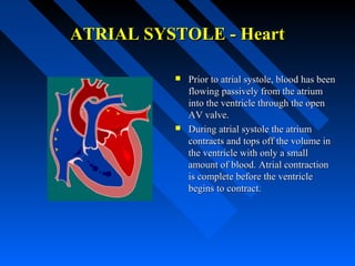
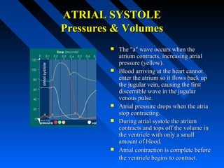
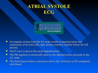
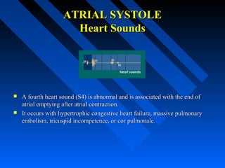
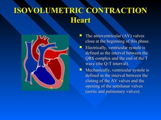
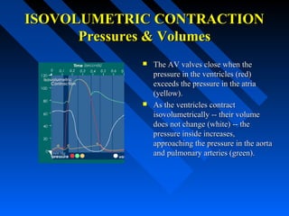
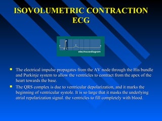
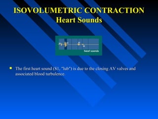
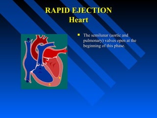
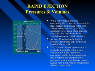
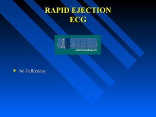
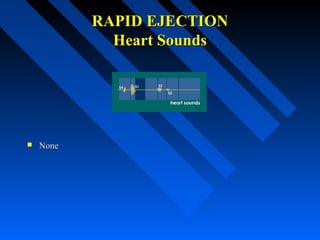
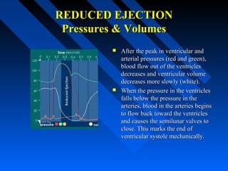
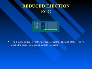
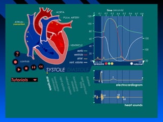
THE CARDIAC CYCLETHE CARDIAC CYCLE
Dr G Bhanu PrakashDr G Bhanu Prakash
ATRIAL SYSTOLEATRIAL SYSTOLE
The end of diastoleThe end of diastole
ATRIAL SYSTOLE - HeartATRIAL SYSTOLE - Heart
 Prior to atrial systole, blood has beenPrior to atrial systole, blood has been
flowing passively from the atriumflowing passively from the atrium
into the ventricle through the openinto the ventricle through the open
AV valve.AV valve.
 During atrial systole the atriumDuring atrial systole the atrium
contracts and tops off the volume incontracts and tops off the volume in
the ventricle with only a smallthe ventricle with only a small
amount of blood. Atrial contractionamount of blood. Atrial contraction
is complete before the ventricleis complete before the ventricle
begins to contract.begins to contract.
ATRIAL SYSTOLEATRIAL SYSTOLE
Pressures & VolumesPressures & Volumes
 The "a" wave occurs when theThe "a" wave occurs when the
atrium contracts, increasing atrialatrium contracts, increasing atrial
pressure (yellow).pressure (yellow).
 Blood arriving at the heart cannotBlood arriving at the heart cannot
enter the atrium so it flows back upenter the atrium so it flows back up
the jugular vein, causing the firstthe jugular vein, causing the first
discernible wave in the jugulardiscernible wave in the jugular
venous pulse.venous pulse.
 Atrial pressure drops when the atriaAtrial pressure drops when the atria
stop contracting.stop contracting.
 During atrial systole the atriumDuring atrial systole the atrium
contracts and tops off the volume incontracts and tops off the volume in
the ventricle with only a smallthe ventricle with only a small
amount of blood.amount of blood.
 Atrial contraction is complete beforeAtrial contraction is complete before
the ventricle begins to contract.the ventricle begins to contract.
ATRIAL SYSTOLEATRIAL SYSTOLE
ECGECG
 An impulse arising from the SA node results in depolarization andAn impulse arising from the SA node results in depolarization and
contraction of the atria (the right atrium contracts slightly before the leftcontraction of the atria (the right atrium contracts slightly before the left
atrium).atrium).
 The P wave is due to this atrial depolarization.The P wave is due to this atrial depolarization.
 The PR segment is electrically quiet as the depolarization proceeds to theThe PR segment is electrically quiet as the depolarization proceeds to the
AV node.AV node.
 This brief pause before contraction allows the ventricles to fill completelyThis brief pause before contraction allows the ventricles to fill completely
with blood.with blood.
ATRIAL SYSTOLEATRIAL SYSTOLE
Heart SoundsHeart Sounds
 A fourth heart sound (S4) is abnormal and is associated with the end ofA fourth heart sound (S4) is abnormal and is associated with the end of
atrial emptying after atrial contraction.atrial emptying after atrial contraction.
 It occurs with hypertrophic congestive heart failure, massive pulmonaryIt occurs with hypertrophic congestive heart failure, massive pulmonary
embolism, tricuspid incompetence, or cor pulmonale.embolism, tricuspid incompetence, or cor pulmonale.
ISOVOLUMETRICISOVOLUMETRIC
CONTRACTIONCONTRACTION
The Beginning of systoleThe Beginning of systole
ISOVOLUMETRIC CONTRACTIONISOVOLUMETRIC CONTRACTION
HeartHeart
 The atrioventricular (AV) valvesThe atrioventricular (AV) valves
close at the beginning of this phase.close at the beginning of this phase.
 Electrically, ventricular systole isElectrically, ventricular systole is
defined as the interval between thedefined as the interval between the
QRS complex and the end of the TQRS complex and the end of the T
wave (the Q-T interval).wave (the Q-T interval).
 Mechanically, ventricular systole isMechanically, ventricular systole is
defined as the interval between thedefined as the interval between the
closing of the AV valves and theclosing of the AV valves and the
opening of the semilunar valvesopening of the semilunar valves
(aortic and pulmonary valves).(aortic and pulmonary valves).
ISOVOLUMETRIC CONTRACTIONISOVOLUMETRIC CONTRACTION
Pressures & VolumesPressures & Volumes
 The AV valves close when theThe AV valves close when the
pressure in the ventricles (red)pressure in the ventricles (red)
exceeds the pressure in the atriaexceeds the pressure in the atria
(yellow).(yellow).
 As the ventricles contractAs the ventricles contract
isovolumetrically -- their volumeisovolumetrically -- their volume
does not change (white) -- thedoes not change (white) -- the
pressure inside increases,pressure inside increases,
approaching the pressure in the aortaapproaching the pressure in the aorta
and pulmonary arteries (green).and pulmonary arteries (green).
ISOVOLUMETRIC CONTRACTIONISOVOLUMETRIC CONTRACTION
ECGECG
 The electrical impulse propagates from the AV node through the His bundleThe electrical impulse propagates from the AV node through the His bundle
and Purkinje system to allow the ventricles to contract from the apex of theand Purkinje system to allow the ventricles to contract from the apex of the
heart towards the base.heart towards the base.
 The QRS complex is due to ventricular depolarization, and it marks theThe QRS complex is due to ventricular depolarization, and it marks the
beginning of ventricular systole. It is so large that it masks the underlyingbeginning of ventricular systole. It is so large that it masks the underlying
atrial repolarization signal. the ventricles to fill completely with blood.atrial repolarization signal. the ventricles to fill completely with blood.
ISOVOLUMETRIC CONTRACTIONISOVOLUMETRIC CONTRACTION
Heart SoundsHeart Sounds
 The first heart sound (S1, "lub") is due to the closing AV valves andThe first heart sound (S1, "lub") is due to the closing AV valves and
associated blood turbulence.associated blood turbulence.
RAPID EJECTIONRAPID EJECTION
HeartHeart
 The semilunar (aortic andThe semilunar (aortic and
pulmonary) valves open at thepulmonary) valves open at the
beginning of this phase.beginning of this phase.
RAPID EJECTIONRAPID EJECTION
Pressures & VolumesPressures & Volumes
 While the ventricles continueWhile the ventricles continue
contracting, the pressure in the ventriclescontracting, the pressure in the ventricles
(red) exceeds the pressure in the aorta(red) exceeds the pressure in the aorta
and pulmonary arteries (green); theand pulmonary arteries (green); the
semilunar valves open, blood exits thesemilunar valves open, blood exits the
ventricles, and the volume in theventricles, and the volume in the
ventricles decreases rapidly (white).ventricles decreases rapidly (white).
 As more blood enters the arteries,As more blood enters the arteries,
pressure there builds until the flow ofpressure there builds until the flow of
blood reaches a peak.blood reaches a peak.
 The "c" wave of atrial pressure is notThe "c" wave of atrial pressure is not
normally discernible in the jugularnormally discernible in the jugular
venous pulse. Right ventricularvenous pulse. Right ventricular
contraction pushes the tricuspid valvecontraction pushes the tricuspid valve
into the atrium and increases atrialinto the atrium and increases atrial
pressure, creating a small wave into thepressure, creating a small wave into the
jugular vein. It is normally simultaneousjugular vein. It is normally simultaneous
with the carotid pulse.with the carotid pulse.
RAPID EJECTIONRAPID EJECTION
ECGECG
 No DeflectionsNo Deflections
RAPID EJECTIONRAPID EJECTION
Heart SoundsHeart Sounds
 NoneNone
REDUCED EJECTIONREDUCED EJECTION
The end of systoleThe end of systole
REDUCED EJECTIONREDUCED EJECTION
HeartHeart
 At the end of this phase theAt the end of this phase the
semilunar (aortic and pulmonary)semilunar (aortic and pulmonary)
valves close.valves close.
REDUCED EJECTIONREDUCED EJECTION
Pressures & VolumesPressures & Volumes
 After the peak in ventricular andAfter the peak in ventricular and
arterial pressures (red and green),arterial pressures (red and green),
blood flow out of the ventriclesblood flow out of the ventricles
decreases and ventricular volumedecreases and ventricular volume
decreases more slowly (white).decreases more slowly (white).
 When the pressure in the ventriclesWhen the pressure in the ventricles
falls below the pressure in thefalls below the pressure in the
arteries, blood in the arteries beginsarteries, blood in the arteries begins
to flow back toward the ventriclesto flow back toward the ventricles
and causes the semilunar valves toand causes the semilunar valves to
close. This marks the end ofclose. This marks the end of
ventricular systole mechanically.ventricular systole mechanically.
REDUCED EJECTIONREDUCED EJECTION
ECGECG
 The T wave is due to ventricular repolarization. The end of the T waveThe T wave is due to ventricular repolarization. The end of the T wave
marks the end of ventricular systole electrically.marks the end of ventricular systole electrically.
REDUCED EJECTIONREDUCED EJECTION
Heart SoundsHeart Sounds
 NoneNone
ISOVOLUMETRICISOVOLUMETRIC
RELAXATIONRELAXATION
The beginning of DiastoleThe beginning of Diastole
ISOVOLUMETRIC RELAXATIONISOVOLUMETRIC RELAXATION
HeartHeart
 At the beginning of this phase theAt the beginning of this phase the
AV valves are closed.AV valves are closed.
ISOVOLUMETRIC RELAXATIONISOVOLUMETRIC RELAXATION
Pressures & VolumesPressures & Volumes
 Throughout this and the previousThroughout this and the previous
two phases, the atrium in diastole hastwo phases, the atrium in diastole has
been filling with blood on top of thebeen filling with blood on top of the
closed AV valve, causing atrialclosed AV valve, causing atrial
pressure to rise gradually (yellow).pressure to rise gradually (yellow).
 The "v" wave is due to the back flowThe "v" wave is due to the back flow
of blood after it hits the closed AVof blood after it hits the closed AV
valve. It is the second discerniblevalve. It is the second discernible
wave of the jugular venous pulse.wave of the jugular venous pulse.
 The pressure in the ventricles (red)The pressure in the ventricles (red)
continues to drop.continues to drop.
 Ventricular volume (white) is at aVentricular volume (white) is at a
minimum and is ready to be filledminimum and is ready to be filled
again with blood.again with blood.
ISOVOLUMETRIC RELAXATIONISOVOLUMETRIC RELAXATION
ECGECG
 No DeflectionsNo Deflections
ISOVOLUMETRIC RELAXATIONISOVOLUMETRIC RELAXATION
Heart SoundsHeart Sounds
 The second heart sound (S2, "dup") occurs when the semilunar (aorticThe second heart sound (S2, "dup") occurs when the semilunar (aortic
and pulmonary) valves close. S2 is normally split because the aorticand pulmonary) valves close. S2 is normally split because the aortic
valve closes slightly earlier than the pulmonary valve.valve closes slightly earlier than the pulmonary valve.
RAPID VENTRICULARRAPID VENTRICULAR
FILLINGFILLING
RAPID VENTRICULAR FILLINGRAPID VENTRICULAR FILLING
HeartHeart
 Once the AV valves open, blood thatOnce the AV valves open, blood that
has accumulated in the atria flowshas accumulated in the atria flows
rapidly into the ventricles.rapidly into the ventricles.
RAPID VENTRICULAR FILLINGRAPID VENTRICULAR FILLING
Pressures & VolumesPressures & Volumes
 Ventricular volume (white) increasesVentricular volume (white) increases
rapidly as blood flows from the atriarapidly as blood flows from the atria
into the ventricles.into the ventricles.
RAPID VENTRICULAR FILLINGRAPID VENTRICULAR FILLING
ECGECG
 No DeflectionsNo Deflections
RAPID VENTRICULAR FILLINGRAPID VENTRICULAR FILLING
Heart SoundsHeart Sounds
 A third heart sound (S3) is usually abnormal and is due to rapidA third heart sound (S3) is usually abnormal and is due to rapid
passive ventricular filling. It occurs in dilated congestive heart failure,passive ventricular filling. It occurs in dilated congestive heart failure,
severe hypertension, myocardial infarction, or mitral incompetence.severe hypertension, myocardial infarction, or mitral incompetence.
REDUCED VENTRICULARREDUCED VENTRICULAR
FILLINGFILLING
(Diastasis)(Diastasis)
REDUCED VENTRICULAR FILLINGREDUCED VENTRICULAR FILLING
HeartHeart
 Rest of blood that has accumulatedRest of blood that has accumulated
in the atria flows slowly into thein the atria flows slowly into the
ventricles.ventricles.
REDUCED VENTRICULAR FILLINGREDUCED VENTRICULAR FILLING
Pressures & VolumesPressures & Volumes
 Ventricular volume (white) increasesVentricular volume (white) increases
more slowly now. The ventriclesmore slowly now. The ventricles
continue to fill with blood until theycontinue to fill with blood until they
are nearly full.are nearly full.
REDUCED VENTRICULAR FILLINGREDUCED VENTRICULAR FILLING
ECGECG
 No DeflectionsNo Deflections
REDUCED VENTRICULAR FILLINGREDUCED VENTRICULAR FILLING
Heart SoundsHeart Sounds
 NoneNone
THE COMPLETE PICTURETHE COMPLETE PICTURE
Cardiac cycle Detailed

Cardiac cycle Detailed

  • 1.
    THE CARDIAC CYCLETHECARDIAC CYCLE Dr G Bhanu PrakashDr G Bhanu Prakash
  • 2.
    ATRIAL SYSTOLEATRIAL SYSTOLE Theend of diastoleThe end of diastole
  • 3.
    ATRIAL SYSTOLE -HeartATRIAL SYSTOLE - Heart  Prior to atrial systole, blood has beenPrior to atrial systole, blood has been flowing passively from the atriumflowing passively from the atrium into the ventricle through the openinto the ventricle through the open AV valve.AV valve.  During atrial systole the atriumDuring atrial systole the atrium contracts and tops off the volume incontracts and tops off the volume in the ventricle with only a smallthe ventricle with only a small amount of blood. Atrial contractionamount of blood. Atrial contraction is complete before the ventricleis complete before the ventricle begins to contract.begins to contract.
  • 4.
    ATRIAL SYSTOLEATRIAL SYSTOLE Pressures& VolumesPressures & Volumes  The "a" wave occurs when theThe "a" wave occurs when the atrium contracts, increasing atrialatrium contracts, increasing atrial pressure (yellow).pressure (yellow).  Blood arriving at the heart cannotBlood arriving at the heart cannot enter the atrium so it flows back upenter the atrium so it flows back up the jugular vein, causing the firstthe jugular vein, causing the first discernible wave in the jugulardiscernible wave in the jugular venous pulse.venous pulse.  Atrial pressure drops when the atriaAtrial pressure drops when the atria stop contracting.stop contracting.  During atrial systole the atriumDuring atrial systole the atrium contracts and tops off the volume incontracts and tops off the volume in the ventricle with only a smallthe ventricle with only a small amount of blood.amount of blood.  Atrial contraction is complete beforeAtrial contraction is complete before the ventricle begins to contract.the ventricle begins to contract.
  • 5.
    ATRIAL SYSTOLEATRIAL SYSTOLE ECGECG An impulse arising from the SA node results in depolarization andAn impulse arising from the SA node results in depolarization and contraction of the atria (the right atrium contracts slightly before the leftcontraction of the atria (the right atrium contracts slightly before the left atrium).atrium).  The P wave is due to this atrial depolarization.The P wave is due to this atrial depolarization.  The PR segment is electrically quiet as the depolarization proceeds to theThe PR segment is electrically quiet as the depolarization proceeds to the AV node.AV node.  This brief pause before contraction allows the ventricles to fill completelyThis brief pause before contraction allows the ventricles to fill completely with blood.with blood.
  • 6.
    ATRIAL SYSTOLEATRIAL SYSTOLE HeartSoundsHeart Sounds  A fourth heart sound (S4) is abnormal and is associated with the end ofA fourth heart sound (S4) is abnormal and is associated with the end of atrial emptying after atrial contraction.atrial emptying after atrial contraction.  It occurs with hypertrophic congestive heart failure, massive pulmonaryIt occurs with hypertrophic congestive heart failure, massive pulmonary embolism, tricuspid incompetence, or cor pulmonale.embolism, tricuspid incompetence, or cor pulmonale.
  • 7.
  • 8.
    ISOVOLUMETRIC CONTRACTIONISOVOLUMETRIC CONTRACTION HeartHeart The atrioventricular (AV) valvesThe atrioventricular (AV) valves close at the beginning of this phase.close at the beginning of this phase.  Electrically, ventricular systole isElectrically, ventricular systole is defined as the interval between thedefined as the interval between the QRS complex and the end of the TQRS complex and the end of the T wave (the Q-T interval).wave (the Q-T interval).  Mechanically, ventricular systole isMechanically, ventricular systole is defined as the interval between thedefined as the interval between the closing of the AV valves and theclosing of the AV valves and the opening of the semilunar valvesopening of the semilunar valves (aortic and pulmonary valves).(aortic and pulmonary valves).
  • 9.
    ISOVOLUMETRIC CONTRACTIONISOVOLUMETRIC CONTRACTION Pressures& VolumesPressures & Volumes  The AV valves close when theThe AV valves close when the pressure in the ventricles (red)pressure in the ventricles (red) exceeds the pressure in the atriaexceeds the pressure in the atria (yellow).(yellow).  As the ventricles contractAs the ventricles contract isovolumetrically -- their volumeisovolumetrically -- their volume does not change (white) -- thedoes not change (white) -- the pressure inside increases,pressure inside increases, approaching the pressure in the aortaapproaching the pressure in the aorta and pulmonary arteries (green).and pulmonary arteries (green).
  • 10.
    ISOVOLUMETRIC CONTRACTIONISOVOLUMETRIC CONTRACTION ECGECG The electrical impulse propagates from the AV node through the His bundleThe electrical impulse propagates from the AV node through the His bundle and Purkinje system to allow the ventricles to contract from the apex of theand Purkinje system to allow the ventricles to contract from the apex of the heart towards the base.heart towards the base.  The QRS complex is due to ventricular depolarization, and it marks theThe QRS complex is due to ventricular depolarization, and it marks the beginning of ventricular systole. It is so large that it masks the underlyingbeginning of ventricular systole. It is so large that it masks the underlying atrial repolarization signal. the ventricles to fill completely with blood.atrial repolarization signal. the ventricles to fill completely with blood.
  • 11.
    ISOVOLUMETRIC CONTRACTIONISOVOLUMETRIC CONTRACTION HeartSoundsHeart Sounds  The first heart sound (S1, "lub") is due to the closing AV valves andThe first heart sound (S1, "lub") is due to the closing AV valves and associated blood turbulence.associated blood turbulence.
  • 12.
  • 13.
    RAPID EJECTIONRAPID EJECTION HeartHeart The semilunar (aortic andThe semilunar (aortic and pulmonary) valves open at thepulmonary) valves open at the beginning of this phase.beginning of this phase.
  • 14.
    RAPID EJECTIONRAPID EJECTION Pressures& VolumesPressures & Volumes  While the ventricles continueWhile the ventricles continue contracting, the pressure in the ventriclescontracting, the pressure in the ventricles (red) exceeds the pressure in the aorta(red) exceeds the pressure in the aorta and pulmonary arteries (green); theand pulmonary arteries (green); the semilunar valves open, blood exits thesemilunar valves open, blood exits the ventricles, and the volume in theventricles, and the volume in the ventricles decreases rapidly (white).ventricles decreases rapidly (white).  As more blood enters the arteries,As more blood enters the arteries, pressure there builds until the flow ofpressure there builds until the flow of blood reaches a peak.blood reaches a peak.  The "c" wave of atrial pressure is notThe "c" wave of atrial pressure is not normally discernible in the jugularnormally discernible in the jugular venous pulse. Right ventricularvenous pulse. Right ventricular contraction pushes the tricuspid valvecontraction pushes the tricuspid valve into the atrium and increases atrialinto the atrium and increases atrial pressure, creating a small wave into thepressure, creating a small wave into the jugular vein. It is normally simultaneousjugular vein. It is normally simultaneous with the carotid pulse.with the carotid pulse.
  • 15.
    RAPID EJECTIONRAPID EJECTION ECGECG No DeflectionsNo Deflections
  • 16.
    RAPID EJECTIONRAPID EJECTION HeartSoundsHeart Sounds  NoneNone
  • 17.
    REDUCED EJECTIONREDUCED EJECTION Theend of systoleThe end of systole
  • 18.
    REDUCED EJECTIONREDUCED EJECTION HeartHeart At the end of this phase theAt the end of this phase the semilunar (aortic and pulmonary)semilunar (aortic and pulmonary) valves close.valves close.
  • 19.
    REDUCED EJECTIONREDUCED EJECTION Pressures& VolumesPressures & Volumes  After the peak in ventricular andAfter the peak in ventricular and arterial pressures (red and green),arterial pressures (red and green), blood flow out of the ventriclesblood flow out of the ventricles decreases and ventricular volumedecreases and ventricular volume decreases more slowly (white).decreases more slowly (white).  When the pressure in the ventriclesWhen the pressure in the ventricles falls below the pressure in thefalls below the pressure in the arteries, blood in the arteries beginsarteries, blood in the arteries begins to flow back toward the ventriclesto flow back toward the ventricles and causes the semilunar valves toand causes the semilunar valves to close. This marks the end ofclose. This marks the end of ventricular systole mechanically.ventricular systole mechanically.
  • 20.
    REDUCED EJECTIONREDUCED EJECTION ECGECG The T wave is due to ventricular repolarization. The end of the T waveThe T wave is due to ventricular repolarization. The end of the T wave marks the end of ventricular systole electrically.marks the end of ventricular systole electrically.
  • 21.
    REDUCED EJECTIONREDUCED EJECTION HeartSoundsHeart Sounds  NoneNone
  • 22.
  • 23.
    ISOVOLUMETRIC RELAXATIONISOVOLUMETRIC RELAXATION HeartHeart At the beginning of this phase theAt the beginning of this phase the AV valves are closed.AV valves are closed.
  • 24.
    ISOVOLUMETRIC RELAXATIONISOVOLUMETRIC RELAXATION Pressures& VolumesPressures & Volumes  Throughout this and the previousThroughout this and the previous two phases, the atrium in diastole hastwo phases, the atrium in diastole has been filling with blood on top of thebeen filling with blood on top of the closed AV valve, causing atrialclosed AV valve, causing atrial pressure to rise gradually (yellow).pressure to rise gradually (yellow).  The "v" wave is due to the back flowThe "v" wave is due to the back flow of blood after it hits the closed AVof blood after it hits the closed AV valve. It is the second discerniblevalve. It is the second discernible wave of the jugular venous pulse.wave of the jugular venous pulse.  The pressure in the ventricles (red)The pressure in the ventricles (red) continues to drop.continues to drop.  Ventricular volume (white) is at aVentricular volume (white) is at a minimum and is ready to be filledminimum and is ready to be filled again with blood.again with blood.
  • 25.
  • 26.
    ISOVOLUMETRIC RELAXATIONISOVOLUMETRIC RELAXATION HeartSoundsHeart Sounds  The second heart sound (S2, "dup") occurs when the semilunar (aorticThe second heart sound (S2, "dup") occurs when the semilunar (aortic and pulmonary) valves close. S2 is normally split because the aorticand pulmonary) valves close. S2 is normally split because the aortic valve closes slightly earlier than the pulmonary valve.valve closes slightly earlier than the pulmonary valve.
  • 27.
  • 28.
    RAPID VENTRICULAR FILLINGRAPIDVENTRICULAR FILLING HeartHeart  Once the AV valves open, blood thatOnce the AV valves open, blood that has accumulated in the atria flowshas accumulated in the atria flows rapidly into the ventricles.rapidly into the ventricles.
  • 29.
    RAPID VENTRICULAR FILLINGRAPIDVENTRICULAR FILLING Pressures & VolumesPressures & Volumes  Ventricular volume (white) increasesVentricular volume (white) increases rapidly as blood flows from the atriarapidly as blood flows from the atria into the ventricles.into the ventricles.
  • 30.
    RAPID VENTRICULAR FILLINGRAPIDVENTRICULAR FILLING ECGECG  No DeflectionsNo Deflections
  • 31.
    RAPID VENTRICULAR FILLINGRAPIDVENTRICULAR FILLING Heart SoundsHeart Sounds  A third heart sound (S3) is usually abnormal and is due to rapidA third heart sound (S3) is usually abnormal and is due to rapid passive ventricular filling. It occurs in dilated congestive heart failure,passive ventricular filling. It occurs in dilated congestive heart failure, severe hypertension, myocardial infarction, or mitral incompetence.severe hypertension, myocardial infarction, or mitral incompetence.
  • 32.
  • 33.
    REDUCED VENTRICULAR FILLINGREDUCEDVENTRICULAR FILLING HeartHeart  Rest of blood that has accumulatedRest of blood that has accumulated in the atria flows slowly into thein the atria flows slowly into the ventricles.ventricles.
  • 34.
    REDUCED VENTRICULAR FILLINGREDUCEDVENTRICULAR FILLING Pressures & VolumesPressures & Volumes  Ventricular volume (white) increasesVentricular volume (white) increases more slowly now. The ventriclesmore slowly now. The ventricles continue to fill with blood until theycontinue to fill with blood until they are nearly full.are nearly full.
  • 35.
    REDUCED VENTRICULAR FILLINGREDUCEDVENTRICULAR FILLING ECGECG  No DeflectionsNo Deflections
  • 36.
    REDUCED VENTRICULAR FILLINGREDUCEDVENTRICULAR FILLING Heart SoundsHeart Sounds  NoneNone
  • 37.
    THE COMPLETE PICTURETHECOMPLETE PICTURE